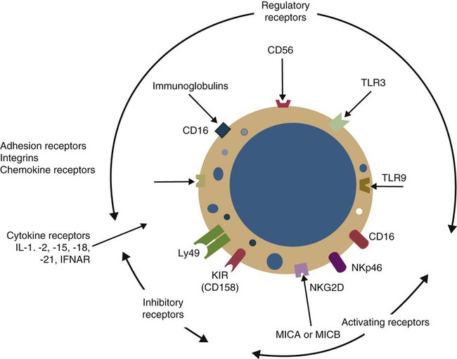
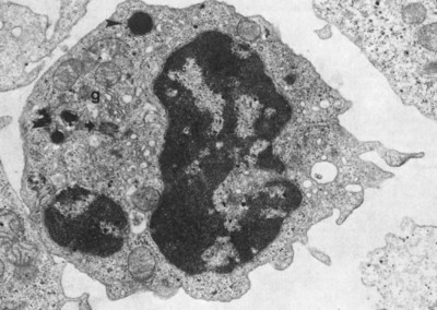
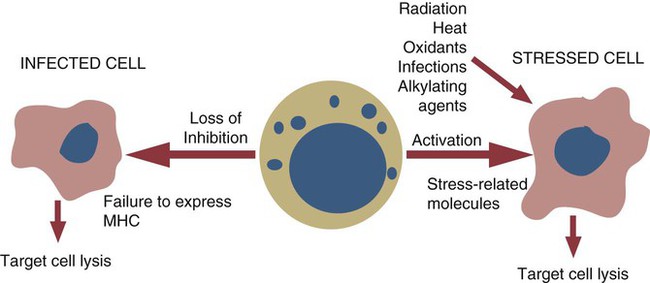
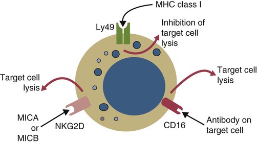
• There exists a third major population of lymphocytes, distinct from B cells and T cells, that plays a key role in the innate immune responses to some infections and cancers. These lymphocytes are called natural killer (NK) cells. • NK cells can kill virus-infected target cells, tumor cells, stressed cells, and some bacteria without prior activation. • NK cells act as a first line of defense against pathogens such as viruses. • In some species, NK cells are large granular lymphocytes. • NK cells use several different types of receptors to recognize their targets. • NK cells employ the “missing-self” recognition strategy. That is, they recognize and attack target cells that fail to express MHC class I molecules. • NK cells also recognize and attack target cells that express certain stress-associated molecules. • The most important mechanism of destruction of spontaneous tumors probably involves killing by NK cells. • Natural killer T (NKT) cells are a population of cells that, as their name suggests, share properties with both NK and T cells. Most lymphocytes participate in adaptive immune responses. These cells, the T and B cells, are responsible for cell-mediated and antibody-mediated immunity, respectively (Chapters 18 and 15). There is, however, a third major population of lymphocytes called natural killer (or NK) cells. NK cells constitute an important subsystem engaged in innate immunity. As such they serve as a first line of defense against pathogens such as viruses, some bacteria, and parasites. They eliminate stressed or damaged cells and play a major role in immunity to tumors. In most mammals, NK cells are large, granular, nonphagocytic lymphocytes (Figure 19-1). In cattle, NK cells are large cells, although they may not contain large intracytoplasmic granules. There is debate about NK cell morphology in the pig. Some investigators claim that they are large granular lymphocytes, whereas others believe that they are small lymphocytes without obvious cytoplasmic granules (Box 19-1). NK cells recognize and kill abnormal cells using totally different mechanisms than do T or B cells. T and B cells use a strategy requiring the recognition of new and foreign antigens. NK cells, in contrast, employ two distinct strategies. One is a “missing-self” strategy. MHC class I molecules expressed on the surface of healthy normal cells can block NK cell killing by sending inhibitory signals. Thus normal cells are not killed. If, however, even a single MHC class I allele is missing, these inhibitory signals are no longer generated, and the target cells are killed. Viruses may suppress MHC class I expression in an attempt to hide from cytotoxic T cells, and tumor cells often fail to express MHC class I. Such cells are prime targets for NK cell attack. Their second strategy involves the use of activating receptors that can recognize that cells are in distress by the presence of stress-induced proteins on their surfaces. By binding to these proteins, NK cells receive signals that cause them to kill their targets (Figure 19-2). There are three families of these NK cell receptors: the killer cell immunoglobulin (Ig)-like receptor (KIR or CD158) proteins classically expressed in primates, two families of C-type lectin receptors—Ly49 primarily expressed in rodents, and NKG2D receptors expressed in both rodents and primates. All three receptor families contain both inhibitory and activating receptors (Figure 19-3). NK cells also express CD2, CD16 (FcγRIII), CD178 (CD95L or Fas ligand), CD40L (CD154), toll-like receptors (TLR3 and TLR9), and leukocyte function-associated antigen-1 (LFA-1) (Figure 19-4). NK cells do not express conventional rearranged V-region antigen receptors such as the B cell antigen receptors (BCRs) or T cell antigen receptors (TCRs), nor do they express a CD3 complex. The third family of MHC-binding receptors on NK cells are activating molecules belong to the NKG2 receptor system. The NKG2 proteins are also C-type lectins. NKG2D, found on all NK cells, recognizes nonclassical MHC class I proteins produced by stressed cells. Two of the most important of these ligands are polymorphic MHC class I–like molecules called MICA (major histocompatibility complex, class I chain-related A) and MICB coded for by MHC class Ic genes (Chapter 11). Unlike normal class I molecules, these are not associated with antigenic peptides. In addition, they have limited tissue distribution and are minimally expressed on normal, healthy cells but are expressed in large amounts on stressed cells. Stresses may include DNA damage due to ionizing radiation or alkylating agents, heat shock, and oxidative stress (see Figure 19-2). MICA and MICB are especially overexpressed in tumor cells and virus-infected cells. When these ligands are engaged, NKG2D overrides the inhibitory effects of conventional MHC class I molecules and triggers NK cytotoxicity. NKG2D is also expressed on activated γ/δ and α/β T cells, suggesting that they too may have a role in innate immunity. It may be that on surfaces, the combination of γ/δ T cells and NK cells kills tumors, whereas within the body, a combination of α/β T cells and NK cells is most effective.
The Third Lymphocyte Population
Natural Killer Cells
Natural Killer Cells
Morphology
Target Cell Recognition

Receptors
KIR receptors
NKG2 receptors
![]()
Stay updated, free articles. Join our Telegram channel

Full access? Get Clinical Tree


The Third Lymphocyte Population: Natural Killer Cells
Only gold members can continue reading. Log In or Register to continue


