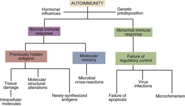
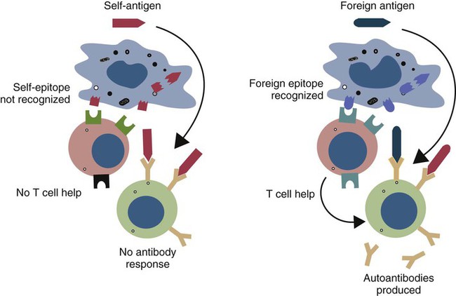
Autoimmunity: General Principles


Autoimmunity


General Principles


Autoimmune diseases are relatively common. They occur in about 5% of humans and probably in a similar proportion of domestic mammals. Most represent the emergence of clones of “rogue” lymphocytes that are directed against normal body components. The fact that they tend to develop late in life suggests that they, like cancer, probably represent the result of multiple random mutations. Thus while one mutation alone may be insufficient to permit an autoimmune response, multiple such mutations may eventually permit self-reactive lymphocytes to develop. Lymphocytes, the key cells of the immune system, are triggered to proliferate by antigens. Given the ubiquity of microorganisms and environmental antigens, lymphocytes are under constant pressure to proliferate. The presence of a constant supply of self-antigens is especially significant. Much of the complexity of the immune system is determined by the need to keep this constant lymphocyte growth in check.


The suppression of lymphocyte growth is managed by multiple mechanisms. These include negative selection within the thymus, the requirement for multiple costimulatory signals, lymphocyte cooperation, and the activities of regulatory cell populations. These regulatory mechanisms often overlap so that the development of self-reactive rogue clones does not occur suddenly. It likely takes multiple accumulated mutations, leading to subtle changes in regulatory pathways, and the loss of control of lymphocyte proliferation. Similar considerations apply to the development of lymphoid tumors. It would be very unusual for autoimmunity to develop as a result of a single mutation in a key molecule.


An inescapable hazard associated with adaptive immunity is the development of autoimmunity. By developing a defense system that can recognize any possible microbial antigenic determinant, vertebrates also developed the potential for self-destruction. The random generation of antigen-binding receptors ensures that many lymphocytes are produced that can bind and respond to self-antigens. It has been estimated that 20% to 50% of T cell receptors (TCRs) and B cell receptors (BCRs) generated in this way will bind to self-antigens with high affinity. These self-reactive cells are usually rigorously suppressed so that only a few animals develop autoimmune disease. However, the reasons why these individuals develop autoimmune diseases are still unclear. Many factors influence susceptibility to autoimmunity. These include sex and age, genetic background, and virus infections. We also know that the development of autoantibodies is a relatively common event that by itself does not inevitably lead to autoimmune disease. Indeed, some autoantibodies serve a physiological function.


Because we do not know precisely what causes autoimmune disease, this chapter reviews some of the many different predisposing factors that have been identified or proposed as well as the mechanisms by which autoimmunity causes tissue damage and disease. As with other immune functions, both B and T cells can mediate autoimmunity. Thus in some autoimmune diseases, the disease is mediated by autoantibodies alone. In others, the damage may be mediated by T cells alone or by some combination of autoantibodies and T cells.


Induction of Autoimmunity


Autoimmune diseases appear to develop spontaneously, and predisposing causes are rarely obvious. Nevertheless they fall into two major categories: they can result from a normal immune response to an unusual or abnormal antigen, or they can result from an abnormal immune response to a normal antigen (Figure 34-1). The second category is probably the most common. In these cases, the mechanisms that normally prevent the development of self-responsive T and B cells fail. Many different environmental factors and genes contribute to this failure, and the failure may not always be complete. Autoimmune diseases may result from an aberrant response to a single specific antigen; alternatively, they may be due to a general defect in the regulation of B or T cell functions.


Normal Immune Responses


Many autoimmune responses simply reflect a normal immune response to an antigen that has been previously hidden or are a result of cross-reactivity between an infectious agent and a normal body component. Many naturally occurring autoantibodies play a role in homeostasis and regulation. They are usually low-titer, low-affinity immunoglobulin M (IgM) or IgG antibodies directed against protein fragments, or proteins damaged by oxidation or enzymes.


Antigens Hidden in Cells or Tissues (Cryptic Antigens)


Many autoimmune responses are triggered when nontolerant T cells meet previously hidden autoantigens. After all, T cells can only be made tolerant to autoantigens if the T cells are first exposed to these antigens. There are many autoantigens that do not induce tolerance because they remain hidden within cells or tissues.


Although the control of the immune system requires that most self-reactive cells be eliminated, one should not assume that all autoimmune responses are bad or even cause disease. Indeed, some autoimmune responses have physiological functions. For example, red blood cells must be removed from the blood once they reach the end of their life span. This process is accomplished by autoantibodies. As red cells age, an anion transport protein called CD233 (or band 3 protein) is gradually oxidized, and a new epitope is generated. This new epitope is recognized by IgG autoantibodies. These autoantibodies bind to aged red cells and trigger their phagocytosis by splenic macrophages. CD233 is found on many cell types, and it may be that its exposure on aged cells and their subsequent opsonization is a major elimination pathway.


Many autoantigens are found in places where they never encounter circulating lymphocytes. For example, in the testes, new antigens may appear only at puberty—long after the T cell system has developed and become tolerant to autoantigens. Injury to the testes may permit proteins from damaged tissues to reach the bloodstream, encounter antigen-sensitive cells, and stimulate autoimmunity. Hidden antigens may also be found within cells. For example, after a heart attack, autoantibodies may be produced against the mitochondria of cardiac muscle cells. In chronic hepatitis in dogs, animals develop antibodies to liver membrane proteins. In diseases such as trypanosomiasis or tuberculosis in which widespread tissue damage occurs, autoantibodies to many different tissue antigens may be detected in serum.


Antigens Generated by Molecular Changes


The production of some autoantibodies may be triggered by the development of completely new epitopes on normal proteins. Two examples of autoantibodies generated in this way are the rheumatoid factors (RFs) and the immunoconglutinins (IKs, after the German spelling).


RFs are autoantibodies directed against other immunoglobulins. When an antibody binds to an antigen, the shape of the immunoglobulin molecule changes in such a way that new epitopes are exposed on its Fc region. These new epitopes may stimulate RF formation. RFs are produced in diseases in which large amounts of immune complexes are generated. These include the autoimmune disease of joints called rheumatoid arthritis and a disease called systemic lupus erythematosus (SLE), in which B cells respond to many different autoantigens.


IKs are autoantibodies directed against the complement components C2, C4, and especially C3. The epitopes that stimulate IK formation are exposed when these complement components are activated. The level of IKs in serum reflects the amount of complement activation; this, in turn, is a measure of the antigenic stimulation to which an animal is subjected. IK levels are thus nonspecific indicators of the prevalence of infectious disease within an animal population. Their physiological role is unclear, but they may enhance complement-mediated opsonization.


Receptor Editing


Both B and T cell antigen receptors are generated by random gene rearrangement. This process inevitably results in the generation of both nonfunctional and autoreactive antigen receptors. Once a complete antigen receptor is formed, however, rearrangement of the receptor gene segments continues. Thus if an immature B cell produces a receptor that binds to a self-antigen, the continuing development of that B cell is blocked while its light chain receptor chains continue to undergo recombination. This is an active process driven by the autoantigen. This replacement of one light chain by another leads to changes in receptor specificity and eventually makes the cells no longer autoreactive. Receptor editing only occurs in immature B cells. Mature B cells that bind autoantigens do not undergo receptor editing but rather are triggered to undergo apoptosis.


Abnormal Immune Responses


Failure of Regulatory Control


Although autoimmunity may be triggered by hidden epitopes, a sustained autoimmune response is necessary for disease to develop. This may result from a failure of the normal control mechanisms of the immune system and can be demonstrated simply by injecting mice with rat red blood cells. Following such an injection, mice not only make antibodies to the rat cells but also develop a self-limited and transient autoimmune response to their own red blood cells. This autoimmune response is rapidly controlled by regulatory cells and lasts for only a few days. If, however, regulatory cell activity in these mice is impaired, as occurs in New Zealand Black (NZB) mice, for example, these autoantibodies will persist to cause red blood cell destruction and anemia.


It is common to find autoimmune diseases associated with lymphoid tumors. For example, myasthenia gravis, an autoimmune disease involving the neuromuscular junction, is commonly associated with the presence of a thymic carcinoma. In humans, there is a fourfold increase in the incidence of rheumatoid arthritis in patients with malignant lymphoid tumors, and there is evidence for a similar association in other mammals. Since many lymphoid tumors result from a failure in immunological control mechanisms, a simultaneous failure in self-tolerance may also occur. Alternatively, some tumors may represent the development of a forbidden clone of cells producing autoantibodies. It is also possible that some lymphoid tumors may develop as a result of prolonged stimulation of the immune system by autoantigens.


Potentially harmful, self-reactive lymphocytes are normally destroyed in the thymus by apoptosis triggered through CD95 (Fas) (Chapter 18). Defects in CD95 or its ligand CD154 (CD95L) cause autoimmunity by permitting abnormal T cells to survive and cause disease. This is well demonstrated in the lpr strain of mice. These animals have a mutation that alters the structure of the intracellular domain of CD95 and blocks its functioning. A mutation (called gld) in CD95L has a similar effect. Both lpr and gld mice develop multiple autoimmune lesions accompanied by lymphoproliferation. Some investigators have suggested that mutations in CD95 may contribute to the pathogenesis of lupus in other mammals. The AIRE (autoimmune regulator) gene permits multiple self-antigens to be expressed in thymic epithelial cells. T cells that respond to these self-antigens are destroyed. Thus humans with a defective AIRE gene develop autoimmunity against multiple endocrine organs, the skin, and other tissues.


Infection-Induced Autoimmunity


Autoimmune diseases are triggered by many environmental factors, and infectious agents are among the most important. Given, however, that infections are very common and autoimmune diseases fairly rare, they clearly cannot account for the entire autoimmune process. For example, mice infected with certain reoviruses develop an autoimmune polyendocrine disease characterized by diabetes mellitus and retarded growth. These reovirus-infected mice make autoantibodies against normal pituitary, pancreas, gastric mucosa, nuclei, glucagon, growth hormone, and insulin. Likewise, in NZB mice, persistent infection with a type C retrovirus leads to the production of autoantibodies against nucleic acids and red blood cells. Bacteria such as Streptococcus pyogenes, Borrelia burgdorferi, and Leptospira interrogans may trigger autoimmune heart disease, arthritis, and uveitis, respectively. The protozoan parasite Trypanosoma cruzi triggers an autoimmune cardiomyopathy.


The situation with spontaneous autoimmune disease is less clear. Many attempts have been made to isolate viruses from patients with autoimmune disease but with mixed results. For example, SLE of dogs and humans has been associated with either a type C retrovirus or paramyxovirus infection. Small quantities of the Epstein-Barr virus genome can be found in the salivary glands of humans with Sjögren’s syndrome. Moreover, epidemiological evidence points to some form of a viral trigger for diseases such as multiple sclerosis, rheumatoid arthritis, and insulin-dependent diabetes mellitus in children.


Just how viruses can induce autoimmunity is unclear, but three major mechanisms are recognized; molecular mimicry, epitope spreading, and bystander activation.


Molecular Mimicry


Autoimmunity may result from molecular mimicry, a term used to describe the sharing of epitopes between an infectious agent or parasite and an autoantigen (Figure 34-2). B cells may be triggered by a foreign epitope that cross-reacts with an autoantigen. However, they will only respond to this epitope if they receive T cell help. If nearby Th cells also recognize these microbial epitopes as foreign, they may trigger a response that permits the self-reactive B cells to make autoantibodies. Once a B cell response is triggered in this way, the infectious agent may be removed while the autoimmune response continues—a “hit-and-run” process.


< div class='tao-gold-member'>

Stay updated, free articles. Join our Telegram channel

Jul 18, 2016 | Posted by in PHARMACOLOGY, TOXICOLOGY & THERAPEUTICS | Comments Off on Autoimmunity: General Principles

Full access? Get Clinical Tree

Get Clinical Tree app for offline access