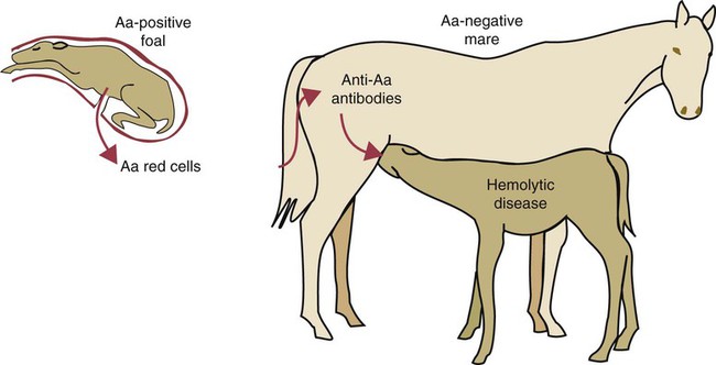
Red Cell Antigens and Type II Hypersensitivity


Red Cell Antigens and Type II Hypersensitivity


Red cells, like nucleated cells, express cell surface molecules that can act as antigens. However, unlike the major histocompatibility complex (MHC) molecules, red cell surface antigens are not involved in antigen processing, although they do influence graft rejection (allografts between blood group–incompatible animals are rapidly rejected.) Most red cell surface antigens are either glycoproteins or glycolipids and are integral functional components of the cell membrane. For example, the ABO antigens in humans are anion and glucose transporter proteins, whereas the antigens of the M and C systems of sheep red cells are associated with the membrane potassium pump and amino acid transport, respectively.


If blood is transfused from one animal to another, genetically different individuals, the red cell antigens will stimulate an antibody response in the recipient. These antibodies cause the rapid elimination of the transfused red cells as a result of intravascular hemolysis by complement and of extravascular destruction through opsonization and removal by the mononuclear phagocyte system. Cell destruction by antibodies in this way is classified as a type II hypersensitivity reaction.


Blood Groups


The antigens expressed on the surface of red blood cells are called blood group antigens or erythrocyte antigens (EAs). There are many different blood group antigens, and they vary in their antigenicity, some being more potent and therefore of greater importance than others. The expression of blood group antigens is controlled by genes and inherited in conventional fashion. For each blood group system, there exists a variable number of alleles. (If blood group alleles are invariably inherited together in groups of two or more, they are called phenogroups.) The alleles, in turn, control a variable number of EAs. The complexity of erythrocyte blood group systems varies greatly. They range from simple systems like the L system of cattle, which consists of two alleles controlling a single antigen, to the highly complex B system of cattle. The B system contains several hundred alleles or phenogroups that, together with the other cattle blood groups, may yield millions of unique blood group combinations. Although most blood group antigens are integral cell membrane components, some are soluble molecules found free in serum, saliva, and other body fluids and passively adsorbed onto red cell surfaces. Examples of such soluble antigens include the J antigens of cattle, the R antigens of sheep, the A antigens of pigs, and the DEA 7 antigens of dogs.


Animals may possess antibodies against foreign blood group antigens even though they may never have been exposed to foreign red cells. For example, J-negative cattle have anti-J antibodies in their serum, and A-negative pigs have anti-A antibodies. These “natural” antibodies (or isoantibodies) are derived not from previous contact with foreign red cells but from exposure to cross-reacting epitopes that are commonly encountered in nature (see Figure 9-8). Many blood group antigens are also common structural components of plants, the intestinal microbiota, protozoa, or helminths. The presence of these natural antibodies is not, however, a uniform phenomenon, and not all blood group antigens are accompanied by the production of natural antibodies to their alternative alleles.


Blood Transfusion and Incompatible Transfusions


Blood is easily transfused from one animal to another. If the donor red cells are identical to those of the recipient, no immune response results. If, however, the recipient possesses preexisting antibodies to donor red cell antigens, they will be attacked immediately. These preexisting antibodies are usually of the immunoglobulin M (IgM) class. When these antibodies bind red cell antigens, they may cause agglutination or hemolysis, or stimulate opsonization and phagocytosis of the transfused cells. In the absence of preexisting antibodies, the transfused red cells will stimulate an immune response in the recipient. The transfused cells will circulate until antibodies are produced and will then be eliminated. A second transfusion with identical foreign cells results in their immediate destruction.


The rapid destruction of large numbers of foreign red cells can lead to serious illness. The severity of transfusion reactions ranges from a mild febrile response to rapid death and depends mainly on the amount of incompatible blood transfused. Early recognition of a problem may avert the most severe consequences. The most severe reactions occur when large amounts of incompatible blood are transfused to a sensitized recipient. This results in complement activation and hemolysis of the transfused cells. Large amounts of free hemoglobin escape, resulting in hemoglobinemia and hemoglobinuria. Large numbers of lysed red cells may trigger blood clotting and disseminated intravascular coagulation. Complement activation also results in anaphylatoxin production, mast cell degranulation, and the release of vasoactive molecules and cytokines. These molecules provoke circulatory shock with hypotension, bradycardia, and apnea. The animal may show sympathetic responses such as sweating, salivation, lacrimation, diarrhea, and vomiting. This may be followed by a second stage in which the animal is hypertensive, with cardiac arrhythmia as well as increased heart and respiratory rates.


If a reaction is suspected, the transfusion must be stopped immediately. It is important to maintain urine flow with fluids and a diuretic because accumulation of hemoglobin in the kidneys may cause renal tubular destruction. Recovery follows elimination of the foreign red cells.


Transfusion reactions can be almost totally prevented by prior testing of the recipient’s serum for antibodies against the donor’s red cells. The test is called cross-matching. Blood from the donor is centrifuged and the plasma discarded. The red cells are then resuspended in saline and recentrifuged. This washing procedure is repeated (usually three times), and eventually a 2% to 4% suspension of red cells in saline is made. These donor red cells are mixed with recipient serum and then incubated at 37° C for 15 to 30 minutes. If the red cells are lysed or agglutinated by the recipient’s serum, no transfusion should be attempted with those cells. It is occasionally found that the donor’s serum may react with the recipient’s red cells. This is not of major clinical significance because transfused donor antibodies are rapidly diluted within the recipient. Nevertheless, blood giving such a reaction is best avoided.


Hemolytic Disease of the Newborn


Female animals may become sensitized to foreign red cells not only by incompatible blood transfusions given for clinical purposes but also by leakage of fetal red cells into their bloodstream through the placenta during pregnancy. In sensitized females, these anti–red cell antibodies may then be concentrated in their colostrum. When a newborn animal suckles, these colostral antibodies are absorbed through the intestinal wall and reach its circulation. These antibodies, directed against the blood group antigens of the newborn, cause rapid destruction of their red cells. The resulting disease is called hemolytic disease of the newborn (HDN) or neonatal isoerythrolysis.


Four conditions must be met for HDN to occur: The young animal must inherit a red cell antigen from its sire that is not present in its mother. The mother must be sensitized to this red cell antigen. The mother’s response to this antigen must be boosted repeatedly by transplacental hemorrhage or repeated pregnancies. Finally, a newborn animal must ingest colostrum containing high-titered antibodies to its red cells.


Blood Groups, Blood Transfusion, and Hemolytic Disease in Domestic Animals
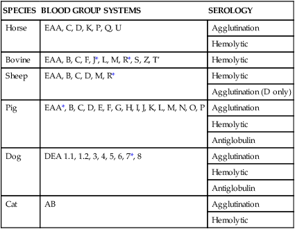

All mammals possess red cell antigens that can affect blood transfusions and on occasion cause HDN in newborn animals (Table 29-1). Although historically they were named alphabetically in order of their discovery, there is a growing tendency to add the prefix EA (erythrocyte antigen) to reduce confusion with MHC antigens.


Horses


Horses possess seven internationally recognized blood group systems (EAA, EAC, EAD, EAK, EAP, EAQ, and EAU.) Some, such as EAC, EAK, and EAU, are simple, one-factor, two-allele, two-phenotype systems. On the other hand, the EAD system is very complex, with at least 25 alleles identified to date. Their major significance lies in the fact that HDN in foals is relatively common (Figure 29-1). In mules, in which the antigenic differences between dam and sire are great, about 8% to 10% of foals may be affected. In thoroughbreds and standardbreds, the prevalence is considerably less, ranging from 0.05% to 2% of foals. This is despite of the fact that in up to 14% of pregnancies, the mare and the stallion have incompatible red cells.


HDN may occur in foals from mares that have been sensitized by previous blood transfusions or by administration of vaccines containing equine tissues. Most commonly, however, mares are sensitized by exposure to fetal red cells as a result of repeated pregnancies. The mechanism of this sensitization is unclear, but fetal red cells are assumed to gain access to the maternal circulation as a result of transplacental hemorrhage. Mares have been shown to respond to fetal red cells as early as day 56 after conception. The greatest leakage probably occurs during the last month of pregnancy and during foaling as a result of the breakdown of placental blood vessels.


Maternal sensitization is usually minimal following a first pregnancy. However, if repeated pregnancies result in exposure to the same red cell antigens, the maternal response will be boosted. Hemolytic disease is therefore usually only a problem in mares that have had several foals. The most severe form of the disease results from the production of antibodies directed against the Aa antigen of the EAA system. Anti-Qa (EAQ system) produces a less severe disease of slower onset. All in all, 90% of clinical cases are attributable to anti-Aa and -Qa, although other minor antigens, such as Pa, Ab, Qc, Ua, Dc, and Db, have also been implicated. Mares that lack Aa and Qa are therefore most likely to produce affected foals. Pregnant mares may also produce antibodies to Ca (EAC system), but these are rarely associated with clinical disease. Indeed preexisting antibodies to Ca may reduce sensitization by Aa. The presence of this anti-Ca in a mare may eliminate foal red cells that enter her bloodstream and prevent further sensitization.


Antibodies produced by mares do not cross the placenta but reach the foal through the colostrum. Affected foals are therefore born healthy but sicken several hours after suckling. The severity of the disease is determined by the amount of antibody absorbed and by the sensitizing antigen. The earliest signs are weakness and depression. The mucous membranes of affected foals may be pale and may eventually show a distinct jaundice. Some foals sicken by 6 to 8 hours and die from shock so rapidly that they may not have time to develop jaundice. More commonly the disease presents as lethargy and weakness between 12 and 48 hours of age, although it may be delayed for as long as 5 days. Icterus of the mucous membranes and sclera is consistent in foals that survive for at least 48 hours. Hemoglobinuria, although uncommon, is diagnostic in a newborn foal. As a result of anoxia, some foals in the terminal stages of the disease may convulse or become comatose. The most common causes of death in these foals are liver failure, brain damage, and bacterial sepsis.


Hemolytic disease is readily diagnosed by clinical signs alone. Hematological examination is of little diagnostic use but may be of assistance in indicating appropriate treatment. Definitive diagnosis requires that immunoglobulin be demonstrated on the surface of the red cells of the foal. In the case of anti-Aa or anti-Qa, addition of a source of complement (fresh normal rabbit serum) causes rapid hemolysis. If hemolytic disease is anticipated, the serum of a pregnant mare may be tested for antibodies by an indirect antiglobulin test. By using red cells from horses with a major sensitizing blood group, it is possible to show that the antibody titer increases significantly in the month before parturition when sensitization is occurring.


A test that may be useful for detecting the presence of antierythrocyte antibodies in colostrum is the jaundiced foal agglutination test. This involves making serial dilutions of colostrum in saline. A drop of anticoagulated foal blood is added to each tube, and the tubes are centrifuged so that the red cells form pellets at the bottom. In the presence of antibodies, the cells clump tightly, and the pellets remain intact when the tubes are emptied. Nonagglutinated red cells, in contrast, flow down the side of the tube. Concentrated colostrum is viscous and tends to induce rouleaux formation that mimics agglutination. However, if the mare’s blood is used as a negative control, this can be accounted for. The foal’s blood should also be diluted in saline to ensure that the foal has not already absorbed antibodies and that false-positive results are not obtained.


Mildly affected foals, with a packed cell volume (PCV) of 15% to 25% and a red cell count greater than 4 × 10⁶, will continue to nurse. Those with a PCV of less than 10% will stop nursing and become recumbent. Marked icterus is suggestive of HDN in foals, but mild icterus may be seen in septicemia despite the fact that septic foals are not anemic.


The prognosis of uncomplicated hemolytic disease is good provided the condition is diagnosed sufficiently early and the appropriate treatment instituted rapidly. Management of HDN includes prevention of further antibody absorption, adequate nutrition, oxygen therapy, fluid and electrolyte therapy, and maintenance of the acid-base balance. Warmth, adequate hydration, and antimicrobial therapy are also critically important. In acute cases, blood transfusion is necessary. A red cell count of less than 3 × 10⁶/µL or a PCV of less than 15% warrants a blood transfusion. Transfused equine red cells have a half-life of only 2 to 4 days, so transfusion is only a temporary life-saving measure. Compatible blood may be difficult to find because of the high prevalence of Aa and Qa in the normal equine population. A donor should not only be Aa or Qa negative but should also lack antibodies to these antigens. Exchange transfusion, although efficient, requires a donor capable of providing at least 5 L of blood as well as a double intravenous catheter and an anesthetized foal. A much simpler procedure that avoids many difficulties is transfusion of washed cells from the mare. About 3 to 4 L of blood is collected in sodium citrate and centrifuged, after which the plasma is discarded. The red cells are washed once in saline and transfused slowly into the foal. The blood is usually given in divided doses about 6 hours apart. Milder cases of hemolytic disease may require only careful nursing.


If hemolytic disease is anticipated as a result of either a rising antibody titer or the previous birth of a hemolytic foal, stripping off the mare’s colostrum and giving the foal colostrum from another mare may prevent its occurrence. The foal should not be allowed to suckle its mare for 24 to 36 hours. Once suckling is permitted, the foal should only be allowed to take small quantities at first and should be observed carefully for adverse side effects.


Neonatal thrombocytopenia has been recorded in the foal. Immunoglobulins can be identified on the foal’s platelets, and antibodies to these platelets can be found in the mare’s serum.


< div class='tao-gold-member'>

Stay updated, free articles. Join our Telegram channel

Jul 18, 2016 | Posted by in PHARMACOLOGY, TOXICOLOGY & THERAPEUTICS | Comments Off on Red Cell Antigens and Type II Hypersensitivity

Full access? Get Clinical Tree

Get Clinical Tree app for offline access