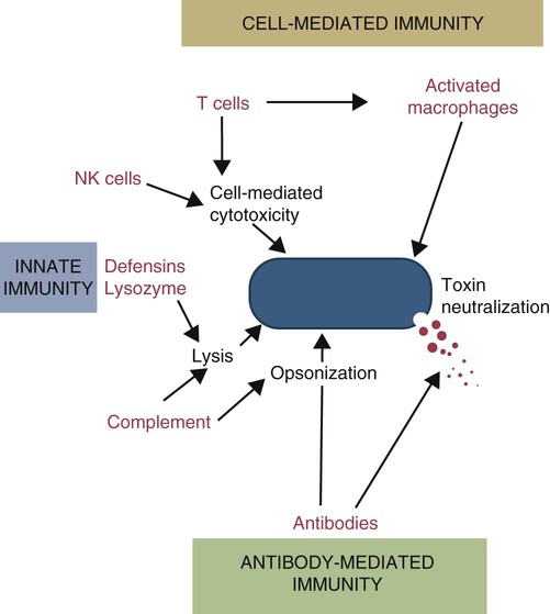
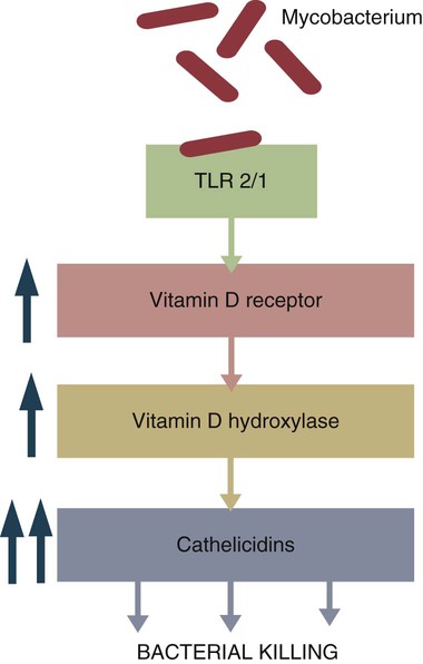
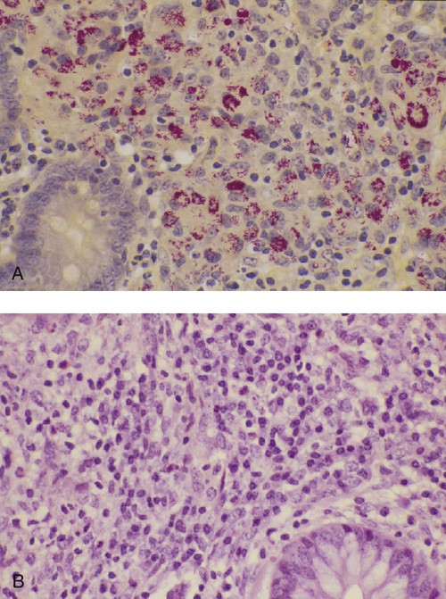
Immunity to Bacteria and Fungi


Immunity to Bacteria and Fungi


Although animals live in environments densely populated with bacteria, most of these organisms neither invade animal tissues nor cause disease. This is unsurprising for several reasons. First, the combined efforts of the innate and adaptive immune systems are sufficient to prevent invasion. Second, even organisms that successfully invade the animal body gain very little by harming their host. On the contrary, illness or death of the host animal may well reduce the survival of the bacteria and is therefore normally avoided. Indeed, many bacteria are essential for the animal’s well-being since they maintain an environment on body surfaces that is hostile to other potential invaders. They also assist in the digestion of foods such as celluloses and promote the normal development of the immune system. Nevertheless, many commensal bacteria are also pathobionts. For example, Clostridium tetani and Clostridium perfringens are commonly found among the intestinal flora of horses, and Bordetella bronchiseptica is found in the nasopharynx of healthy swine. Bacterial disease is not, therefore, an inevitable consequence of the presence of pathogenic organisms on the body surface. The development of disease is related to many other factors, including the response of the host, the presence of damaged tissues, the location of the bacteria within the body, and the disease-producing power (or virulence) of the bacteria. Only when the balance between host immunity and bacterial virulence is upset will disease or death result.


The adaptation of bacteria to a host depends on factors that enable the bacteria to survive and grow—virulence factors. Many of these virulence factors are encoded on mobile genetic elements that can be transmitted between species (e.g., plasmids). These virulence factors permit the bacteria to adapt to a specific environment and promote their transmission between hosts. Depending on their niche within the body, bacteria can use virulence factors to penetrate surface epithelia, to bind to cell surfaces, to acquire iron, to evade immune responses, to hide within cells, and to promote transmission to another host. Some of the strategies adopted by bacteria are associated with damage to host tissues and must be counteracted by the immune system.


Innate Immunity


Antimicrobial immunity consists of an early innate response followed by a sustained adaptive response. Recognition of invading bacteria through toll-like receptors (TLRs) and other receptors induces inflammation, cytokine release, and complement activation. If this is insufficient to eliminate the invaders, adaptive immune mechanisms take over. Thus dendritic cells and macrophages ingest invading bacteria and initiate adaptive immunity by secreting cytokines and triggering both T and B cell responses. The importance of these innate defenses is emphasized by the observation that the resistance of chickens to Salmonella enterica Typhimurium appears to be linked to allelic variations in TLR4, whereas the resistance of foals to Rhodococcus equi depends on TLR2.


TLRs are responsible in large part for the initial recognition of invading bacteria. Binding of microbial pathogen-associated molecular patterns (PAMPs) to TLRs triggers a signal cascade that activates genes that are critical in host defense.


The production of cytokines by horse neutrophils following exposure to R. equi provides an example of these responses. Thus after exposure to R. equi, neutrophils express increased amounts of interleukin-23 (IL-23). This IL-23 promotes Th17 cell differentiation. Driven by transforming growth factor-β (TGF-β) and IL-6, the Th17 cells then participate in inflammatory reactions. Th17 cells confer protection against extracellular bacteria and fungi, especially at epithelial surfaces. Not only do these Th17 cells produce IL-17 but also IL-6, GM-CSF, G-CSF, chemokines, and metalloproteases. They trigger inflammation and coordinate early neutrophil recruitment to infection sites. Type I interferons are also readily produced in response to bacterial PAMPS. IFN-α/β boosts macrophage responses enhancing their production of IFN-γ, nitric oxide, and TNF-α.


Natural killer (NK) cells play a protective role in some bacterial, protozoan, and fungal infections. For example, some bacteria may activate NK cells by upregulating expression of NKG2D ligands on cells. Activated NK cells produce a large amount of IFN-γ that in turn activates both macrophages and dendritic cells.


Although many bacteria are destroyed by phagocytosis, others are killed when free in the circulation. Bacteria can be destroyed by complement acting through the alternate or lectin pathways. Bacterial cell walls, lacking sialic acid, inactivate factor H and stabilize the alternate C3 convertase (C3bBbP). As a result, these bacteria are either opsonized or lysed. Activation of the terminal complement components leads to the development of terminal complement complexes (TCCs). These TCCs may be unable to insert themselves into the complex carbohydrates of the microbial cell wall. However, lysozyme in the blood may digest the cell wall and enable the TCCs to gain access to the lipid bilayer of the inner bacterial membrane.


Antimicrobial peptides are critical for the defense against some bacteria such as the mycobacteria (Box 25-1). Suppression of bacterial growth by withholding iron is discussed in Chapter 6.


Box 25-1   Vitamin D and Immunity


When an intracellular bacterium such as Mycobacterium tuberculosis interacts with TLR1 or TLR2 on the surface of macrophages, it upregulates many different genes and enhances their antimicrobial activity. In mice, this is mainly mediated by nitric oxide. In humans, however, nitric oxide is not elevated, and other mechanisms are involved (Figure 25-2). One gene activated by TLR1/2 signaling in humans is that coding for the vitamin D receptor. This receptor is therefore upregulated on activated macrophages. Binding of vitamin D to its receptor upregulates expression of the gene for the antibacterial peptide cathelicidin. The cathelicidin, in turn, can kill intracellular M. tuberculosis. It is no coincidence, therefore, that resistance to tuberculosis is directly related to serum vitamin D levels and that humans with a deficiency of vitamin D show significantly decreased resistance to this infection. It is of interest to recall that sanatorium treatment of tuberculosis classically involved exposure to fresh air and sunlight, a procedure that would be expected to increase vitamin D levels in human patients. Conversely, mice are nocturnal mammals that would not be expected to have high vitamin D levels and must rely on other pathways.


Adaptive Immunity


There are five basic mechanisms by which the adaptive immune responses combat bacterial infections (Figure 25-1): (1) neutralization of toxins or enzymes by antibody; (2) killing of bacteria by the classical complement pathway; (3) opsonization of bacteria by antibodies and complement, resulting in their phagocytosis and destruction; (4) destruction of intracellular bacteria by activated macrophages; and (5) direct killing of bacteria by cytotoxic T cells and NK cells. The relative importance of each of these processes depends on the species of bacteria involved and on the mechanisms by which they cause disease.


Immunity to Invasive Bacteria


Protection against invasive bacteria is usually mediated by antibodies directed against their surface antigens. Efficient phagocytosis requires that the bacteria be coated with opsonins that can be recognized by phagocytic cells. These opsonins include antibodies and C3b in addition to the innate opsonins such as MBL. Antibodies not only are effective opsonins in their own right but also increase the binding of C3b by activating the classical complement pathway. Antibodies directed against capsular (K) antigens may neutralize the antiphagocytic properties of bacterial capsules, thus permitting their destruction by phagocytic cells. In bacteria lacking capsules, antibodies directed against O antigens act as opsonins. Protection also results when antibodies are produced against the Escherichia coli pilus antigens F4 (K88) and F5 (K99). The antibodies may interfere with the expression of pili. Once the adherence pili are suppressed, these strains of E. coli cannot bind to the intestinal wall and thus are no longer pathogenic.


The importance of bacterial capsules in immunity is seen in anthrax. B. anthracis organisms possess both a capsule and an exotoxin. Antitoxic immunity is protective but slow to develop. In addition, toxin production tends to be prolonged since the organism is encapsulated and phagocytic cells have difficulty eliminating it. As a result, death is usually inevitable in unvaccinated animals. The vaccine commonly employed against animal anthrax contains an unencapsulated but toxigenic strain of B. anthracis. Given in the form of spores that can germinate, the unencapsulated bacteria are eliminated by phagocytic cells before dangerous amounts of toxin are synthesized but not before antitoxic immunity is established.


Molecule for molecule, immunoglobulin M (IgM) is about 500 to 1000 times more efficient than IgG in opsonization and about 100 times more potent than IgG in sensitizing bacteria for complement-mediated lysis. During a primary immune response, therefore, the quantitative deficiency of the IgM response is compensated for by its quality, ensuring early and efficient protection.


The traditional view of antibodies has been that they alone could not kill microorganisms, but simply could mark microorganisms for destruction. That view is now known to be incorrect. Many antibodies have direct antimicrobial activities. Antibodies against E. coli may be bacteriostatic since they interfere with production of the iron-binding protein enterochelin and thus prevent bacterial iron scavenging. IgM and IgG antibodies against Borrelia burgdorferi damage surface proteins on the bacteria and are bactericidal in the absence of complement. There is also evidence that antibodies are able to generate oxidants and may kill bacteria directly.


Heat-Shock Protein Response


Many new proteins are induced in bacteria by stressors such as a heat, starvation, and exposure to oxidants; toxins such as heavy metals; protein synthesis inhibitors; and viral infections. The heat-shock proteins (HSPs) are the best understood of these new proteins. HSPs are present in all bacteria at very low levels at normal temperatures. Mild stress such as a low-grade fever will induce HSP production. For example, HSP levels climb from 1.5% to 15% of the total protein in stressed E. coli. There are three major bacterial HSPs: HSP 90, HSP 70, and HSP 60. (The number refers to their molecular weight.) When a bacterium is phagocytosed and exposed to the neutrophil respiratory burst, the resulting stress triggers the production of bacterial HSP. As a result, HSP 60 is the dominant antigen in infections caused by mycobacteria, Coxiella burnetii, legionella, treponema, and Borrelia species. These HSPs are highly antigenic for several reasons. First, they are produced in abundance within the infected host; second, they are readily processed by antigen-presenting cells; and third, the immune system may possess unusually large numbers of cells capable of responding to HSPs. In addition, some γ/δ T cells may preferentially recognize bacterial HSPs. Thus anti-HSP responses may induce significant protection against many bacterial pathogens.


Immunity to Intracellular Bacteria


As discussed in Chapter 18, some bacteria such as Brucella abortus, Mycobacterium tuberculosis, Campylobacter jejuni, R. equi, Listeria monocytogenes, Corynebacterium pseudotuberculosis, C. burnetii, and some serotypes of S. enterica can grow readily inside macrophages. In addition, L. monocytogenes can travel from cell to cell without exposure to the extracellular fluid through cytoskeletal membrane protrusions.


Autophagy, as described in Chapter 4, is also a key component of the destruction of intracellular bacteria. The same cellular machinery used to destroy unwanted organelles can be employed to eliminate intracellular organisms. Autophagy (or more correctly, xenophagy) may also play a key role in delivering microbial antigens to the appropriate major histocompatibility complex (MHC) molecules.


Protection against intracellular bacteria is mediated by macrophages activated through the M1 pathway. Classically activated M1 macrophages are responsive to inflammatory cytokines and microbial products (Chapter 5). Although macrophages from unimmunized animals cannot usually destroy these bacteria, this ability is acquired about 10 days after onset of infection once the macrophages are activated (Chapter 18). IFN-γ, especially in association with TNF-α, greatly enhances the production of cytokines such as TNF-α, IL-6, IL-1β and IL-12, enzymes such as indoleamine 2,3-dioxygenase (IDO) and nitric oxide synthase 2 (NOS2), and the release of reactive oxygen and nitrogen intermediates. M1 polarization has been shown to be important in resistance to L. monocytogenes, S. enterica Typhi and Typhimurium, mycobacteria, and chlamydia. For example, IFN-γ and TNF-α produced by primed T cells generate M1 macrophages, acidify their phagosomes, and kill mycobacteria. Uncontrolled M1 activation by organisms such as streptococci and E. coli, however, can contribute to pathology by inducing, for example, sepsis, tissue damage, and organ failure. The response of these activated macrophages tends to be nonspecific, particularly in listerial infections, and M1 macrophages are able to destroy many normally resistant bacteria. Thus an animal recovering from an infection with L. monocytogenes develops increased resistance to infection by M. tuberculosis. The development of M1 macrophages often coincides with the appearance of delayed (type IV) hypersensitivity responses to intradermally administered antigen (Chapter 31).


Both CD4+ and CD8+ cells are also involved in immunity to Listeria. CD8+ cytotoxic T cells lyse listeria or mycobacteria-infected cells and complement the Th1 cells that activate the macrophages. R. equi–infected macrophages are recognized and killed by CD8+ T cells in a MHC class I unrestricted manner.


It has been observed that protective immunity against intracellular bacteria cannot be induced by vaccines containing killed bacteria. Only vaccines containing living bacteria are protective. This is because of the differential stimulation of helper T cell populations by live and dead bacteria. Infection of mice with live B. abortus stimulates Th1 cells to secrete IFN-γ. Conversely, immunization of these mice with Brucella protein extracts induces Th2 cells to secrete IL-4. Likewise, live but not dead L. monocytogenes or B. abortus organisms induce macrophage secretion of TNF-α. Killed Brucella organisms stimulate IL-1 production to a greater extent than live bacteria. Resistance to these intracellular bacteria is generally short lived, persisting for only as long as viable bacteria remain in the body. (Tuberculosis is an exception, in which case memory is prolonged.)


If, in a bacterial disease, it is observed that dead vaccines do not give good protection, that serum cannot confer protection, that antibody levels do not relate to resistance, and that delayed hypersensitivity reactions can be elicited to the bacterial antigens, the possibility that cell-mediated immunity may play an important role in resistance to the causative organism should be considered, and the use of vaccines containing living bacteria should be contemplated.


Modification of Bacterial Disease by Immune Responses


The immune response clearly influences the course and severity of an infection. At best, it will result in a cure. In the absence of a cure, however, the infection may be profoundly modified. Much depends on whether a cell-mediated or antibody response is generated. Thus the type of helper T cells induced during infection may affect the course of disease. As described in Chapter 18, cell-mediated responses are required to control intracellular bacteria since only activated macrophages can prevent their growth. Macrophage activation requires that Th1 cells produce IFN-γ. Once activated, these M1 cells can localize or cure these infections. If an animal mounts an inappropriate Th2 response, cell-mediated immunity fails to develop, M2 macrophages are generated, and chronic progressive disease may result. This is readily seen in mycobacterial diseases. For example, in humans, leprosy occurs in two distinct forms called tuberculoid and lepromatous leprosy. Tuberculoid (or paucibacillary [PB]) leprosy is characterized by an intense cell-mediated immune response dominated by Th1 cells and M1 macrophages. Lesions of this form of the disease contain very few organisms. Lepromatous (or multibacillary [MB]) leprosy, in contrast, is characterized by very high antibody levels and poor cell-mediated responses. Humans with lepromatous leprosy employ Th2 cells secreting IL-4 and IL-10. The IL-10 reduces the production of IL-12, which in turn decreases IFN-γ secretion by Th1 cells and generates M2 macrophages. This reduces the patient’s ability to control Mycobacterium leprae, and their lesions contain enormous numbers of bacteria. The prognosis of lepromatous leprosy is much poorer than for tuberculoid leprosy.


A similar diversity of lesions is seen in Johne’s disease of sheep. Some animals develop MB disease, in which their intestinal lesions contain enormous numbers of bacteria (Figure 25-3) and little histological evidence of a cell-mediated response. Their granulomas tend to lack organization, with large numbers of bacteria-laden macrophages intermixed with lymphocytes. In contrast, other sheep may develop PB disease, in which the lesions contain very few bacteria but large numbers of lymphocytes. These are organized nodular lesions with epithelioid cells and multinucleated giant cells at the center surrounded by fibrous connective tissue. The two forms of the disease are associated with differential expression of cytokine and chemokine receptors. Thus animals with the PB disease have increased numbers of CD25+ T cells that produce more IL-2 and much more IFN-γ than sheep with the MB form of the disease (Figure 25-4). In contrast, sheep with the MB disease have higher antibody levels and a lack of cellular immune responses. It is likely, therefore, that sheep with PB lesions mount an immune response in which Th1 cells predominate, whereas those with MB disease use Th2 cells.


< div class='tao-gold-member'>

Stay updated, free articles. Join our Telegram channel

Jul 18, 2016 | Posted by in PHARMACOLOGY, TOXICOLOGY & THERAPEUTICS | Comments Off on Immunity to Bacteria and Fungi

Full access? Get Clinical Tree

Get Clinical Tree app for offline access