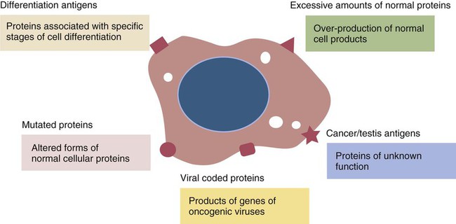
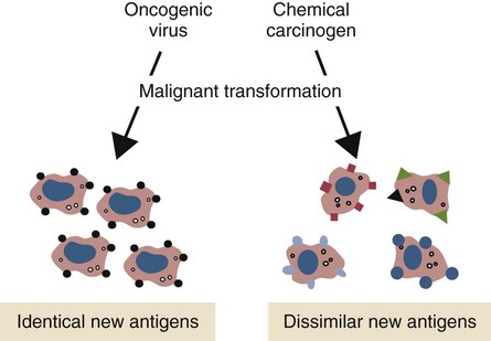
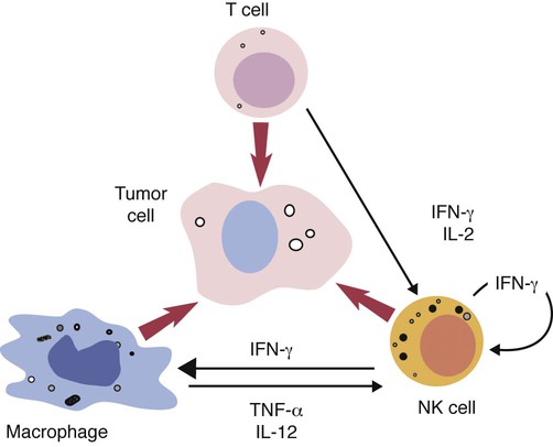
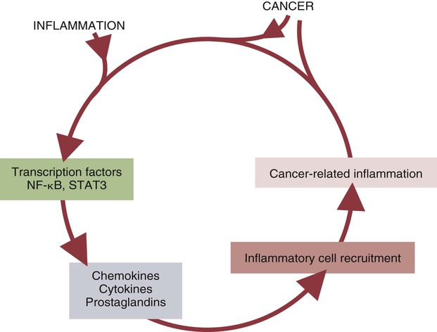
Resistance to Tumors


Resistance to Tumors


Normal cellular functions depend on the careful regulation of cell division. When cells multiply, it is essential that they do so only as and when required. Unfortunately, as a result of mutations triggered by chemicals, radiation, or viruses, cells may occasionally break free of the constraints that regulate cell division. A cell that is proliferating in an uncontrolled fashion will give rise to a growing clone of cells that eventually develops into a tumor or neoplasm. If these cells remain clustered together at a single site, the tumor is said to be benign. Benign tumors can usually be removed by surgery. In some cases, however, tumor cells break off from the main tumor mass and are carried by the blood or lymph to distant sites, where they lodge and continue to grow. This form of tumor is said to be malignant. The secondary tumors that arise in these distant sites are called metastases. Surgical treatment of malignant tumors may be very difficult because it may be impossible to remove all metastases. Malignant tumors are subdivided according to their tissue of origin. Tumors arising from epithelial cells are called carcinomas; those arising from mesenchymal cells, such as muscle, lymphoid, or connective tissue cells, are called sarcomas. A leukemia is a tumor derived from hematopoietic stem cells.


The essential difference between a normal cell and a tumor cell is a loss of control of cell growth as a result of multiple mutations. These mutations may also result in tumor cells expressing abnormal proteins on their surface.


Tumors as Allografts


When organ transplantation became a common procedure as a result of the development of potent immunosuppressive drugs, it was found that patients with prolonged graft survival were many times more likely to develop certain cancers than were nonimmunosuppressed individuals. It was also observed that some immunodeficient patients had an increased tendency to develop malignant tumors. It was therefore suggested that the immune system was responsible for the prevention of cancer. From this suggestion the immune surveillance theory emerged. This theory held that the body constantly produces neoplastic cells but that in a healthy individual the immune system rapidly recognizes and eliminates these cells. The theory suggested that progressive cancer would result if the cancer cells somehow evaded recognition by T cells.


The immune surveillance theory soon ran into problems. Common human cancers such as those of the lung or breast do not develop more frequently in immunodeficient individuals. Likewise, nude (nu/nu) mice, although T cell deficient, are no more susceptible than normal mice to chemically induced or spontaneous tumors (Chapter 37). Finally, it became clear that many tumor antigens induce tolerance in a manner similar to normal self-antigens. Thus most evidence has failed to support the idea that the immune system distinguishes between tumor cells and normal, healthy cells.


Notwithstanding this, there are situations in which the immune system can recognize and kill cancer cells. For example, some immunodeficient mouse strains, which are “cleaner” subjects than nude mice, show an increased prevalence of spontaneous cancer. (Nude mice have some persistent T and B cell function and intact innate defenses.) These include recombinase-activating gene (RAG) knockout mice that cannot produce functional T or B cells and STAT-1 knockout mice that lack both adaptive and innate responses by being unresponsive to interferon-γ (IFN-γ). RAG knockout mice suffer from an increased incidence of spontaneous tumors of the intestinal epithelium, whereas RAG/STAT-1 knockout mice develop mammary cancers.


It is perhaps therefore more appropriate to think of the immune system as “immunoediting” cancers. Under some circumstances the immune system can indeed eliminate some cancer cells. If, however, the cancer cells evade destruction, they may survive indefinitely or be selected by the immune responses to produce new tumor cell variants. Eventually, however, some of these variants may escape any control by the immune system and grow to produce clinical cancers.


If a metastasizing tumor does not invade lymphoid organs, it may escape immune surveillance. On the other hand, tumors that invade lymph nodes can be divided into strongly and weakly immunogenic types. Strongly immunogenic tumors elicit a strong T cell response following processing by dendritic cells. Weakly immunogenic tumors tend to grow as walled-off nodules that may not be processed in sufficient amounts to trigger immune responses. These are the most common tumors in humans. It is possible that tumor cells that trigger inflammation in tissues trigger dendritic cell activation and processing. On the other hand, tumors that fail to generate inflammation may simply be ignored by the immune system. Alternatively, it is possible that the antigens of the tumor may be tolerated by the immune system. If successful tumor therapy is to be achieved, these two states must be distinguished.


Most humans who develop “spontaneous” cancer have a normal immune system. Immunosuppressed individuals such as allograft recipients and patients with AIDS develop a very different spectrum of cancers from that of the general population. The only cancers to which they are at greater risk are those caused by viruses, such as Kaposi’s sarcoma. Immunosuppressed individuals are at no more likely than the general population to develop the common cancers, such as those of breast, lung, or colon.


Although the original surveillance hypothesis has had to be drastically modified, it is clear that under some circumstances the immune system may destroy tumor cells and that this response may be enhanced to protect an individual against some cancers. However, there is a great difference between the strong and effective cell-mediated immune response triggered by foreign organ grafts and the much weaker responses to the antigens associated with tumor cells.


The outcome of the interaction between a tumor cell and the immune system can thus have one of three results: first, elimination of the cancer; second, cancer equilibrium whereby the more immunogenic cells are destroyed but permitting the less immunogenic cells to survive (immunoediting); or third, tumor escape, whereby the tumor cells are unaffected by any immune responses they may trigger.


Tumor Antigens


Spontaneous cancer cells develop as a result of multiple mutations in regulatory genes. These mutations may generate molecules that are unique to the tumor cells (tumor-specific antigens) or, more commonly, abnormal or unusual molecules (tumor-associated antigens). To distinguish between normal and tumor cells, host T cells must recognize tumor cell antigens. Five major types of tumor antigen have been identified. First, there are differentiation antigens associated with specific stages in the development of a cell type. For example, some tumor cells may express the products of developmental genes that are turned off in adult cells and are normally only expressed early in an individual’s development. These proteins are called oncofetal antigens. Examples include tumors of the gastrointestinal tract that produce a glycoprotein called carcinoembryonic antigen (CEA; also called CD66e), normally found only in the fetal intestine. The presence of detectable amounts of CEA in serum may indicate the presence of a colon or rectal adenocarcinoma. α-Fetoprotein produced by hepatoma cells is normally found only in the fetal liver. Likewise, squamous cell carcinoma cells may possess antigens normally restricted to fetal liver and skin. These oncofetal antigens are usually poor immunogens and do not provoke protective immunity. However, their detection in blood may be useful for diagnosis and for monitoring the progress of the tumor.


Second, there are mutated forms of normal cellular proteins. For example, melanoma cells may express the products of mutated oncogenes on their surface (Figure 33-1). Some tumor antigens are recognized because they are abnormally glycosylated. Chemically induced tumors may express mutated surface antigens unique to the tumor and not to the inducing chemical (Figure 33-2). Because carcinogenic chemicals can produce many different mutations, tumors induced by a single chemical in different animals may be antigenically different. Even within a single chemically induced tumor mass, antigenically distinct subpopulations of cells exist. As a result, immunity to one chemically induced tumor does not prevent growth of a second tumor induced by the same chemical.


Third, normal proteins are produced in excessive amounts. A good example is the production of prostate-specific antigen (PSA) by prostate carcinomas of humans. PSA is a protease exclusively produced by the prostate epithelium. Increased blood levels of this protein indicate excessive prostate activity. One cause of this is the growth of a carcinoma.


Fourth, cancer/testis (CT) antigens are a group of tumor antigens only expressed in the testes and in various malignancies. Their function is unknown.


Fifth, tumors caused by viruses express antigens characteristic of the inducing virus or of other endogenous retroviruses. These antigens, although coded for by a viral genome, are not part of a virion. Examples include the FOCMA antigens found on the neoplastic lymphoid cells of cats infected with feline leukemia virus and Marek’s tumor-specific antigens found on Marek’s disease tumor cells in chickens. (Both of these are virus-induced, naturally occurring, T cell tumors.)


Inflammation and Tumors


The tumor microenvironment plays a large part in determining the behavior of a tumor. The tumor cells communicate extensively with nearby cells. Among the most important of these are fibroblasts and inflammatory cells. As a result, the elimination of tumor cells by immune mechanisms is determined in part by the presence of inflammation. Inflammatory diseases increase the risk for developing many types of cancer; conversely, the use of nonsteroidal antiinflammatory drugs reduces tumor susceptibility (Figure 33-4). Mutations in oncogenes such as ras and myc are closely linked to the inflammatory pathways. Inflammatory cytokines, chemokines, and cells are present in the microenvironment of early tumors. Blocking of inflammatory mediators, key transcription factors, or inflammatory cells decreases the incidence and spread of cancer. Conversely, adoptive transfer of inflammatory cells may promote tumor development. More than half of the mass of a tumor may consist of supporting cells, including fibroblasts, macrophages, and vascular endothelial cells. Cancers cannot spread and metastasize without the support of these cells. The process by which these stromal cells are recruited is closely related to inflammation. Inflammatory cytokines, such as tumor necrosis factor-α (TNF-α), and macrophages are often required for tumor development and spread.


Cancer and inflammation are linked by two pathways. The intrinsic pathway is activated by mutations leading to neoplasia. These include the activation of various oncogenes, chromosomal rearrangements, or the inactivation of tumor suppressor genes. Cells transformed in this way generate transcription factors, produce inflammatory mediators, and generate an inflammatory microenvironment around tumor cells. The extrinsic pathway involves the development of inflammation by inflammatory or infectious disease. Toll-like receptor (TLR) ligands or interleukin-1β (IL-1β) generate transcription factors such as NF-κB by acting through the MyD88 pathway. These transcription factors may be generated not only in inflammatory cells but also in any nearby tumor or stromal cells. NF-κB is an important endogenous tumor promoter. It stimulates inflammatory cytokine production and promotes the survival of tumor cells by reducing expression of the antiapoptotic gene bcl-2 (Chapter 18).


Tumor cells exploit signals from their microenvironment. Stromal cells such as fibroblasts, macrophages, and endothelial cells can all generate IL-6, a cytokine that promotes tumor growth and angiogenesis. Inflammatory cells such as macrophages, mast cells, and tumor-infiltrating lymphocytes can promote tumor growth by remodeling tissues, stimulating angiogenesis and suppressing immune responses. This polarization may result from the activities of regulatory T (Treg) cells within the tumors secreting transforming growth factor-β (TGF-β) and IL-10.


Cellular Defenses


Natural Killer Cells


Natural killer (NK) cells are the subject of Chapter 19. These are cytotoxic cells that respond to abnormal or stressed cells without prior priming and belong to the innate immune system. They have two major types of receptors: inhibitory receptors that can recognize the presence of major histocompatibility complex (MHC) class I molecules on a target cell surface and in so doing are prevented from killing their targets; and activating receptors that can recognize the presence of certain stress-induced proteins on cell surfaces and as a result are activated and kill their targets. Thus NK cells effectively kill two types of cellular targets: cells that fail to express MHC class I molecules and cells that express certain stress-related proteins. Both conditions commonly apply to cancer cells. As a result, NK cells play a key role in the destruction of tumors.


Failure of Immunity to Tumor Cells


The fact that tumors are so readily induced and are so relatively common testifies to the inadequacies of the immunological protective mechanisms. Studies of tumor-bearing animals have indicated several mechanisms by which immune systems fail to reject tumors.


Immunosuppression


It is commonly observed that tumor-bearing animals are immunosuppressed. This suppression is most clearly seen in animals with lymphoid tumors; for example, tumors of B cells tend to suppress antibody formation, whereas tumors of T cell origin suppress cell-mediated immune responses and NK cell activity. Mechanisms of immunosuppression can include defects in antigen recognition, in co-stimulation, and in cytokine production. Immunosuppression in animals with chemically induced tumors is due in part to production of immunosuppressive molecules such as prostaglandins by tumor cells or tumor-associated macrophages. The presence of actively growing tumor cells represents a severe protein drain on an animal. This protein loss may also be immunosuppressive.


Some tumor-derived molecules may redirect macrophage activities so that they promote tumor growth. Thus tumor-derived IL-4, IL-6, IL-10, TGF-β, prostaglandin E2, and macrophage colony-stimulating factor can deactivate or suppress the activation of macrophages and Th1 responses. TGF-β can convert antitumor effector cells into Treg cells. Many tumors produce indolamine 2,3-dioxygenase (IDO), a potent immunosuppressive agent and a suppressor of NK cell function (Chapter 20).


Tumor cells can suppress macrophage cytokine production and circumvent macrophage cytotoxicity. Tumors may also evade T cell responses as a result of their failure to trigger inflammation and other innate responses. Interferon-induced signaling is impaired in the B and T cells of many patients with cancer. Once tumors have effectively immunosuppressed the host, they enter the escape phase during which their growth is uncontrolled.


Lymphocyte phenotypes have been measured in normal and tumor-bearing dogs. In addition to having higher leukocyte counts than normal dogs, tumor-bearing animals tend to show reduced numbers of CD4 and CD8 T cells. This decline in T cell numbers increases as the disease progresses. Tumor-bearing dogs may also have elevated plasma IL-6 and α1-acid glycoprotein levels relative to normal animals.


CD95 Ligand Expression


CD95 ligand (CD95L) is normally expressed on both cytotoxic T cells and NK cells. When it binds to the death receptor CD95 on target cells, it triggers their apoptosis. CD95L, however, also has been detected on some leukemic T cells and NK cells, colon adenocarcinoma cells, melanomas, and hepatocellular carcinomas. Since cytotoxic T cells may also express CD95, it is possible that cytotoxicity may work in reverse and that these CD95L+ tumor cells may kill the T cells. At the same time, these cancer cells may downregulate their own CD95 so that they become resistant to cell-mediated cytotoxicity. It is interesting to note that the anticancer drug doxorubicin enhances expression of both CD95 and CD95L on tumor cells and may permit these molecules to interact with T cells, thus killing themselves by apoptosis. Some tumor cells such as those in lung carcinomas may secrete decoy receptors for CD95L. These decoy receptors bind to CD95L and prevent it from binding to CD95. Thus tumor cells that downregulate CD95 while upregulating their decoy receptor expression may be resistant to T cell cytotoxicity.

< div class='tao-gold-member'>

Stay updated, free articles. Join our Telegram channel

Jul 18, 2016 | Posted by in PHARMACOLOGY, TOXICOLOGY & THERAPEUTICS | Comments Off on Resistance to Tumors

Full access? Get Clinical Tree

Get Clinical Tree app for offline access