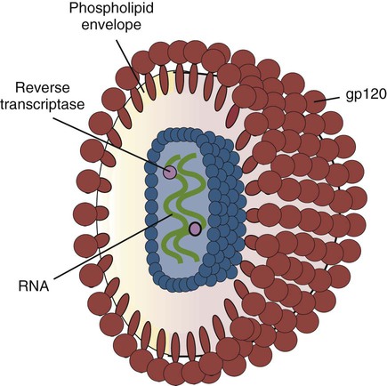
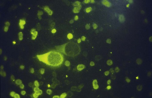
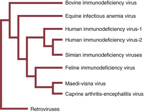
Secondary Immunological Defects


Secondary Immunological Defects


The immune system, like any body system, is subject to destruction and dysfunction as a result of attacks by pathogenic and environmental agents. Among the most important of these agents are microorganisms, especially viruses, toxins, stress of various types, old age, and malnutrition.


Virus-Induced Immunosuppression


Viruses that affect the immune system may be divided into those that affect primary lymphoid tissues and those that affect secondary lymphoid tissues. Both types of virus can cause profound immunodeficiencies. For example, in chickens, the infectious bursal disease virus (IBDV) destroys lymphocytes in the bursa of Fabricius. IBDV is not completely specific for bursal cells; it also destroys cells in the spleen and thymus. These tissues usually recover, whereas the bursa atrophies. The resulting immunosuppression, as might be predicted, is most evident in young birds infected soon after hatching, at a time when the bursa is actively engaged in generating B cells.


One of the most important animal viruses that destroys secondary lymphoid organs is canine distemper. Canine distemper virus, although it can multiply in many different cell types, has a predilection for lymphocytes, epithelia, and nervous tissue. Its primary cellular receptor is CD150, expressed on activated B and T cells. The distemper virus spreads from its initial invasion sites in the tonsils and bronchial lymph nodes to the bloodstream, where it kills both T and B cells and causes a lymphopenia. Subsequently it invades secondary lymphoid organs such as the spleen, lymph nodes, mucosal lymphoid tissues, and bone marrow, where it destroys more cells. The virus also invades and destroys the thymus. The shedding of infected cells from these lymphoid organs enables the virus to reach epithelial tissues and the brain. Canine distemper virus triggers lymphocyte and macrophage apoptosis and produces profound immunosuppression. It also suppresses production of interleukin-1 (IL-1) and IL-2 while stimulating prostaglandin release by macrophages. As a result, lymphocyte responses to mitogens are depressed, immunoglobulin levels fall, and skin allograft rejection is suppressed. This immunosuppression accounts, in large part, for the clinical signs of canine distemper. For example, many dogs with distemper develop Pneumocystis pneumonia. (Pneumocystis is a fungus that occurs in the lungs. It does not cause disease in immunocompetent animals but produces a severe pneumonia in animals with suppressed immune function. Indeed, the development of Pneumocystis pneumonia is evidence of a significant immunodeficiency.) If germ-free dogs are infected by virulent distemper virus, they develop a relatively mild disease, presumably because secondary infection cannot occur.


Loss of lymphocytes is common in virus infections since viral survival and persistence may require immunosuppression (Figure 38-1). Thus a lymphopenia occurs in feline panleukopenia, canine parvovirus-2 infection, feline leukemia, and African swine fever. Bovine virus diarrhea virus (BVDV) causes destruction of both B and T cells in the lymph nodes, spleen, thymus, and Peyer’s patches. As in canine distemper, surviving B cells fail to make immunoglobulins and respond poorly to mitogens. Viral destruction of Peyer’s patches causes intestinal ulceration and leads to secondary bacterial invasion. Both persistently infected cattle and normal cattle infected with cytopathic BVDV have depressed neutrophil functions, and bacterial clearance from the blood is impaired. A related virus, border disease virus, preferentially infects CD8+ T cells and interferes with their cytotoxic and immunoregulatory functions.


Herpesviruses are also immunosuppressive. For example, equine herpesvirus-1 causes a drop in T cell numbers and depresses cell-mediated responses in foals. Bovine herpesvirus-1 (BHV-1) also causes a drop in T cells and in the responses to T cell mitogens. Although BHV-1 stimulates bovine alveolar macrophages to express increased amounts of MHC class II and promotes antibody-mediated phagocytosis, it also depresses macrophage-mediated cytotoxicity and IL-1 synthesis. Parainfluenzavirus-3 and infectious bovine rhinotracheitis viruses have long been known to interfere with alveolar macrophage function. They inhibit phagosome-lysosome fusion, paving the way for secondary infections with Mannheimia hemolytica in stressed calves. Porcine reproductive and respiratory syndrome (PRRS) virus in pigs causes destruction of alveolar macrophages and predisposes affected animals to severe enzootic pneumonia. It also kills dendritic cells, a feature that may account for the ability of PRRS virus to persist in pigs for up to 6 months.


The effect of some viruses on the immune system may be relatively complex or anomalous. In maedi-visna, a neurological disease of sheep caused by a retrovirus, cell-mediated immune responses such as graft rejection are suppressed, whereas B cell responses are enhanced (Chapter 26). Some leukemia viruses may be selectively immunosuppressive, so that depression of the IgG response is greater than that of the IgM response. In equine infectious anemia, the IgG3 response is variably depressed, whereas synthesis of the other immunoglobulin classes remains unaffected.


The results of virus-induced lymphoid tissue destruction are readily seen. Animals are lymphopenic and have reduced lymphocyte responses to mitogens. For example, responses to phytohemagglutinin are depressed in influenza, measles, canine distemper, Marek’s disease, Newcastle disease, feline leukemia, bovine virus diarrhea, and lymphocytic choriomeningitis. Destruction of lymphoid tissues may also result in hypogammaglobulinemia or a reduced response to antigens. Thymic atrophy and lymphopenia are common manifestations of many virus infections, and before any primary immunodeficiency syndrome is diagnosed, rigorous steps must be taken to exclude the possibility that it is, in fact, secondary to a virus infection.


Retrovirus Infections in Primates


More than 40 lentiviruses have been isolated from nonhuman primates, especially African species. These simian immunodeficiency viruses include SIVmac isolated from a rhesus macaque (Macaca mulatta) in a laboratory; SIVagm from an African green monkey (Chlorocebus sabaeus); SIVsm from a sooty mangabey (Cercocebus atys); SIVmnd from a mandrill (Mandrillus sphinx); and most important, SIVcpz from a chimpanzee (Pan troglodytes). SIVcpz is the ancestor of HIV-1, whereas SIVsm is the ancestor of HIV-2. All these isolates selectively invade CD4+ T cells. When SIVmac infects rhesus macaques and other Asian species, it stimulates a strong but ineffective immune response. Viral replication continues and eventually causes an immunodeficiency syndrome similar to human AIDS. The infection is believed to be transmitted sexually. Clinical disease progression is slow, but the animals eventually develop lymphadenopathy, severe weight loss, chronic diarrhea, lymphomas, neurological lesions, and opportunistic infections by organisms such as Pneumocystis, Mycobacterium avium-intracellulare, Candida albicans, and Cryptosporidium parvum. The macaques are immunosuppressed as a result of depletion of CD4+ T cells, macrophages, and dendritic cells. The virus invades both T cells and macrophages using two cellular receptors, CD4 and either the receptor for the chemokine CCR5 or for CXCR4. About 25% of infected animals do not mount a significant response to SIV and die within 3 to 5 months as a result of a severe SIV encephalitis. Macaques that mount an immune response usually die 1 to 3 years after infection. Spontaneous recovery does not occur. The other SIVs cause persistent viremia but rarely result in disease in African primates. This is because they rapidly develop an antiinflammatory response that prevents chronic T cell activation and apoptosis.


Type D Simian Retroviruses


An acquired immunodeficiency syndrome (AIDS) develops in primates infected with one of several endogenous type D simian retroviruses (SRVs). These viruses, much more common than the lentiviruses, are transmitted by biting; vertical transmission rarely occurs. SRVs have a much broader tissue tropism than the SIVs and, in addition to lymphocytes and macrophages, can also infect fibroblasts, epithelial cells, and the central nervous system. The SRVs destroy both B and T cells, leading to death from opportunistic infections. The syndrome is associated with a profound drop in serum IgG and IgM levels and a severe lymphopenia. Monocyte function is unimpaired, but surviving lymphocytes do not respond to mitogens. Affected monkeys are also profoundly neutropenic. On necropsy, the monkeys have a generalized lymphadenopathy, hepatomegaly, and splenomegaly. There is a loss of lymphocytes from the T-dependent areas of the secondary lymphoid organs. B cell areas show an initial hyperplasia of the secondary follicles followed by the loss of these follicles and an absence of plasma cells. These histological changes are very similar to those seen in AIDS in humans. In many cases, normally innocuous agents such as Pneumocystis, cytomegalovirus, C. parvum, and C. albicans cause infection. Some affected monkeys develop tumors such as fibrosarcomas. About half of the infected animals develop neutralizing antibodies and survive the disease. The others die from septicemia or diarrhea with wasting.


Retrovirus Infections in Cats


Feline Leukemia


Feline leukemia virus (FeLV) is an oncogenic retrovirus that can cause both proliferative and degenerative diseases in cats (Figure 38-2). Three naturally occurring viral variants are recognized based on the structure of their gp70 protein. FeLV-A is the predominant, naturally transmitted variant. It is present in all FeLV-infected cats. The other variants are only found in association with FeLV-A. When FeLV-A combines with endogenous retroviral sequences (sequences that are stably integrated into the cat genome and are normally never expressed but are genetically transmitted), FeLV-B is formed. FeLV-B is found in about 50% of viremic cats and has a greater propensity than FeLV-A to cause tumors. FeLV-C is found in about 1% to 2% of infected cats and arises from FeLV-A through a mutation in the envelope gene. It is much more suppressive for bone marrow than FeLV-A.


FOCMA


A unique surface protein, called feline oncornavirus cell membrane antigen (FOCMA), is expressed on FeLV-infected cells. It is coded for by endogenous retroviral genes within the cat genome. It is not expressed on normal cells but rather on cells infected with FeLV. It was originally believed that the presence of FOCMA on a cell membrane identified the cell as an FeLV-induced tumor cell. Of those cats that fail to make neutralizing antibodies to FeLV and remain viremic, about 80% develop antitumor activity by making antibodies against FOCMA. A cat that makes antibodies to FOCMA can usually destroy virus-induced tumor cells. Unfortunately, antibodies to FOCMA do not confer protection against the FeLV-induced degenerative diseases, and viremic cats that fail to produce anti-FOCMA antibodies are fully susceptible to all the FeLV syndromes, including lymphosarcoma. Some feline lymphosarcoma cells may express FOCMA in the absence of evidence of FeLV infection.


Pathogenesis


Once FeLV infects a cat, the virus first grows in the lymphoid tissues of the pharynx and tonsils. This is followed by a transient viremia as it spreads throughout the body and infects the other lymphoid organs. A mild lymphopenia occurs 1 to 2 weeks after infection. This is of variable duration but lasts longer in young cats than in adults. There is also a variable neutropenia. Antibodies develop between 7 and 42 days after the onset of infection, and the virus is cleared between 28 and 42 days. Virus can be found in the thymus at day 1, in blood between 2 and 145 days, and in lymphoid organs between 3 and 28 days. The presence of both antibodies and virus results in the production of immune complexes and the development of a membranoproliferative glomerulonephritis. Some cats may lose their viremia but remain latently infected. In latently infected animals, the virus persists in the bone marrow, but there is no virus in the blood, and virus-neutralizing antibodies are present. Treatment with steroids or culturing bone marrow cells in vitro allows productive re-expression of the virus. Stress (e.g., crowding, shipping) may cause a recurrence of viremia in 5% to 10% of cats. The presence of neutralizing antibodies does not correlate well with the disease state, and challenging recovered cats may not provoke a secondary antibody response. Prenatal or early infection of kittens with FeLV can also result in a persistent viremia.


Immunosuppression


T Cell Defects

FeLV develops T cell tropic variants as a result of mutations in their envelope genes. These immunodeficiency-inducing variants replicate to high numbers in T cells. They enter T cells by binding to two receptors. One receptor is a phosphate transporter protein (Pit1). The second is a novel cell surface protein called FeLIX (“FeLV infectivity X-essory protein”). The lymphopenia in FeLV-infected cats is due to a loss of CD4+ T cells. CD8+ T cells may also drop in the early stages of the disease so that the CD4/CD8 ratio may remain within normal limits. (The CD4/CD8 ratio in normal cats ranges from about 0.4 to 3.5, with a median value of about 1.9.) CD8+ T cell numbers eventually recover, and the CD4/CD8 ratio then drops. B cell numbers may also be depressed, but this depends on the severity of secondary infections. Kittens infected with FeLV develop a wasting syndrome associated with thymic atrophy and recurrent infections. Depending on the severity of the secondary infections, this may be associated with either lymphoid atrophy or lymphoid hyperplasia. In cats without secondary infection, lymphoid atrophy is associated with loss of cells from the paracortical areas of lymph nodes. Changes in the spleen are less marked but may result in a reduction in the entire white pulp. As a result of T cell loss, FeLV infected cats have depressed cell-mediated immunity. This depression is probably due to the effects of p15e, the immunosuppressive envelope protein of the FeLV virus, which is produced in very large quantities by dying cells. The p15e suppresses the responses of cats to FOCMA, suppresses lymphocyte mitogen responses, and blocks the responses of T cells to IL-2. As a result, FeLV-infected cats may carry skin allografts for about twice as long as normal cats (24 days compared with 12). Leukocytes of infected cats produce significantly less IL-2 than leukocytes from normal cats. This decline is especially marked in cats with leukemia or lymphosarcoma arising in the thymus. This immunosuppression also predisposes viremic cats to secondary diseases such as feline infectious peritonitis, mycoplasmosis, toxoplasmosis, septicemia, and fungal infections. Bone marrow stem cells are also inhibited by p15e, preventing production of erythroid cells and causing a nonregenerative anemia.


B Cell Defects

In contrast to the severe T cell dysfunction, B cell activities in FeLV-infected cats are only mildly impaired. There may be poor responses to low doses of antigen as well as reduced IgM production, but serum IgG levels remain normal. Because B cell function and antibody production are relatively normal in chronically infected cats, antibodies to the virus are produced in large quantities. These antibodies combine with circulating virions or soluble proteins to form immune complexes. The immune complexes are deposited in the renal glomeruli and cause severe mesangioproliferative glomerulonephritis, leading to hypoproteinemia, edema, uremia, and death. Viral antigens binding to erythrocytes can also cause an antiglobulin-positive hemolytic anemia. Immune complexes also activate the classical complement pathway. As a result, complement will be consumed, and FeLV cats may have very low levels of complement. This loss may reduce resistance to tumors since normal cat serum infused into leukemic cats can cause tumor regression.


FeLV-AIDS


During natural FeLV infection, a highly immunosuppressive form of the virus may develop. Called FeLV-AIDS, this causes fatal immunodeficiency in nearly 100% of infected cats. The isolate consists of two virus populations. One, designated 61E, is replication competent but does not induce immunodeficiency disease by itself. The other, 61C, is replication defective, but when inoculated together with 61E, it induces a fatal immunodeficiency syndrome.


The immunodeficiency syndrome is characterized by progressive weight loss and lymphoid hyperplasia followed by severe lymphoid depletion, chronic diarrhea, and opportunistic infections. The onset of disease is preceded by an inability to respond to T-independent antigens. As early as 9 weeks after infection, the CD4+ T cells produce lower levels of B cell stimulatory cytokines. This is followed by a drastic drop in CD4+ T cells, whereas CD8+ T cell and B cell numbers remain normal. The clinical defect in FeLV-AIDS is an inability to mount antibody responses, although in vitro B cell function appears to be normal. The immunodeficiency is associated with mutations in a 34-amino acid sequence at the C-terminus of viral gp70. The mutation changes the conformation of the surface glycoprotein and prevents the virus from blocking infection by additional virions, leading to subsequent cell killing.


Immunity


About 40% of cats infected with FeLV do not mount an adequate immune response against the virus and become persistently infected. Persistently infected cats remain viremic. The remaining 60% of infected cats mount a strong immune response. These cats develop virus-neutralizing antibodies to the major envelope glycoprotein, gp70. Immune cats also develop virus-specific cytotoxic T cells to viral gag/pro antigens. These prevent the virus from invading cells, and these cats become strongly immune. Antibodies against antigens other than gp70 may also play a role in immunity.


Three types of effective vaccines are currently available against FeLV. One type contains supernatant fluid from a cell line persistently infected with FeLV. This fluid contains several of the major protein antigens of FeLV. The second type of FeLV vaccine consists of inactivated whole virions from tissue culture, which are usually administered with a powerful adjuvant. The third type of FeLV vaccine is a canarypox vectored recombinant product that can be administered without an adjuvant. Widespread vaccination has significantly reduced the prevalence of this disease in the United States. These vaccines do appear to differ in their ability to prevent latent infections, although they are effective in preventing the development of clinical disease.


Diagnosis


The introduction of sensitive molecular diagnostic techniques to replace serologic assays has changed our views on persistent FeLV infections. Real-time and reverse-transcriptase polymerase chain reaction (PCR) assays are much more sensitive and specific than virus isolation, antigen detection, or immunofluorescence. These tests have shown that many cats may have FeLV DNA integrated into their cells but never develop an antigenemia. Vaccines may be able to prevent the development of clinical disease but not proviral integration. These latent infections may persist for years, and viremia or disease develops occasionally. On the other hand, this integrated viral DNA may also be required for long-term protection. Other cats may have both detectable viral nucleic acids and antigenemia (i.e., active infections). FeLV antigenemia may be detected by an antigen-capture ELISA, by the membrane filter technique, or by rapid immunochromatography of blood or serum. A direct immunofluorescent test on a buffy coat smear using antibodies to group-specific antigen can detect cell-associated antigen and hence intracellular viremia (Figure 38-3). Alternative testing methods include testing saliva or tears using material collected on a swab or filter paper strips.


Feline Immunodeficiency Virus


Feline immunodeficiency virus (FIV) was originally isolated from cats with clinical immunodeficiency. The virus is an enveloped, single-stranded RNA virus belonging to the lentivirus subgroup of retroviruses. It is differentiated from FeLV (a γ-retrovirus) by the biochemical requirements of its reverse transcriptase. (FIV reverse transcriptase requires magnesium, whereas FeLV requires manganese.) FIV is related to HIV, the cause of AIDS (Figure 38-4). FeLV and FIV are distinctly different viruses, and antibodies made against one do not react with the other. Nevertheless, approximately 12% to 33% of FIV-infected cats may also be infected with FeLV, an especially potent immunosuppressive mixture. At least five different genetic clades (or subtypes) of FIV have been identified. Subtype variations may account for differences in pathogenicity, tissue tropism, and clinical disease.


< div class='tao-gold-member'>

Stay updated, free articles. Join our Telegram channel

Jul 18, 2016 | Posted by in PHARMACOLOGY, TOXICOLOGY & THERAPEUTICS | Comments Off on Secondary Immunological Defects

Full access? Get Clinical Tree

Get Clinical Tree app for offline access