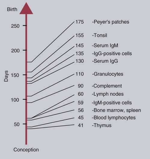
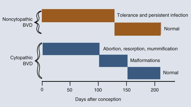
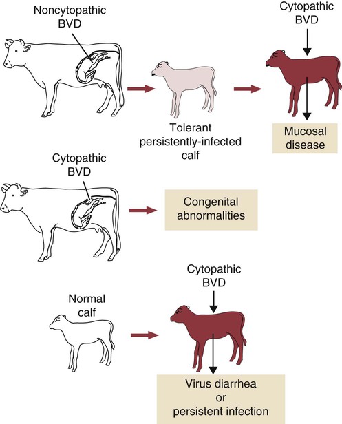
Immunity in the Fetus and Newborn


Immunity in the Fetus and Newborn


When a mammal is born, it emerges from the sterile uterus into an environment where it is immediately exposed to a host of microorganisms. Its surfaces, such as the gastrointestinal tract, acquire a complex microbial flora within hours. If it is to survive, the newborn animal must be able to control this microbial invasion. In practice, the adaptive immune system takes some time to become fully functional, and innate mechanisms are responsible for the initial resistance to infection. In some species with a short gestation period, such as mice, the adaptive immune system may not even be fully developed at birth. In animals with a long gestation period, such as the domestic mammals, the adaptive immune system is fully developed at birth but cannot function at adult levels for several weeks. The complete development of adaptive immunity depends on antigenic stimulation. The proper development of B cells and B cell receptor (BCR) diversity requires clonal selection and antigen-driven cell multiplication (Chapter 15). Thus, newborn mammals are vulnerable to infection for the first few weeks of life. They need assistance in defending themselves at this time. This temporary help is provided by the mother in the form of antibodies and possibly T cells. The passive transfer of immunity from mother to newborn is essential for survival.


Development of the Immune System


The development of the immune system in the mammalian fetus follows a consistent pattern. The thymus is the first lymphoid organ to develop, followed closely by the secondary lymphoid organs. B cells appear soon after the development of the spleen and lymph nodes, but antibodies are not usually found until late in fetal life, if at all (Box 21-1). The ability of the fetus to respond to antigens develops very rapidly after the lymphoid organs appear, but all antigens are not equally capable of stimulating fetal lymphoid tissue. The immune system develops in a series of steps, each step permitting the fetus to respond to more antigens. These steps are driven by a gradual increase in the use of gene conversion or somatic mutation to increase antibody diversity. The ability to mount cell-mediated immune responses develops at the same time as antibody production. T cell receptor (TCR) diversity is also limited in the fetus and neonate, and their cytokine production may be low. This may simply be due to their lack of exposure to foreign antigens.


Specific Animal Immune Systems


Foal


The gestation period of the mare is about 340 days. Lymphocytes are seen first in the thymus at about 60 to 80 days postconception. They are found in the mesenteric lymph node and intestinal lamina propria at 90 days and in the spleen at 175 days. Blood lymphocytes appear at about 120 days. A few plasma cells may be seen at 240 days. Graft-versus-host disease, a cell-mediated response, has developed in immunodeficient foals transplanted with tissues from a 79-day-old fetus. The equine fetus can respond to coliphage T2 at 200 days postconception and to Venezuelan equine encephalitis virus at 230 days. Newborn foals have detectable quantities of IgM and IgG and occasionally IgG3 in their serum, but IgE production in the horse does not begin until foals are 9 to 11 months of age. Like other large herbivores, the foal has a well-developed ileal Peyer’s patch that serves as a primary lymphoid organ and eventually involutes. Major B cell markers are expressed by 90 to 120 days gestation. IGHM and IGLC transcripts are expressed in liver, bone marrow, and spleen at all ages. The expression of essential B cell genes demonstrates that gene recombination and immunoglobulin class switching occur during equine fetal life. As a result, small amounts of IgM and IgG are detectable at birth. Despite this competence, B cell functions may be actively suppressed by regulatory T (Treg) cells during the first few months of a foal’s life.


Calf


Although the gestation period of the cow is 280 days, the fetal thymus is recognizable by 40 days postconception. The bone marrow and spleen appear at 55 days. Lymph nodes are found at 60 days, but Peyer’s patches do not appear until 175 days (Figure 21-1). Blood lymphocytes are seen in fetal calves by day 45, IgM+ B cells by day 59, and IgG+ B cells by day 135. The time of appearance of serum antibodies depends on the sensitivity of the techniques used. It is therefore no accident that the earliest detectable immune responses are those directed against viruses, using highly sensitive virus neutralization tests. Fetal calves have been reported to respond to rotavirus at 73 days, to parvovirus at 93 days, and to parainfluenza 3 virus at 120 days. Fetal blood lymphocytes can respond to mitogens between 75 and 80 days, but this ability is temporarily lost near the time of birth as a result of high serum steroid levels. T cell subpopulations are present in calves at levels comparable to adults, but B cell numbers increase significantly during the first 6 months after birth.


Lamb


The gestation period of the ewe is about 145 days. Major histocompatibility complex (MHC) class I–positive cells can be detected by day 19, and MHC class II positive cells can be found by day 25. The thymus and lymph nodes are recognizable by 35 and 50 days postconception, respectively. Gut-associated follicles appear in the colon at 60 days, jejunal Peyer’s patches at about days 75 to 80, and ileal Peyer’s patches at days 110 to 115. Blood lymphocytes are seen in fetal lambs by day 32, and CD4+ and CD8+ cells appear in the thymus by 35 to 38 days. B cells are detectable at 48 days in the spleen and by that time have already begun to rearrange their IGLV genes. C3 receptors appear by day 120, but Fc receptors do not appear until the animal is born. Fetal liver lymphocytes can respond to phytohemagglutinin by 38 days. Lambs can produce antibodies to phage ϕX174 at day 41 and reject skin allografts by day 77. Some fetal lambs can produce antibodies to Akabane virus by as early as 50 days postconception. Antibodies to Cache Valley virus can be provoked by day 76, to SV40 virus by day 90, to T4 phage by day 105, to bluetongue virus by day 122, and to lymphocytic choriomeningitis virus by day 140. The proportions of α/β and γ/δ T cells change as lambs mature. Thus, 1 month before birth, 18% of blood T cells are γ/δ positive. By 1 month after birth, they constitute 60% of blood T cells.


Piglet


The gestation period of the sow is about 115 days. B cells appear in the yolk sac at day 20, progress to the fetal liver by day 30, and progress to the bone marrow by day 45. The first SWC3+ leukocytes can be found in the yolk sac and liver on day 17. The thymus develops by 40 days postconception and is colonized by two waves of T cell progenitors beginning on day 38. γ/δ T cells appear first in the thymus and in peripheral blood about 10 days later. α/β T cells develop by day 55, but their numbers grow rapidly so that they predominate late in gestation. The intestinal lymphoid tissues are devoid of T cells at birth. CD4+ T cells appear in the intestine at 2 weeks of age, and CD8+ T cells appear at 4 weeks. Their proliferation appears to be driven by the intestinal microflora. IgM+ B cells can be found in liver at 40 days, spleen by day 50, and bone marrow by day 60. Fetal piglets can produce antibodies to parvoviruses at 58 days and can reject allografts at about the same time. Blood lymphocytes can respond to mitogens between 48 and 54 days. Natural killer (NK) cell activity does not develop until several weeks after birth, although cells with an NK phenotype can be identified at 45 days’ gestation in spleen and umbilical blood.


B cells are the first lymphocytes to appear in peripheral blood. The number of circulating B cells rises significantly between 70 and 80 days’ gestation. The response to antigens in the fetus is of the IgM type, but newborn and fetal piglets also produce a small immunoglobulin that may not have light chains. It is interesting to note that B cells can be found in the thymus of newborn pigs.


The molecular development of the antibody repertoire has been followed in the developing pig. Thus, VDJ rearrangement is first seen in the fetal liver at day 30. However, the fetal piglet does not initially use all its IGHV or IGHD genes. Likewise, N-region addition does not occur before day 40, suggesting that the onset of terminal deoxynucleotidyltransferase activity occurs after that time. IgM, IgA, and IgG transcripts are present from 50 days in all major lymphoid organs. Piglets are thus born with relatively limited B cell diversity. B cell numbers increase for the first 4 weeks after birth, but their antigen-binding repertoire does not begin to expand until 4 to 6 weeks of age. Similar studies on rabbits have shown that the fetal immunoglobulin repertoire does not diversify until after birth, and this appears to be triggered by bacterial colonization of the gastrointestinal tract.


Chick


Stem cells arise in the yolk sac membrane and migrate to the thymus and bursa at 5 to 7 days’ incubation. These cells differentiate within the bursa, and follicles develop by day 12. Lymphocytes with surface IgM may be detected in the bursa by day 14, and antibodies to keyhole limpet hemocyanin and to sheep erythrocytes may be produced by 16 and 18 days’ incubation, respectively. Lymphocytes with surface IgY develop on day 21 around the time of hatching, whereas IgA-positive cells first appear in the intestine 3 to 7 days after hatching. Vaccination of 18-day embryonated eggs is commonly employed in the modern poultry industry. The major in ovo vaccine is against the Marek’s disease herpesvirus, but others against Newcastle disease and coccidiosis are available, and those against infectious bronchitis and infectious bursal disease are under development.


The Immune System and Intrauterine Infection


Although a fetus is not totally defenseless, it is less capable than an adult of combating infection. Its adaptive immune system is not fully functional; as a result, some infections may be mild or unapparent in the mother but severe or lethal in the fetus. Examples include bluetongue, infectious bovine rhinotracheitis [bovine herpesvirus 1 (BHV-1)], bovine viral diarrhea, rubella in humans, and toxoplasmosis. Fetal infections commonly trigger an immune response as shown by lymphoid hyperplasia and elevated immunoglobulin levels. For this reason the presence of any immunoglobulins in the serum of a newborn, unsuckled animal suggests infection in utero.


In general, the response to these viruses is determined by the state of immunological development of the fetus. For example, if live bluetongue virus vaccine, which is nonpathogenic for normal adult sheep, is given to pregnant ewes at 50 days postconception, it causes severe lesions in the nervous system of fetal lambs, including hydranencephaly and retinal dysplasia, whereas if it is given at 100 days postconception or to newborn lambs, only a mild inflammatory response is seen. Bluetongue vaccine virus given to fetal lambs between 50 and 70 days postconception may be isolated from lamb tissues for several weeks, but if given after 100 days, reisolation is not usually possible. Akabane virus acts in a similar fashion in lambs. If given before 30 to 36 days postconception, it causes congenital deformities. If given to older fetuses, it provokes antibody formation and is much less likely to cause malformations. Piglets that receive parvovirus before 55 days postconception will usually be aborted or stillborn. After 72 days, however, piglets will normally develop high levels of antibodies to the parvovirus and survive. Prenatal infection of calves with BHV-1 results in a fatal disease, in contrast to postnatal infections, which are relatively mild. The transition between these two types of infection occurs during the last month of pregnancy.


The effects of the timing of viral infection are well seen with bovine viral diarrhea virus (BVDV). If a cow is infected early in pregnancy (up to 50 days), she may abort. On the other hand, infections occurring between 50 and 120 days, before the fetus develops immune competence, lead to asymptomatic persistent infection because the calves develop tolerance to the virus (Figure 21-2). These calves are viremic yet, because of their tolerance, fail to make antibodies or T cells against the virus. Some of these calves may show minor neurologic problems and failure to thrive, but many are clinically normal. If the cow is infected with BVDV between 100 and 180 days postconception, calves may be born with severe malformations involving the central nervous system and eye, as well as jaw defects, atrophy, and growth retardation. Vaccines containing modified live BVDV may have a similar effect if administered at the same time. Calves infected after 150 to 180 days’ gestation are usually clinically normal.


Since they are specifically tolerant to BVDV, persistently infected calves shed large quantities of virus in body secretions and excretions and so act as a major source of infectious virus. The persistently infected calves may also produce neutralizing antibodies if immunized with a live BVDV vaccine of a serotype different from that of the persistent virus. Despite this, the original virus will persist in these animals. These persistently infected calves grow slowly and often die of opportunistic infections such as pneumonia before reaching adulthood. (BVDV has a tropism for lymphocytes and is immunosuppressive.) Their neutrophil phagocytic and bactericidal functions are also depressed.


BVD viruses occur in two distinct biotypes: cytopathic and noncytopathic. (The name derives from their behavior in cell culture, not their pathogenicity in animals.) Noncytopathic strains do not trigger type I interferon (IFN) production and therefore can survive in calves and cause persistent infections. Cytopathic strains induce IFN production and cannot cause persistent infection. These cytopathic strains, however, do cause mucosal disease (MD), a severe enteric disease leading to profuse diarrhea and death (Figure 21-3). Mucosal disease develops as a result of a mutation in a nonstructural viral gene that changes the BVDV biotype from noncytopathic to cytopathic while the animal fails to produce neutralizing antibodies or T cells. The cytopathic strain can spread between tolerant animals and lead to a severe mucosal disease outbreak. Both cytopathic and noncytopathic viruses can be isolated from these animals. Recombination may also occur between persistent noncytopathic strains and cytopathic strains in vaccines and lead to MD outbreaks. Although some of the lesions in MD are attributable to the direct pathogenic effects of BVDV, glomerulonephritis and other immune complex–mediated lesions also develop. The reasons for this are unclear but may reflect superinfection or the production of non-neutralizing antibodies. Because persistently infected calves can reach adulthood and breed, it is possible for BVD infection to persist indefinitely within carrier animals and their progeny. Epidemiological studies suggest that between 0.4% and 1.7% of cattle in the United States are persistently infected in this way.


Immune Response of Newborn Mammals


After developing in the sterile environment of the uterus, newborn mammals first encounter a diverse population of microbes at the time of birth. They must be able to combat any attempted invasion immediately. The young mammals are capable of mounting both innate and adaptive immune responses at birth. However, any adaptive immune response mounted by a newborn must be a primary response with a prolonged lag period and low concentrations of antibodies. Innate immune responses are therefore critical for survival in the first weeks of life.


Role of the Intestinal Microflora


The development of the newborn immune system is largely driven by the intestinal microflora (Chapter 22). In its absence, “germ-free” mammals fail to fully develop their mucosal lymphoid tissues. The commensal flora generates a complex mixture of pathogen-associated molecular patterns (PAMPs) that act through epithelial cell toll-like receptors (TLRs). Likewise, microbial antigens are taken up by dendritic cells and presented to CD4+ T cells. These signals collectively promote the functional development of the immune system. The intestinal microflora also plays a key role in determining any Th1 or Th2 bias in immune function. This is the basis of the “hygiene hypothesis,” the idea that the development of allergies is influenced by microbial exposure early in life (Chapter 28).


Innate Immunity


Newborns can produce a diverse array of antimicrobial molecules, including lectins such as the pentraxins and collectins, peptides such as the defensins, and lactoferrin and lysozyme. Surfactant proteins A and D as well as β-defensin 1 and TLR4 are produced in the preterm lamb lung. As a result, invaders can be killed relatively efficiently. TLRs are present and functional in the newborn. In the fetal pig, neutrophils at 90 days postconception are fully capable of phagocytosing bacteria such as Staphylococcus aureus. However, they are deficient in bactericidal activity, which only reaches adult levels 10 days later. Near birth, the phagocytic and bactericidal capacity of these neutrophils declines as a result of increased steroid levels. The neutrophils of newborn foals move relatively slowly compared with their dams. The serum of newborn mammals, however, is deficient in some complement components, resulting in a poor opsonic activity. Serum C3 increases rapidly after birth in newborn piglets and reaches adult levels by 14 days of age.


After birth, macrophages are able to support the growth of some viruses that macrophages from adult animals do not. Virucidal activity is gradually acquired. Changes also occur in the distribution of macrophages. Newborn piglets have few pulmonary intravascular macrophages. During the first few days after birth, blood monocytes adhere to the pulmonary capillary endothelium and differentiate into macrophages. In the newborn piglet, 75% of particles are removed from the blood by the liver and spleen, but by 2 months of age, 75% are removed by the lungs. The alveolar macrophages of newborn pigs have poor phagocytic activity, but this is effectively acquired by 7 days.


Newborn calves have fewer NK cells than adults, but these respond more strongly to simulation with interleukin-2 (IL-2) or IL-15 and are more cytotoxic! Age-dependent changes occur in the level of acute-phase proteins in newborn calves. Serum amyloid A (SAA), lipopolysaccharide-binding protein, haptoglobin and α1-acid glycoprotein are present in high concentrations immediately after birth but gradually decline by 21 days. In developing piglets, IL-8 and tumor necrosis factor-α (TNF-α) production by blood monocytes increased significantly during the postnatal period, whereas monocyte production of IL-1β was unchanged.

< div class='tao-gold-member'>

Stay updated, free articles. Join our Telegram channel

Jul 18, 2016 | Posted by in PHARMACOLOGY, TOXICOLOGY & THERAPEUTICS | Comments Off on Immunity in the Fetus and Newborn

Full access? Get Clinical Tree

Get Clinical Tree app for offline access