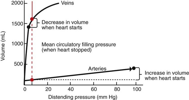
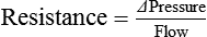
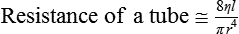
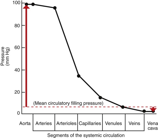
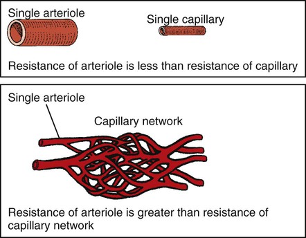
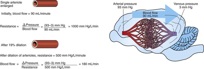
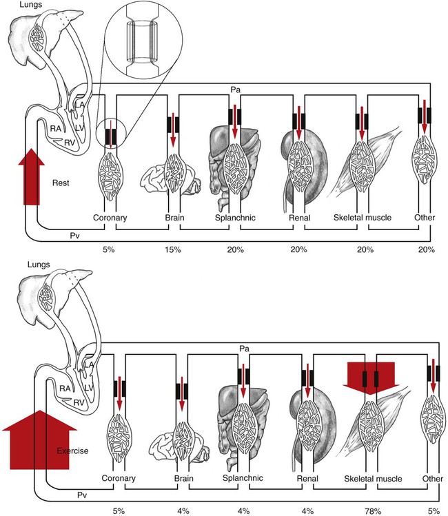
The Systemic and Pulmonary Circulations


The Systemic and Pulmonary Circulations


Blood Pressure Represents a Potential Energy That Propels Blood Through the Circulation


The systemic circulation has the aorta as its inlet point and the venae cavae as its outlet. The remainder of the circulation (i.e., right heart, pulmonary circuit, and left heart) is, by definition, the central circulation. Blood enters the central circulation from the venae cavae and leaves the central circulation through the aorta.


Figure 22-1 shows the normal pressure profile in the systemic circulation. This figure portrays the pressures that would be measured if a miniature pressure gauge were inserted into the various vessels that blood passes through in its journey through the systemic circulation. The blood pressure is highest in the aorta (typically, mean aortic pressure is 98 mm Hg) and lowest in the venae cavae (typically, 3 mm Hg). The difference between these pressures (i.e., 95 mm Hg) constitutes the driving force for the movement of blood, by bulk flow, through the systemic circulation. As discussed in Chapter 18, such a pressure difference between the inlet and outlet of a tube (or system of tubes) is called perfusion pressure difference (or more commonly, just perfusion pressure).


Aortic blood pressure can be thought of as the potential energy available to move blood; the decrease in pressure in the sequential segments of the systemic circuit represents the amount of this potential energy that is “used up” in moving blood through each segment. Pressure energy is used up through friction, which is generated as the molecules and cells of blood rub against each other and against the walls of the blood vessels. The energy used up through friction is converted to heat, although the actual increase in the temperature of the blood and blood vessels as a result of friction is very small.


The amount of the blood pressure energy used up in each of the sequential segments of the systemic circulation depends on the amount of friction or resistance that the blood encounters. The aorta and large arteries offer very little resistance to blood flow (very little friction), so the blood pressure decreases only a little in these vessels (from 98 to about 95 mm Hg). The greatest pressure decrease (greatest loss of pressure energy through friction) occurs as blood flows through arterioles; that is, the resistance to blood flow is greater in the arterioles than in any other segment of the systemic circulation. The capillaries and the venules offer a substantial resistance to blood flow, but the resistance (and therefore the pressure decrease) is not as great in these vessels as it is in the arterioles. The large veins and the venae cavae are low-resistance vessels, so little pressure energy is expended in driving the blood flow through these vessels.


The pumping of blood by the heart maintains the pressure difference between the aorta and the venae cavae. If the heart stops, blood continues to flow for a few moments from the aorta toward the venae cavae. As this blood leaves the aorta, the aortic walls become less distended, and the blood pressure inside the aorta decreases. As extra blood accumulates in the venae cavae, they become more distended than before, and the blood pressure inside the venae cavae increases. Soon, there is no pressure difference between the aorta and the venae cavae. Blood flow in the systemic circuit ceases, and the pressure everywhere in the systemic circulation is the same. It has been demonstrated experimentally that this eventual pressure is about 7 mm Hg. This pressure, in a static circulation, is called the mean circulatory filling pressure. The mean circulatory filling pressure is greater than zero (i.e., above atmospheric pressure), because there is a “fullness” to the circulation; that is, even if the heart stops, blood still distends the vessels that contain it. The vessel walls, being elastic, recoil (“push back”) against this distention, which accounts for the persistence of pressure in the circulation even if the heart stops. If a transfusion of blood is administered to an animal with the heart stopped, the vessels become more distended, and the mean circulating filling pressure rises above 7 mm Hg. Conversely, if blood is removed from an animal with the heart stopped, the pressure everywhere falls to a level below 7 mm Hg.


Consider what happens if the heart is restarted in an animal after the pressure has equalized everywhere at 7 mm Hg. With each heartbeat, the heart takes some blood out of the venae cavae and this volume of blood is transferred (via the pulmonary circulation) into the aorta. The volume of blood in the venae cavae decreases, so the venae cavae become less distended and vena caval pressure drops below 7 mm Hg. The volume of blood in the aorta increases, so the aorta becomes more distended and aortic pressure rises above 7 mm Hg. As illustrated in Figure 22-1, the vena caval pressure drops about 4 mm Hg (from 7 to 3 mm Hg), and the aortic pressure rises about 91 mm Hg (from 7 to 98 mm Hg). It is important to understand why the pressure decreases only a little in the venae cavae but increases so much in the aorta, even though the volume of blood removed from the venae cavae with each heartbeat is the same as the volume of blood added to the aorta. The reason is that the veins are much more compliant (distensible) than the arteries; one can add or remove blood from veins without changing the venous pressure very much, whereas the addition or removal of blood from arteries changes the arterial pressure a great deal.


A compliant vessel readily distends when pressure or volume is added. It yields to pressure. By definition, compliance is the change in the volume within a vessel or a chamber divided by the associated change in distending (transmural) pressure, as follows:


Compliance=ΔVolumeΔTransmural pressure


image

Compliance corresponds to the slope of a volume-versus-pressure graph. As illustrated in Figure 22-2, veins are about 20 times more compliant than arteries (over the range of pressures typically encountered in the circulation). Therefore, veins can accept or give up a large volume of blood without incurring much of a change in pressure. Veins readily expand or contract to accommodate the changes in blood volume that occur with fluid intake (e.g., drinking) or fluid loss (e.g., sweating). Veins thus function as the major blood volume reservoirs of the body. In contrast, arteries function as pressure reservoirs, providing the temporary storage site for the surge of pressure energy that is created with each cardiac ejection. Arteries are tough vessels, with low compliance. Therefore, arteries can accommodate a large increase in pressure during a cardiac ejection and then sustain the pressure high enough between cardiac ejections to provide a continuous flow of blood through the systemic circulation.


image
FIGURE 22-2 Typical relationships between volume (of blood) and distending pressure for veins and arteries. Veins are more compliant (easier to distend) than arteries, so they hold a greater volume of blood for a given distending pressure. This concept is illustrated for a distending pressure of 7 mm Hg (vertical dashed red line), which is a normal value for the mean circulatory filling pressure (the pressure that would exist in the circulation if the heart were stopped, as shown in Figure 22-1). For a distending pressure of 7 mm Hg, the veins contain about 1600 mL of blood and the arteries only 125 mL (red circles). When the heart is restarted, the venous volume decreases, and the arterial volume increases (black circles). Because the veins are much more compliant than the arteries, the venous pressure changes very little (decreases from 7 to 3 mm Hg), whereas the arterial pressure changes greatly (increases from 7 to 98 mm Hg).


Vascular Resistance Is Defined as Perfusion Pressure Divided by Flow


Everyday experience tells us that it is easier to force fluid through a large tube than through a small tube. For example, it is easier to drink a milk shake through a large-diameter straw than through a small-diameter straw. For a given driving force (perfusion pressure difference), the flow is higher in the large tube because it offers less resistance to flow (less friction) than the small tube. The precise definition of resistance is:


Resistance=ΔPressureFlow


image

Where Δ Pressure is perfusion pressure difference, or simply perfusion pressure (i.e., the pressure at the tube inlet minus the pressure at its outlet). Figure 22-3 presents these concepts in pictorial and graphic form. The dashed lines in this figure indicate that a perfusion pressure of 60 mm Hg causes a flow of 1600 milliliters per minute (mL/min) through the large tube. Thus the resistance of the large tube is 37.5 mm Hg/liter per minute (L/min). The same perfusion pressure (60 mm Hg) causes a flow of only 100 mL/min through the small tube. The resistance of the small tube is therefore 600 mm Hg/L/min. The resistance of the small tube is 16 times greater than the resistance of the large tube.


In the late 1800s the French physician J.L.M. Poiseuille demonstrated the dominant effect of radius on the resistance of a tube. He showed the following:


Resistance ofa tube8ηlπr4


image

Where l is the length of the tube, r is the radius, η is the viscosity of the fluid flowing through the tube, and π has its usual meaning.


This equation (Poiseuille’s law) emphasizes that radius (r) is the dominant factor influencing the resistance of a tube; resistance varies inversely with the fourth power of radius. Doubling the radius of a tube decreases its resistance by a factor of 16 (24). This explains why using a larger diameter straw makes it so much easier to drink a milk shake. Resistance is also influenced by the length (l) of the tube; it is harder to force fluid through a long tube than through a short tube of the same radius. The final determinant of resistance is the viscosity (η) of the fluid. The higher the viscosity of the fluid, the higher is the resistance to its flow through a tube. For example, honey is more viscous than water, so a tube offers a higher resistance to the flow of honey than to the flow of water.


As already described, the arterioles are the segment of the systemic circulation with the highest resistance to blood flow (see Figure 22-1). It may seem paradoxical that the arterioles are the site of highest resistance when the capillaries are smaller vessels. After all, Poiseuille’s law and Figure 22-3 emphasize that a smaller tube has a much higher resistance than a larger tube. The resolution of this paradox is presented in Figure 22-4. It is true that each capillary has a smaller radius and therefore a greater resistance than each arteriole. However, each arteriole in the body distributes blood to many capillaries, and the net resistance of all those capillaries is less than the resistance of the single arteriole that delivers blood to them. It is only because each arteriole delivers blood to so many capillaries that the net resistance of the capillaries is less than the resistance of the arteriole.


Arterioles are the site not only of the highest resistance in the circulation, but also of adjustable resistance. Variation in arteriolar resistance is the main factor that determines how much blood flows through each organ in the body; an increase in arteriolar resistance in an organ decreases the blood flow through that organ, and vice versa. Arterioles change their resistance, moment to moment, by changing their radius. (The length of an arteriole does not change, at least not over the short term.) The walls of arterioles are relatively thick and muscular. Contraction of the arteriolar smooth muscle decreases the radius of arterioles, and this vasoconstriction substantially increases resistance to blood flow. Relaxation of the smooth muscle allows the radius of the vessels to increase, and this vasodilation substantially reduces the resistance to blood flow.


Figure 22-5 illustrates that a small change in the radius of arterioles in an organ brings about a large change in resistance and therefore in blood flow. In this example the arterial pressure is 93 mm Hg and the venous pressure is 3 mm Hg, so the perfusion pressure is 90 mm Hg. The brain blood flow is initially observed to be 90 mL/min. Based on the mathematical definition of resistance, the resistance of the brain blood vessels is 1000 mm Hg/L/min. Most of this resistance is provided by the brain arterioles. Next, consider the consequence of a slight vasodilation, such that the radius of the arterioles increases by 19% (e.g., from a radius of 1.00 to a radius of 1.19). Recall from Poiseuille’s law that the resistance varies inversely as the fourth power of the radius. Because 1.194 equals 2.00, a 19% increase in radius cuts the resistance in half. Decreasing the brain’s resistance by half (to 500 mm Hg/L/min) would double the brain blood flow (to 180 mL/min).


The Net Resistance of the Systemic Circulation Is Called the Total Peripheral Resistance


As with any other resistance, systemic vascular resistance (SVR), also called total peripheral resistance (TPR), is defined as a pressure difference (perfusion pressure) divided by a flow. In a calculation of the resistance of the systemic circulation, the perfusion pressure is the pressure in the aorta minus the pressure in the venae cavae. The flow is the total amount of blood that flows through the systemic circuit, which is equal to the cardiac output:


TPR=(Mean aortic pressureMean vena caval pressure)Cardiac output


image

For a typical dog at rest, the mean aortic pressure is 98 mm Hg, the mean vena caval pressure is 3 mm Hg, and the cardiac output is 2.5 L/min. Under these conditions, TPR is 38 mm Hg/L/min, which means that it takes a driving pressure of 38 mm Hg to force 1 L/min of blood through the systemic circuit.


Because the pressure in the venae cavae is usually close to zero, it is sometimes ignored in the calculation of TPR. The resultant simplified equation states that TPR is approximately equal to mean aortic pressure divided by the cardiac output. Usually, this equation is rearranged to form the statement that the mean aortic blood pressure (Pa) is approximately equal to the cardiac output (CO) multiplied by TPR:


PaCO×TPR


image

This equation expresses one of the central concepts in cardiovascular physiology, namely that mean aortic blood pressure is determined by two, and only two, factors. Thus, if the aortic pressure is increased, it must be because the cardiac output increased, the TPR increased, or both. There are no other possibilities.


Arterial Pressure Is Determined by the Cardiac Output and the Total Peripheral Resistance


Three examples illustrate the application of the concept that the mean aortic blood pressure is determined by cardiac output and TPR. First, in the most common form of human essential hypertension, the cardiac output is normal. The blood pressure is elevated because of excessively constricted systemic arterioles, which increases TPR above normal. What remains unclear about human essential hypertension is why the arterioles are constricted. High blood pressure is a serious health problem in human medicine, because patients with uncontrolled hypertension often develop cardiac hypertrophy, and they are at high risk for cardiac arrhythmias, myocardial infarction, renal failure, and stroke. Naturally occurring hypertension is rare in veterinary species, although several techniques have been developed to induce hypertension in laboratory animals for research purposes.


Severe hemorrhage or dehydration is another condition in which the arterial pressure becomes abnormal, and it provides several distinct contrasts to chronic hypertension. For example, hemorrhage and dehydration are often encountered in veterinary medicine. Also, the arterial pressure is reduced in these conditions, not elevated. The cause of the decreased pressure is a decreased cardiac output. Hemorrhage or dehydration characteristically reduces the cardiac preload, which reduces the stroke volume and cardiac output. TPR is actually increased above normal because the body constricts the arterioles in the kidneys, splanchnic circulation, and resting skeletal muscle. Vasoconstriction in these organs minimizes the fall in arterial pressure and diverts the available cardiac output to the organs that are most critical for moment-to-moment survival, which include the brain, exercising skeletal muscle, and the heart (i.e., coronary circulation).


The response to vigorous exercise provides a third application of the concept that the mean aortic blood pressure is determined by the cardiac output and TPR. As in hemorrhage, exercise causes the cardiac output and TPR to change in opposite directions. In exercise, however, the cardiac output is elevated, and TPR is decreased. TPR decreases because the arterioles in the exercising skeletal muscle dilate, which increases skeletal muscle blood flow. During vigorous exercise, TPR decreases to about one-fourth of its resting value. The cardiac output increases about fourfold. The result is that the aortic pressure is negligibly changed. Figure 22-6 depicts the cardiovascular adjustments to vigorous exercise.


image
FIGURE 22-6 Cardiac output and its distribution compared during rest (top) and vigorous exercise (bottom) in a typical large dog. The width of the red arrows denotes the amount of blood flow. The flow of blood into the right side of the heart (which is equal to the cardiac output) is represented by the very wide arrows on the left. The cardiac output is 2.5 L/min at rest and increases to 10.0 L/min during exercise (fourfold increase). The entire cardiac output passes through the lungs and then is pumped by the left ventricle (LV) into the systemic arterial system (horizontal tube across top). The systemic arteries deliver blood to each of the systemic vascular beds, which are grouped here into Coronary, Brain, Splanchnic, Renal, Skeletal muscle, and Other. In each systemic organ, blood must pass through high-resistance arterioles (heavy bars) before reaching the capillaries. The arterioles act as adjustable cuffs or constrictors (see magnified view, top). The proportion of the total cardiac output that passes through each organ is indicated by a percentage at the bottom. Because each organ is exposed to the same arterial pressure (Pa) and venous pressure (Pv), the proportion of cardiac output that each organ receives is determined by its resistance. Resistance is determined primarily by the arteriolar diameter, which is indicated by the size of the opening between the heavy bars. During vigorous exercise, skeletal muscle arterioles dilate maximally, and the blood flow to the exercising muscles increases sixteenfold (from 0.5 L/min at rest to 7.8 L/min). Coronary arterioles also dilate, and the coronary blood flow increases about fourfold, which meets the increased demand by the heart muscle for oxygen. Vasoconstriction causes a small decrease in blood flow to the splanchnic and renal circulations. Blood flow to the brain is basically unchanged, although the percentage of total cardiac output received by the brain decreases. RV, Right ventricle; LA, left atrium; RA, right atrium.
< div class='tao-gold-member'>

Stay updated, free articles. Join our Telegram channel

Jul 18, 2016 | Posted by in PHARMACOLOGY, TOXICOLOGY & THERAPEUTICS | Comments Off on The Systemic and Pulmonary Circulations

Full access? Get Clinical Tree

Get Clinical Tree app for offline access