The Molecular and Cellular Bases of Physiological Regulation


The Molecular and Cellular Bases of Physiological Regulation


Key Points


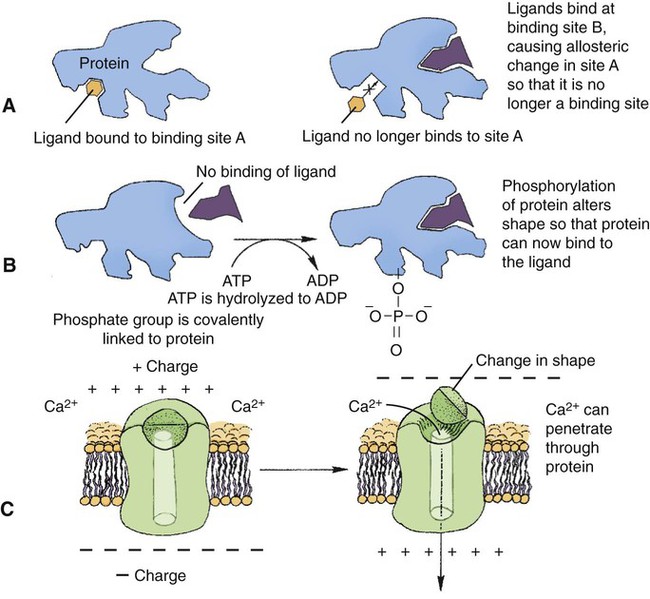
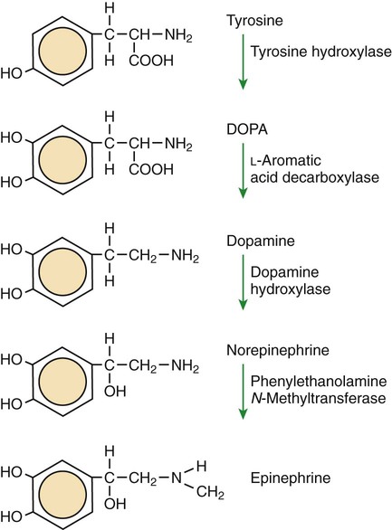
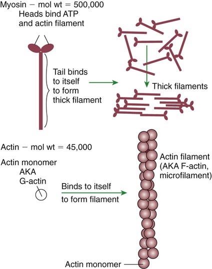
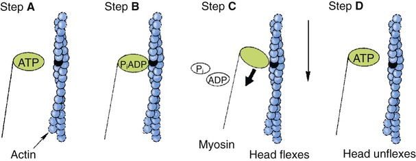
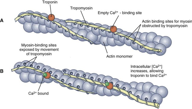
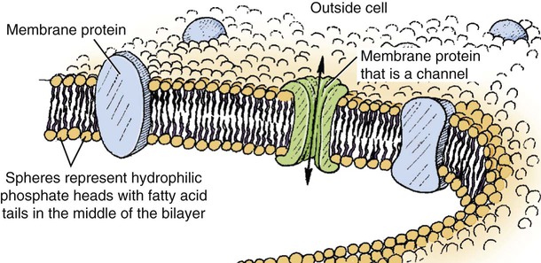
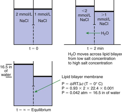
1. All physiological change is mediated by proteins.


2. Protein function depends on protein shape and shape changes.


3. A series of enzymatic reactions converts tyrosine into the signaling molecules dopamine, norepinephrine, and epinephrine.


4. Muscle contraction and its initiation and cessation depend on the binding specificity and allosteric properties of proteins.


5. Biological membranes are a mosaic of proteins embedded in a phospholipid bilayer.


Transport


1. Only small, uncharged molecules and oily molecules can penetrate biomembranes without the aid of proteins.


2. Molecules move spontaneously from regions of high free energy to regions of lower free energy.


3. Important transport equations summarize the contributions of the various driving forces.


4. Starling’s hypothesis relates fluid flow across the capillaries to hydrostatic pressure and osmotic pressure.


5. Membrane proteins that serve the triple functions of selective transport, catalysis, and coupling can pump ions and molecules to regions of higher free energy.


6. Many membrane proteins selectively facilitate the transport of ions/molecules from high to low electrochemical potential.


7. Passive transport of K+ across the plasma membrane creates an electrical potential.


8. Spatial organization of active and passive transport proteins enables material to pass completely through the cell.


9. Membrane fusion allows for a combination of compartmentalization and transport of material.


Information transmission and transduction


1. Cell signaling often occurs by a lengthy chain of sequential molecular interactions.


2. Signaling pathways begin with the binding of an extracellular molecule to a receptor.


3. Specific physiological information is inherent in the receptor/ligand complex, not in the hormone/neurotransmitter molecule.


4. G-protein–coupled receptors are the largest family (a superfamily) of receptors and help regulate almost all physiological processes.


5. Most G-protein–linked information is sent to the cytoplasm by second messengers.


6. Ca2+ transport across plasma and intracellular membranes is an important second messenger.


7. Cyclic AMP is produced by activation of a membrane-bound enzyme in response to hormone/neurotransmitter binding to receptors.


8. The receptor-mediated hydrolysis of a rare phospholipid of the plasma membrane produces two different second messengers with different actions.


9. Steroid hormones and other lipid signals interact with nuclear receptors, which are transcription factors within the cell.


Physiology is the study of the regulation of change within organisms, in this case higher animals. Our understanding of physiology has changed dramatically in the past 30 years as a result of insight into the molecular basis of biological regulation. This chapter summarizes (and simplifies considerably) our current understanding of the molecular and cellular basis of that regulation. Most of the principles in this chapter apply to all animal cells. The approach taken is one of functional molecular anatomy. That is, the molecular structure of the cell is examined with particular emphasis on the physiological function, in the intact animal, of the molecules and supramolecular structures responsible for the function. Only those aspects of cell function that illuminate the medical physiology of the higher animals are discussed; the reader is referred to the Bibliography for more complete coverage of the cell. Some review of basic concepts and vocabulary is presented. However, the discussion assumes that the reader is familiar with the cell and its constituent molecules, as presented in courses in general biology and undergraduate courses in biochemistry.


All Physiological Change Is Mediated by Proteins


All physiological change is mediated by a single class of polymeric macromolecules (large molecules), the proteins. Protein function can be subdivided into a number of categories: catalysis, reaction coupling, transport, structure, and signaling.


Catalysis is the ability to increase greatly the rate of a chemical reaction without altering the equilibrium of the reaction. The majority of biochemical reactions occur at a physiologically useful rate only because of protein catalysts, called enzymes. Examples of enzymatic catalysis in the synthesis of a class of physiological regulator molecules, catecholamines, are given later in this chapter.


In reaction coupling, two reactions are joined together with the transfer of energy. Energy from a spontaneous reaction (similar to water flowing downhill) is funneled to a nonspontaneous reaction (e.g., sawing wood) so that the sum of the two reactions is spontaneous. That is, the energy liberated by the “downhill” reaction is used to drive the “uphill” reaction. This is the basic function of a motor; the “downhill” burning of gasoline is coupled with the “uphill” movement of the car. The ability of proteins to couple spontaneous and nonspontaneous reactions allows cells to be chemical motors, using chemical energy to do various jobs of work. One such job of work, the contraction of striated muscle, is discussed later with particular emphasis on the proteins involved.


Proteins provide a pathway for the membrane transport of most molecules and all ions into and out of the cell. Transport and transport proteins are discussed more fully after a discussion of the lipid bilayer membrane, the major obstacle to transport.


Proteins that form filaments and that glue cells to each other and to their environment are responsible for the structure and organization of cells and multicellular assemblies (i.e., the tissues and organs of animals). The internal structure of the muscle cell, as well as its ability to do work, is a result of the properties of the muscle proteins discussed later.


At its most basic level, signaling requires only a controlled change or difference. Human signaling occurs by way of open and closed electrical circuits (telegraphy), puffs of smoke in the air, and complex black marks on a contrasting background (numbers and letters). As discussed next, a fundamental property of proteins is the ability to change shape. The cell can use changes of protein shape directly to send signals, and the function of some proteins is purely informational. That is, all that some proteins do by changing shape is transmit and transduce information. Information can be defined as “any difference that makes a difference,” or more simply, any difference that regulates something. Catalysis, coupling, transport, structural, and signaling functions can be combined on individual protein molecules. As will become apparent, such multifunctional proteins carry out many important physiological functions. Also important is that a change in one or more of these protein functions can be used to carry information, to serve as a signal within the cell. Thus, in addition to proteins specialized exclusively to carry information, changes in enzymatic activity or ion transport can also make a difference, transmitting information and triggering an appropriate response.


Protein Function Depends on Protein Shape and Shape Changes


Protein function is founded on two molecular characteristics: (1) proteins can bind to other molecules very specifically; and (2) proteins change shape, which in turn alters their binding properties and their function. The binding specificity of proteins is the result of their complex three-dimensional structure. Grooves or indentations on the surface of protein molecules, called binding sites, permit specific interactions with a molecule of a complementary shape, called the ligand. This complementary-shape mechanism underlying binding is similar to the shape interaction between a lock and key.


Several aspects of the lock-and-key analogy are worth noting. As with a lock, only a small part of the protein is engaged in binding. The binding is very specific; small changes in the shape of the binding site (keyhole) or the shape of the ligand (key) can cause major changes in protein (lock) behavior. Similar to the lock and key, the complementary-shape interaction serves a recognition function; only those molecules with the right shape affect protein function. This recognition function plays a primary role in information transfer. The protein recognizes a particular signal by binding to it, thus changing the protein’s shape and thus its function. Unlike the majority of locks, however, proteins frequently have multiple binding sites for multiple ligands.


Thus the three-dimensional shape of a protein, its conformation, determines protein function. A major force that stabilizes protein conformation is the hydrophobic interaction. Oily, hydrophobic (water-fearing) amino acids tend to congregate in the middle of a protein away from water, whereas hydrophilic (water-loving) amino acids tend to be found on the protein’s outer surface interacting with the abundant cellular water. The hydrophobic interaction is also important in stabilizing the interaction of proteins with the lipids of biological membranes, as discussed shortly. Protein shape is also stabilized by hydrogen bonding between polar amino acid pairs in the polypeptide (protein) chain.


The same weak forces responsible for protein conformation are also used to hold the ligand in the protein-binding site. The position of the ligand in the binding site is stabilized by hydrogen bonds between the polar groups of the ligand and polar, amino acid side groups lining the binding site, just as hydrogen bonds within the polypeptide chain stabilize the shape of the polypeptide. Precisely because the same forces are responsible for the shape of the protein and for its binding properties, shape influences binding, and in turn, binding can influence protein shape. The ability of proteins to change shape is called allostery (Greek, “other shape”).


Allosteric changes in protein conformation arise in four general ways. One way, just mentioned, is that most proteins change shape depending on which ligands are bound at particular binding sites (Figure 1-1, A). The sequence—specific ligand binding → protein shape change → change in protein-binding properties and protein function → this change regulates something—is a common molecular mechanism underlying physiological control. This method involves no alteration in the covalent structure of the protein.


A second method of producing conformational change, however, occurs as a result of the covalent modification of one or more of the amino acid side groups of the protein (see Figure 1-1, B). By far the most common such change is the covalent addition of a phosphate group to the hydroxyl (—OH) group on the side chain of serine, threonine, or tyrosine residues in the protein. This modification is called phosphorylation. Because the phosphate group is highly charged, phosphorylation of a protein alters hydrogen bonding and other electrostatic interactions within the protein chain, altering its conformation and functional properties.


In a third method, some physiologically important proteins change shape in response to the electrical field surrounding the protein (see Figure 1-1, C). These respond to a voltage change by altering the position of charged amino acids, thus altering protein shape.


The fourth method of protein shape change is the least well understood (not shown). Some proteins change shape in a controlled manner in response to mechanical forces. Although this is not surprising, because all solids and solidlike substances change shape at least slightly in response to force, we know relatively little about mechanosensitive proteins. The best current example is a protein involved in the very early events of hearing that changes its transport of ions in response to the mechanical stimulation by sound (small changes of air pressure in waves).


The significance of binding specificity and allostery can be better appreciated with two examples of their roles in physiological function. The first example is the role of enzymes in synthesizing three small, structurally similar, nonprotein signaling molecules. This example shows how binding specificity is important in catalytic function and how allostery underlies the regulation of the synthesis. The second example is more complex: the role of proteins in the contraction of muscle. The contraction of muscle shows how proteins can exploit the basic properties of specific binding and allosteric shape change to do more than one job of work at the same time; muscle proteins serve a structural role, serve a catalytic function, and couple the “downhill” hydrolysis of adenosine triphosphate (ATP) to do mechanical work, the “uphill” lifting of weight.


A Series of Enzymatic Reactions Converts Tyrosine into the Signaling Molecules Dopamine, Norepinephrine, and Epinephrine


Figure 1-2 is a diagram of the series of reactions by which the amino acid tyrosine is converted into three different signaling molecules: (1) dopamine, a brain neurotransmitter; (2) norepinephrine, a neurotransmitter of the brain and peripheral autonomic nervous system; and (3) epinephrine, an autonomic neurotransmitter and hormone. Dopamine, norepinephrine, and epinephrine share a similar structure. All contain a phenyl (benzene) ring with two hydroxyl groups (i.e., catechol) and an amine group (thus catecholamines). They are among the large number of molecules that function as neurotransmitters. That is, the electrically coded information sent along nerve cells causes the release of a chemical, the neurotransmitter, at the terminal of the neuron, which is next to a target cell, such as another nerve, a muscle, or an endocrine cell. The electrically encoded information of the nerve is transmitted to the target cell by the binding of the neurotransmitter to proteins on the surface of the target cell. Proper neurotransmitter synthesis is crucial to nervous function and physiological regulation.


In the first step of catecholamine biosynthesis, tyrosine binds to the enzyme tyrosine hydroxylase, which catalyzes the addition of another hydroxyl group to the phenyl group to form dihydroxyphenylalanine, almost always called DOPA. This hydroxyl group alters the enzyme-ligand interaction; the key no longer fits the keyhole. DOPA is released from the tyrosine hydroxylase and is then bound by another enzyme, L-aromatic amino acid decarboxylase. As the name implies, this enzyme catalyzes the removal of the carboxyl group, converting DOPA to dopamine. Dopamine is converted into norepinephrine by the activity of dopamine hydroxylase, which adds yet another hydroxyl group, this time to the two-carbon tail of dopamine. Finally, addition of a methyl group to the amino nitrogen by phenylethanolamine N-methyltransferase gives rise to epinephrine (also called adrenalin). Note the binding specificity of the enzymes: whereas the catecholamine structures are all similar to one another, different enzymes bind each one (e.g., epinephrine does not bind to dopamine hydroxylase).


The allosteric properties of one enzyme in this pathway provide an example of physiological regulation. Certain hormones and neurotransmitters cause the phosphorylation of tyrosine hydroxylase, the first enzyme in the pathway, increasing its activity. That is, phosphorylation of the enzyme increases the rate at which it catalyzes the conversion of tyrosine to DOPA. Because this step is the slowest in the pathway, an increase in the activity of this protein increases the net rate of synthesis of all the catecholamines. Regulated decreases in the rate of catecholamine synthesis are achieved by a different allosteric mechanism: binding of end products to the enzyme. Dopamine, norepinephrine, and epinephrine can all bind to tyrosine hydroxylase at a site different than the site for tyrosine. These binding events inhibit the enzymatic activity. The inhibition of the pathway by its own end products makes this a classic case of allosteric control called end-product inhibition. Many substances regulate their own synthesis by inhibiting an initial enzyme in the pathway. If the cell has enough end products, these products inhibit further synthesis by allosteric changes in the enzyme. This is an example of the following sequence: specific binding → protein shape change → change in protein-binding properties and protein function → this change regulates something.


Muscle Contraction and its Initiation and Cessation Depend on the Binding Specificity and Allosteric Properties of Proteins


There are three types of muscle tissue in vertebrates: (1) skeletal muscle, responsible for the animal’s ability to move; (2) cardiac muscle, a muscle type found only in the heart but structurally similar to skeletal muscle; and (3) smooth muscle, which surrounds hollow organs such as blood vessels, gut, and uterus. All three produce tensile force by contracting and shortening the length of the muscle. All muscle contraction occurs by the binding and the allosteric properties of two proteins, actin and myosin. Starting and stopping the contraction process depends on two additional proteins in skeletal and cardiac muscle, troponin and tropomyosin. Contraction initiation and cessation in smooth muscle depend on a different system with different proteins, and are discussed later in this chapter.


Myosin is a large protein whose shape resembles a two-headed golf club. The elongated tail of the myosin molecule corresponds to the shaft of the golf club, and there are two knobs at one end of the tail that, as with golf clubs, are called heads. Myosin tails bind specifically to other myosin tails, forming bipolar aggregates called thick filaments (Figure 1-3). Myosin heads specifically bind ATP and another muscle protein, actin. Actin binds to itself to form long, thin filaments, called thin filaments in muscle and called F-actin (filamentous actin) in other cell types. Actin filaments play an important architectural role in all animal cells. Although actin is best understood in muscle cells, all animal cells depend on actin filaments for their shape and for their capacity to migrate in their environment. Actin filaments can be “woven” in various ways to produce different structures, such as ropelike bundles and clothlike networks. These actin bundles and actin networks are used to support the cell in particular shapes, similar to ropes holding up the woven cloth of a tent.


In muscle, the interaction of myosin, ATP, and actin produces contraction and force, as shown in Figure 1-4:


Step A: ATP binds to a myosin head; in this conformation, myosin has little ability to bind to actin.


Step B: Enzymatic activity associated with the myosin head, an adenosinetriphosphatase (ATPase), rapidly causes a partial hydrolysis of ATP to adenosine diphosphate (ADP) and inorganic phosphate (Pi), both of which stay bound to the myosin. With ADP and Pi bound, myosin has a slightly different shape that binds avidly to nearby actin filaments.


Step C: When myosin binds to actin, called cross-bridging, the myosin head couples the complete hydrolysis of ATP to a forceful flexing of the myosin head. This allosteric change causes the actin filament to slide past the thick filament. This sliding puts the actin filament under tension, which in turn causes the muscle to contract (shorten) against the load of the muscle (i.e., lifting a weight or pumping out blood). All muscle contraction depends on this sliding filament mechanism of actin and myosin-based filaments. This same allosteric change of myosin also alters myosin-binding properties so that it releases the ADP and Pi.


Step D: The binding of a new ATP molecule to the myosin head again causes myosin to change shape; the head unflexes and loses its affinity for actin, releasing the cross-bridge, and the cycle can start over. Rigor mortis of dead animals is caused by a lack of new ATP to bind to myosin heads. In the absence of ATP, myosin heads remain in Step C (i.e., bound to actin). The muscle is stiff because it is completely cross-bridged together.


This actomyosin motor uses the binding and allosteric properties of proteins to (1) create structural filaments capable of withstanding and transmitting mechanical force, (2) catalyze the hydrolysis of ATP, and (3) couple the “downhill” ATP hydrolysis to the “uphill” contraction to produce force. For just the one protein, myosin, there are a number of examples of the characteristic sequence described earlier: specific binding → protein shape change → change in protein-binding properties and protein function → this change makes a difference.


This system of contractile proteins requires some control so that, for example, the heart beats rhythmically and skeletal muscle contraction is coordinated. At the organismal level, skeletal and cardiac muscle contraction is primarily under control by electrical stimulation from nerves or other electrically active cells (see Chapter 6). The transmission of electrical excitation to the actomyosin system is called excitation-contraction coupling. Excitation-contraction coupling in all types of muscle depends on changes in intracellular calcium ion (Ca2+) concentration. In skeletal and cardiac muscle, but not smooth muscle, two additional thin-filament proteins, troponin and tropomyosin, are required for this coupling. (Excitation-contraction coupling for smooth muscle is discussed later in this chapter.) In striated muscles, troponin binds to tropomyosin and to Ca2+. Tropomyosin is a long, thin protein that binds in the groove of the actin filament in such a way that its positions, high in the groove or snuggled down deep in the groove, allow or prevent the myosin head access to the thin filament (Figure 1-5). Excitation-contraction coupling of striated muscle works as follows:


Step A: Electrical excitation of a striated muscle cell causes an increase in the intracellular concentration of Ca2+.


Step B: The additional Ca2+ binds to troponin, causing an allosteric change in troponin.


Step C: Because Ca2+ is bound to troponin, which in turn is bound to tropomyosin, the Ca2+-induced change in troponin conformation is transmitted to the tropomyosin molecule. When troponin binds Ca2+, tropomyosin changes its binding to actin in such a way that it exposes the actin site for myosin cross-bridging. (Tropomyosin snuggles down deeper in its actin groove, revealing actin to the myosin head.) As long as troponin binds Ca2+, the muscle contracts by the actomyosin cycle outlined earlier.


Step D: When the Ca2+ concentration drops to normal, however, troponin no longer binds Ca2+. This causes tropomyosin to move up in the thin filament groove so that it again blocks the myosin-binding sites on actin. Myosin heads can no longer cross-bridge, and muscle contraction stops.


As with the actomyosin force generation itself, its regulation also shows many examples of the specific binding function. The specific binding of Ca2+ to troponin is a purely informational use of protein binding and shape change; that is, troponin has no catalytic, transport, or structural function, but transmits the “on” signal to the next protein. The binding of tropomyosin to actin serves not only a regulatory role but also a structural role; the actin filament is stabilized by tropomyosin, making it less likely to disassemble into actin subunits. The change in the binding geometry of tropomyosin that directly regulates myosin access to actin is a good example of the importance of allosteric change and the following sequence: specific binding (troponin to tropomyosin) → protein (tropomyosin) shape change → change in protein-binding properties (tropomyosin to actin) → a difference in the position of tropomyosin, which in turn regulates the actomyosin motor.


Biological Membranes Are a Mosaic of Proteins Embedded in a Phospholipid Bilayer


Before continuing the discussion of the cellular basis of physiological control, an additional basic structure must be introduced. This is the phospholipid bilayer of the biomembranes of cells. Phospholipids are molecules that have two long tails of hydrophobic fatty acid and a head containing a charged, hydrophilic phosphate group. Under appropriate aqueous conditions, these molecules spontaneously form an organized membrane structure, similar to the film of a soap bubble. This filmy layer is composed of two layers (a bilayer) of phospholipid molecules. In both layers the hydrophilic heads point outward to hydrogen bond with water, and the oily, fatty-acid tails point inward, toward one another and away from the water. Proteins embedded in this lipid bilayer, called intrinsic membrane proteins or just membrane proteins, produce the fluid mosaic structure of biomembranes shown in Figure 1-6. All biological membranes share this fluid mosaic structure, whether the membrane is the outer plasma membrane separating cytoplasm from extracellular fluid or the membrane surrounding intracellular membranous organelles such as endoplasmic reticulum or lysosomes. It is called a fluid mosaic because of the mosaic of proteins among phospholipids, and because the phospholipid layer is fluid; proteins can move around and diffuse within the plane of the bilayer “like icebergs floating in a phospholipid sea” (the apt phrase of S. J. Singer, one of the originators of the model).


Biological membranes are another crucial molecular structure underlying physiological control. The basic fluid mosaic structure serves four broad functions: (1) compartmentation, (2) selective transport, (3) information processing and transmission, and (4) organizing biochemical reactions in space.


Compartmentation is the ability to separate and segregate different regions by composition and function. For example, the lysosome is a membranous organelle within cells that contains hydrolytic (digestive) enzymes that can potentially digest the cell. The lysosomal membrane compartmentalizes these potentially harmful enzymes, segregating them from the bulk cytoplasm. The rigor mortis, mentioned earlier, that begins shortly after death is transitory because on death the lysosomes begin to break open, releasing their enzymes, and the actomyosin cross-bridges are eventually digested apart.


Clearly, the membrane cannot keep a compartment perfectly sealed; material must enter and leave the cell and its internal compartments. Selective transport results partly from the properties of the phospholipid bilayer but mostly from transport proteins embedded in the membrane. These proteins are characteristically selective in their transport functions; for example, the protein that is the specialized ion channel underlying neuronal signaling is 15 times more permeable to sodium ions (Na+) than to potassium ions (K+). Transport is a major topic of cell physiology and is discussed in more detail later.


If the cells of an organism are to respond to external changes, they must receive information about the state of the outside world. Just as we higher animals have our sensory organs—eyes, ears, nose, and so forth—arrayed on our outside surface, so too do cells have most of their environmental information processing and transmission apparatus on their external surfaces. These are intrinsic membrane proteins of the plasma membrane, called membrane receptors, that serve a purely informational function, as discussed earlier.


At first glance it might seem odd that a fluid membrane could provide spatial organization for biochemical reactions. However, returning to the “icebergs in a phospholipid sea” analogy, random collisions are much more likely for material in the two-dimensional membrane surface than for material moving through the three-dimensional volume of the cytoplasm. (If the Titanic had been able to dive or fly, it would have had additional ways to avoid the iceberg!) This much larger collision probability is exploited by the cell in a number of physiological processes. Membranes can also be fenced off into distinct regions, across which there is limited diffusion of membrane proteins. For example, certain cells in the kidney have two membrane regions that are quite distinct with respect to transport proteins, which is important in the regulation of salt and water balance by the animal.


Transport


Only Small, Uncharged Molecules and Oily Molecules Can Penetrate Biomembranes Without the Aid of Proteins


Charged particles (i.e., ions) do not pass through a pure phospholipid bilayer because of the inner, hydrophobic region of bilayer. Polar molecules (molecules with no net charge but with electrical imbalances) with a molecular weight greater than about 100 daltons are also unable to pass readily through a pure lipid bilayer, thus excluding all sugar molecules (monosaccharides), amino acids, nucleosides, as well as their polymers (polysaccharide, proteins, nucleic acids). On the other hand, some crucially important polar molecules (e.g., water, urea) are small enough to pass through the lipid bilayer. Small, moderate-size, and large molecules that are soluble in oily solvents readily pass through a pure lipid bilayer. Physiologically important molecules in this class include O2, N2, and the steroid hormones (see Chapters 33 and 34). However, many toxic, synthetic molecules, such as insecticides, are also in this category.


Molecules Move Spontaneously from Regions of High Free Energy to Regions of Lower Free Energy


The majority of biochemicals do not pass readily through a phospholipid bilayer. Transport of this molecular majority requires a protein pathway across the biomembrane. Also needed is a force causing movement along the pathway. Before elaborating on membrane proteins as pathways through the lipid bilayer, the energy factors that drive the transport are considered.


Objects fall spontaneously because of gravity. This is a manifestation of the principle that movement occurs to minimize the potential energy of the object. Indeed, all change in the universe (at scales greater than the subatomic particles) occurs to minimize the potential energy, also called the free energy, of the system. The movement of molecules is strongly affected by forces such as concentration, pressure (both part of chemical potential), and voltage (electrical potential). Molecules move spontaneously from a region of higher concentration to lower concentration, from higher to lower pressure, and from higher to lower electrical potential. Each of these factors—concentration, pressure, and electrical potential—is a source of free energy. The transport of a molecule does not depend necessarily on any one factor; rather, the sum of all the free energy contributions is the determinant of transport. The sum of all the free energy contributions on a substance is usually expressed on a per-mole basis as the electrochemical potential. The electrochemical potential is the free energy of the substance, from all sources, per mole of the substance.


For spontaneous transport to occur, there must be a difference in the electrochemical potential of the substance between two regions. The two regions are usually two compartments separated by a membrane. This difference in electrochemical potential is called the driving force. Typically, students have little difficulty understanding the direction of spontaneous flow as long as only one factor contributes to the electrochemical potential, pressure, or concentration or the voltage. However, understanding physiological transport, both across cells and across tissues, requires an understanding of the contribution of each factor to the driving force. For example, the flow of fluid from the capillaries of the vascular system depends on the balance between both the hydrostatic pressure difference and the concentration difference of solutes (osmotic pressure) across the capillary. Similarly, movement of Na+ and K+ ions across the plasma membrane of nerve cells depends on the driving forces contributed by both voltage differences and ion concentration differences across the membrane.


Material moves spontaneously from regions of high electrochemical potential to low electrochemical potential. Such transport is called diffusion or passive transport. Net movement of material (i.e., diffusion) stops when the electrochemical difference between regions equals zero. The state at which the free energy or the electrochemical potential difference is zero is called equilibrium. Equilibrium means “balance,” not equality. Equilibrium is reached when the free energy (electrochemical potential) is balanced; the value on one side is the same as the other. In most cases the source of the free energies on the two sides never becomes equal; the concentrations, the pressure, and the voltages remain different, but their differences “balance out” so that the sum of the free energy differences is zero.


Equilibrium is a particularly important concept because it describes the state toward which change occurs if no work is put into the system. When the system reaches equilibrium, no further net change occurs unless some work is done on the system. The words net change are important. Molecules at equilibrium still move and exchange places, but as much goes in one direction as in the other, so there is no net flow of material.


If the cell requires material to move from low to high electrochemical potential (i.e., in the direction away from equilibrium), thus increasing the difference in free energy between two regions, then some driving force, some work, must be provided by some other decrease in free energy. This type of transport is active transport. Active transport uses proteins that combine transport and reaction coupling functions; the protein couples the “uphill” movement of material to a “downhill” reaction such as ATP hydrolysis.


Important Transport Equations Summarize the Contributions of the Various Driving Forces


It is worthwhile developing some quantitative aspects of transport, beginning with simple examples and developing equations for the effect of more than one driving force. These equations can be seen as summaries of the physical laws. In most cases the equations describe phenomena with which we have experience by living in a technological society. In these equations, c stands for concentration, V for volume, P for pressure, and so forth; these are common concepts. It is important, however, to think about these equations in real-life terms, not as abstract symbols.


One of these equations relates a hydrostatic (pressure) driving force for water movement that just balances a driving force caused by a chemical potential difference. Osmosis is the movement of water across a semipermeable membrane in response to the difference in the electrochemical potential of water on the two sides of the membrane (Figure 1-7). The chemical potential of water is lower in 1 liter (L) of water (H2O) in which is dissolved 2 millimoles (mmol) of sodium chloride (NaCl) than in 1 L of H2O in which is dissolved 1 mmol of NaCl. If these two solutions are separated by a pure lipid bilayer, Na+ and Cl ions cannot move to equilibrate the concentration. Rather, the freely permeable water moves from the side with the higher water potential (low concentration of solute) to the side with the lower water potential (higher concentration of solute). Thus, water follows solute (a good summary of osmosis), and this water movement dilutes the 2 mmol solution. However, water movement never produces equal concentrations of salt. Rather, another driving force appears as the water moves. The hydrostatic pressure of water increases on the side to which the water moves, increasing the electrochemical potential of the water on that side. Net water movement stops when the increase in water potential from hydrostatic pressure exactly balances the decrease in water potential from the dissolved salt, so that the electrochemical potential becomes equal on both sides of the membrane.


The initial potential difference of water shown in Figure 1-7 is caused by the difference in the concentration of material dissolved in the water. A proper explanation of why the water in a solution has a lower chemical potential than pure water (and why water in a concentrated solution has a lower potential than in a dilute solution) is beyond the scope of this chapter. However, readers familiar with the concept of entropy will realize that the disorder of a system increases with the introduction of different particles into a pure substance and with the number of different particles introduced. An analogy would be that a canister with mixed sugar and salt is more disordered, and therefore at higher entropy, than a canister with only pure salt or pure sugar. Also, the disorder of the system increases as more sugar is added to salt (up to 50 : 50); a pinch of sugar in a canister of salt only increases the disorder slightly. Because an increase in entropy causes a decrease in free energy, the free energy of a solution is decreased as the mole fraction of solute increases.


Osmosis is important to cells and tissues because, generally, water can move freely across them, whereas much of the dissolved material cannot. Given a concentration difference of some nonpermeable substances, the van’t Hoff equation relates how much water pressure is required to bring the system to equilibrium, that is, the free energy contributed by a pressure difference across the membrane that exactly balances an opposing free energy contribution caused by a concentration difference.


Π=iRTΔc


image


This equation summarizes a balance of driving forces; P amount of hydrostatic (osmotic) pressure is the same driving force as a particular concentration difference, Δc. The osmotic pressure depends only on the concentration difference of the substance; no other property of the substance need be taken into account. (Those phenomena that depend only on concentration, such as osmotic pressure, freezing-point depression, and boiling-point elevation, are called colligative properties.) The van’t Hoff equation is strictly true only for ideal solutions that are approximated in our less-than-ideal world only by very dilute solutions. Real solutions require a “fudge factor,” called the osmotic coefficient, symbolized by Φ (phi). The osmotic coefficient can be looked up in a table, and then plugged into the equation as follows:


Π=ΦiRTΔc


image

The term Φic for a given substance represents the osmotically effective concentration of that substance and is often called the osmolar or osmotic concentration, measured in osmoles per liter (Osm/L). In general, the osmolar concentration of a substance is approximated by the usual concentration times the number of ions formed by the substance; the osmotic coefficient provides a small correction. The osmolarity of a 100 mmol NaCl solution (0.1 mol) is then 0.93 (Φ for NaCl) × 2(NaCl → Na+ + Cl) × 0.1 mol = 0.186 Osm = 186 mOsm.


The previous equation summarizes a phenomenon crucial for physiological function. The greater the concentration difference of an impermeable substance across a membrane, the greater is the tendency for water to move to the side of high concentration. (Water follows solute.) Indeed, if you plug some numbers into this equation, you may be surprised at the large pressures required to balance modest concentration differences. For example, an NaCl concentration difference of 0.1 mol (5.8 g/L) is equilibrated by a pressure (4.2 atm) equal to a column of water 141 ft high (divers must be wary of the bends when ascending from below 70 ft of water). The importance of this is that a small concentration difference can produce a strong force for moving water. The body makes effective use of this to transport water in many tissues: ions/molecules are transported into or out of a compartment → and water follows by osmosis.


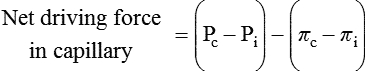
Starling’s Hypothesis Relates Fluid Flow Across the Capillaries to Hydrostatic Pressure and Osmotic Pressure


An excellent practical example of how a balance of driving forces is responsible for the flow of water and permeable substances across a semipermeable membrane is the movement of water and ions across the single layer of cells (endothelial cells) that compose blood capillaries. The single cell layer composes, in effect, a semipermeable membrane with different transport qualities than that of a simple lipid-bilayer membrane. The junctions between cells are sufficiently permeable to allow small molecules and ions to diffuse between compartments. Only large molecules, most importantly proteins, are unable to move through the holes. The difference in protein concentration between the blood and the water solution surrounding tissue cells, called the extracellular fluid (ECF) or interstitial fluid (ISF), creates an osmotic pressure for the movement of water with all its dissolved small molecules and ions. This osmotic pressure resulting from dissolved proteins has a special name: colloid osmotic pressure or oncotic pressure. Protein is more concentrated in the blood than in the interstitial fluid, producing an oncotic pressure of about 0.02 to 0.03 atm = 15 to 25 mm Hg, driving water into the capillary. On the basis of this driving force alone, one would expect the capillaries to fill up with water, thus dehydrating the tissue spaces. However, the heart is a pump that exerts a true hydrostatic pressure on the blood, tending to drive the water (and other permeable molecules) out of the capillaries. The net driving force is the algebraic sum of the oncotic pressure difference and hydrostatic pressure difference between the capillaries and the interstitial fluid, as follows:


Net driving forcein capillary=(PcPi)(πcπi)


image
< div class='tao-gold-member'>

Stay updated, free articles. Join our Telegram channel

Jul 18, 2016 | Posted by in PHARMACOLOGY, TOXICOLOGY & THERAPEUTICS | Comments Off on The Molecular and Cellular Bases of Physiological Regulation

Full access? Get Clinical Tree

Get Clinical Tree app for offline access