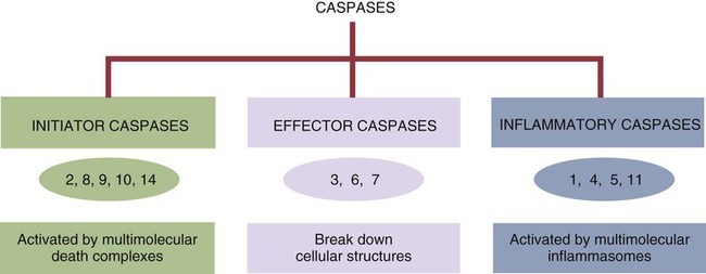
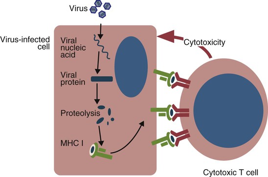
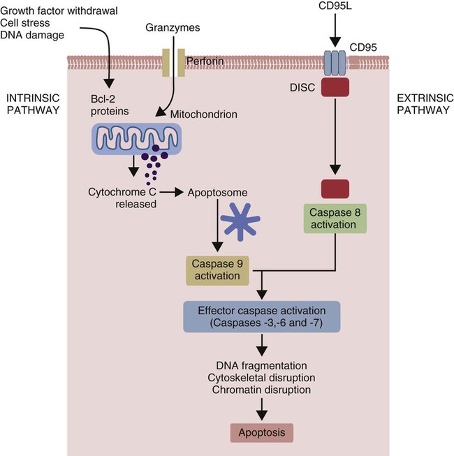
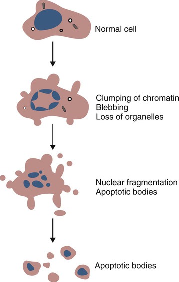
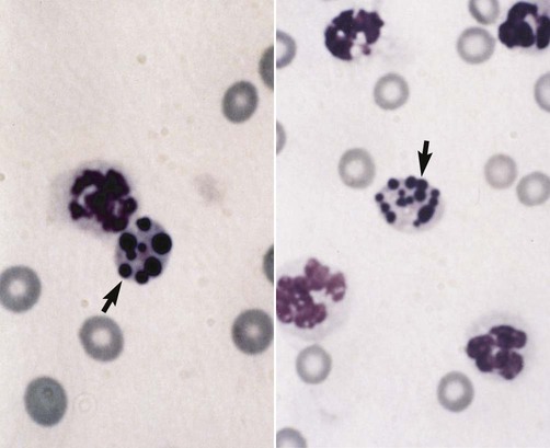
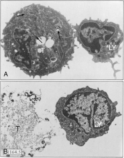
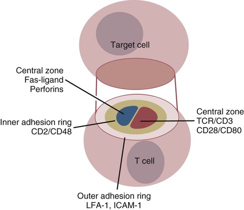
T Cell Function and the Destruction of Cell-Associated Invaders


T Cell Function and the Destruction of Cell-Associated Invaders


Antibodies bind to invading organisms in the circulation or tissue fluids, hastening their destruction. However, not all foreign organisms are found outside cells. All viruses and some bacteria can grow inside cells at sites inaccessible to antibodies. Antibodies are therefore of limited use in defending the body against these invaders. Viruses and other intracellular organisms must be eliminated by other mechanisms. For this, the body uses two different cell-mediated techniques. Either infected cells are killed rapidly so that the invader has no time to grow, or infected cells develop the ability to destroy the intracellular organism. In general, organisms such as viruses that enter the cell cytosol or nucleus are killed by cell destruction, whereas organisms such as bacteria or parasites that reside within endosomes are destroyed through cell activation. T cells mediate both processes. The antigens that trigger these responses arise from intracellular locations and are called endogenous antigens.


Endogenous Antigens


As described in Chapter 10, every time a cell makes a protein, a sample is processed and peptides are carried to the cell surface bound to major histocompatibility complex (MHC) class I molecules (Figure 18-1). If these peptides are not recognized by T cells, no response is triggered. If, however, the peptide-MHC complex binds T cell antigen receptors (TCR), then T cells will be triggered to respond. For example, when a virus infects a cell, T cells may recognize many of the peptides derived from the viral proteins. The T cells that respond to these endogenous antigens are CD8+. They use this CD8 to bind to MHC class I molecules on the target cells, thus promoting intercellular signaling and eventually killing the target cells.


Apoptosis


Cells can kill themselves. Old, surplus, damaged, or abnormal cells that would otherwise interfere with normal tissue functions can be persuaded to die as necessary. This cell suicide is called apoptosis. Apoptosis is carefully regulated and must only be activated when a cell must die. Structurally, apoptosis is characterized by membrane blebbing, nuclear fragmentation, and phagocytosis of the dying cell.


There are two major pathways of apoptosis. The extrinsic or death receptor pathway and the intrinsic or mitochondrial pathway. The death receptor pathway is triggered by cytokines such as tumor necrosis factor-α (TNF-α) acting through specific death receptors such as CD95 (Fas). Death receptors are a family of type 1 cell surface receptors that when activated trigger apoptosis. They all possess an 80-amino acid cytoplasmic sequence called a death domain. The most important of these death receptors are Fas (CD95) and the receptors for tumor necrosis factor (TNFR). Death receptors are activated by ligands commonly expressed on cytotoxic cells. The ligands bind to the death receptors and as a result assemble multiple adaptor proteins into a signaling complex. Once assembled, this complex activates initiator caspases-8 and -10 (Figure 18-2).


The mitochondrial pathway, in contrast, is triggered by noxious stimuli that cause mitochondrial injury. The damaging stimuli (e.g., oxidants, radiation) activate proapoptotic bcl-2 proteins, which then cause the release of cytochrome C from mitochondria (Figure 18-3). The cytochrome C triggers the formation of a large multiprotein complex called an apoptosome. The apoptosome then activates initiator caspase-9.


The initiator caspases activated by either pathway then trigger a cascade of “effector caspases” (caspase-3, -6, and-7) that degrade numerous proteins, activate endonucleases, break down organelles, and result in cell death and disassembly. The DNA of apoptotic cells characteristically breaks into many low-molecular-weight fragments. This fragmentation may be responsible for the characteristic way in which the nuclear chromatin condenses against the nuclear membrane (Figure 18-4). Affected cells shrink and detach from the surrounding cells. Eventually nuclear break-up and cytoplasmic budding form cell fragments called apoptotic bodies (Figure 18-5).


As cells undergo apoptosis, their cell membrane “flips” so that the lipid phosphatidylserine is exposed on their surface. This lipid binds to receptors on macrophages and dendritic cells and triggers phagocytosis of the dying cell. It also triggers the release of anti-inflammatory cytokines such as transforming growth factor-β (TGF-β) while inhibiting the release of proinflammatory cytokines such as TNF-α.


If cells are severely damaged as a result of trauma, toxicity, or microbial invasion, they may die as a result of necrosis. This has been believed to be a largely unregulated process, although a molecular signaling network regulating the process (necroptosis) has been partially defined. Cells killed by necrosis will trigger inflammation. Thus HMGB-1 escaping from necrotic cell nuclei is a potent inflammatory mediator. Likewise, when dendritic cells engulf necrotic cells, they not only process their proteins into MHC-antigen complexes but also express co-stimulatory molecules. T cells that recognize this antigen therefore are activated. Thus a cell killed by a virus through necrosis can trigger inflammation and provoke a T cell response to the viral antigens.


Cell Cooperation


During a primary immune response, CD8+ cytotoxic T cells cannot respond to infected cells alone. There are about 1013 nucleated cells in a human-sized body and possibly several hundred naïve T cells with receptors for each individual viral antigen. Clearly it would be almost impossible for these T cells to find all the cells expressing corresponding viral antigens by themselves. Naïve cytotoxic T cells tend to remain within lymphoid organs, and dendritic cells can carry antigens to them. A subset of dendritic cells processes these endogenous antigens, links them to their MHC class I molecules, and carries them to secondary lymphoid organs where they are presented to CD8+ T cells. To respond fully, these CD8+ cells must also be co-stimulated by CD4+ Th1 cells. Co-stimulation is only effective if both the CD8+ and CD4+ T cells recognize antigen on the same antigen-presenting cell. This happens in a defined sequence. Thus a helper T cell first interacts with an antigen-presenting dendritic cell in the normal way through CD40 and CD154. Immature dendritic cells express low levels of MHC and co-stimulatory molecules and are thus poor T cell stimulators. Helper T cells, however, activate these dendritic cells, upregulate their expression of MHC, and stimulate their production of interleukin-12 (IL-12) and the T cell chemotactic chemokine CCL22. Only when it is fully activated in this way can a dendritic cell successfully trigger a cytotoxic T cell response.


Once activated, dendritic cells present MHC class I–linked peptides to CD8+ T cells. They do this readily if the dendritic cells are themselves infected. However, they can also present peptides from nonreplicating organisms or from dying infected cells. Thus by processing dying cells, dendritic cells can present T cells with endogenous antigens. The cytotoxic T cells require three key signals. The first is IL-12 from activated dendritic cells. The second signal comes from the antigen–MHC class I complex on an abnormal cell. The third signal comes from IL-2 and IFN-γ secreted by the Th1 cells. After all three signals are received, the CD8 T cells can respond.


Different levels of stimulation trigger different responses in CD8 T cells. As with Th cells, the duration of the stimulus is also important. Thus although activated cytotoxic T cells can be triggered by brief exposure to antigen, naïve T cells must be stimulated for several hours before responding. The required stimulation time may be shortened by increasing TCR occupancy or by providing additional co-stimulation. Once activated, cytotoxic T cells divide rapidly.


Cytotoxic T Cell Responses


Once fully activated, CD8+ T cells leave lymphoid organs and seek out infected cells by themselves. When they recognize an antigen expressed on another cell, the T cells will kill their target. Although most cells only undergo apoptosis after receiving very specific signals, cytotoxic T cells can induce apoptosis in any cell they recognize (Figure 18-6).


The density of peptide-MHC complexes on a target cell required to stimulate T cell cytotoxicity is much lower than that needed to stimulate cytokine production. Thus T cell binding to a single peptide-MHC complex may be sufficient to kill a target, whereas binding to 100 to 1000 complexes is required to stimulate cytokine production and clonal expansion. Presumably cytotoxic T cells need to be highly sensitive to viral peptides so that they can kill infected cells as soon as possible. These differences in signal thresholds are probably due to the structure of the immunological synapses formed when a cytotoxic T cell encounters another cell.


When T cells encounter a target, an immunological synapse forms at the point of contact (Figure 18-7). This synapse has two “centers.” One part of the central zone contains the TCR-CD8 complex. The other serves as the portal of entry of T cell cytotoxic molecules into the target cell. Both are surrounded by a pSMAC rich in adhesion molecules that forms a “gasket,” preventing the accidental spill of cytotoxic molecules. Once a synapse forms, cytotoxic T cell killing is highly efficient. Within seconds after contacting a T cell, the organelles and the nucleus of the target show apoptotic changes, and the target is dead in less than 10 minutes. Cytotoxic T cells are also serial killers that can disengage and move on to kill other targets within 5 to 6 minutes.


< div class='tao-gold-member'>

Stay updated, free articles. Join our Telegram channel

Jul 18, 2016 | Posted by in PHARMACOLOGY, TOXICOLOGY & THERAPEUTICS | Comments Off on T Cell Function and the Destruction of Cell-Associated Invaders

Full access? Get Clinical Tree

Get Clinical Tree app for offline access