Systemic Responses to Inflammation


Systemic Responses to Inflammation


In one sense, inflammation is a very local response, focused on sites of tissue damage or microbial invasion, but it also has significant systemic effects on other, distant parts of the body. If the local inflammation is minor, such systemic effects may not even be noticed. If, however, inflammation is extensively affecting multiple organ systems, or if the microbial invader succeeds in spreading throughout the body, these systemic effects become clinically significant. Collectively we call them sickness. It need hardly be pointed out that in veterinary medicine, it is these signs of sickness that commonly draw the attention of an owner to an animal’s illness.


Sickness Behavior


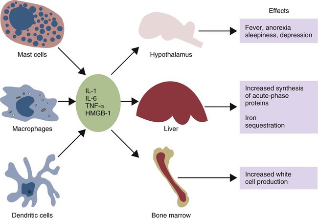
When an animal is invaded by pathogens, a generalized response may occur—a response that we call sickness. The subjective feelings of sickness—malaise, lassitude, fatigue, loss of appetite, and muscle and joint pains—along with a fever, are signs of a systemic innate immune response. They reflect a change in the body’s priorities as it fights off the invaders. Microbial PAMPs acting through the pattern-recognition receptors (PRRs) of phagocytic cells stimulate the production of interleukin-1β (IL-1β), IL-6, and TNF-α. All three of these cytokines signal to the brain (Figure 6-1). They use two routes. One route is through the neurons that serve damaged tissue. IL-1 receptors are expressed on these sensory neurons, especially the vagus nerve. Sensory stimulation by IL-1β through the vagus nerve can trigger afferent signaling to the brain. (IL-1 will not trigger a fever if the vagus nerve is cut.) Lipopolysaccharide (LPS) and TLR4 can also trigger these vagal signals. They therefore trigger fever, nausea, and other sickness responses in the brain. The second route involves cytokines that either diffuse into the brain from the bloodstream or are produced within the brain. Microglial cells express TLR4, whereas neurons express TNF receptors. These cytokines can act on neurons or microglial cells to modify behavior and, for example, alter pain perception. As a result, anti-TNF antibodies can greatly reduce the pain associated with rheumatoid arthritis (Chapter 36).


The most obvious of the brain’s responses to infection is the development of a fever. IL-1, IL-6, and TNF-α all trigger changes in body temperature. These cytokines induce the expression of cyclooxygenase-2 (COX-2) in the hypothalamus that results in prostaglandin production, which causes the body’s thermostatic set-point to rise. In response, animals conserve heat by vasoconstriction and increase heat production by shivering, thus raising their body temperature until it reaches the new set-point. This fever enhances some components of the immune responses. For example, it enhances transendothelial neutrophil migration and chemotaxis and increases neutrophil accumulation within tissues. It also accelerates caspase-dependent neutrophil apoptosis. Raised body temperatures cause dendritic cells to mature; enhance the circulation of lymphocytes; and promote the secretion of IL-2. Fever range temperatures enhance the survival of T cells by inhibiting their apoptosis. In addition to causing a fever, inflammatory cytokines, especially IL-1, promote the release of sleep-inducing molecules in the brain. Increased lethargy is commonly associated with a fever and may, by reducing the energy demands of an animal, increase the efficiency of defense and repair mechanisms. IL-1 also suppresses the hunger centers of the brain and induces the loss of appetite associated with infections. The benefits of this are unclear, but it may permit the animal to be more selective about its food. If the anorexia persists, it can have an adverse effect on animal growth and production.


High-mobility group band protein-1 (HMGB1) (Chapter 3) is a potent sickness-inducing cytokine. Although IL-1, IL-6, and TNF-α have long been known to cause septic shock and sickness behavior, it is now clear that these three molecules induce HMGB1 release from macrophages several hours after initiation of sickness. It enters secretory lysosomes and is then released slowly from the cells. HMGB1 has been implicated in food aversion and weight loss by its actions on the hypothalamic-pituitary axis. It mediates endotoxin lethality, arthritis, and macrophage activation. The inflammation induced by necrotic cells is caused in part by the release of HMGB1 from disrupted nuclei and damaged mitochondria.


Metabolic Changes


In addition to their effects on the nervous and immune systems, IL-1, IL-6, and TNF-α act on skeletal muscle to increase protein catabolism and release amino acids. Although this eventually results in muscle wastage, the newly available amino acids are available for increased antibody and cytokine synthesis. Other systemic responses include the development of a neutrophilia (elevated blood neutrophils) as a result of enhanced stem cell activity, weight loss due to muscle wasting and loss of adipose tissue, and the production of many new proteins (acute-phase proteins) that help fight infection.


Animals exposed to chronic, low doses of TNF-α lose weight and become anemic and protein depleted. This occurs because TNF-α inhibits the synthesis of enzymes necessary for the uptake of lipids by preadipocytes and causes mature adipocytes to lose stored lipids. TNF-α is thus responsible for the weight loss seen in animals with cancer or chronic parasitic and bacterial diseases. Weight loss is a common response to infection (and sometimes to vaccination) and is therefore of considerable significance to livestock producers.


Acute-Phase Proteins


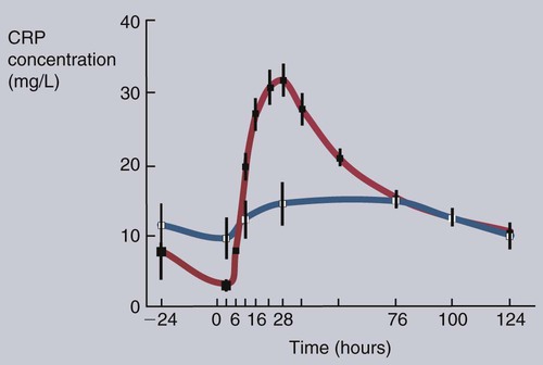
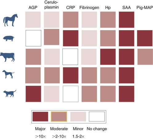
Under the influence of IL-1β, TNF-α, and especially IL-6, liver hepatocytes increase protein synthesis and secretion. New proteins may also be synthesized in lymph nodes, tonsils, and spleen as well as in blood leukocytes. This increase begins about 90 minutes after injury or systemic inflammation and subsides within 48 hours (Figure 6-2). It may also occur following prolonged stress such as road transportation or confinement. Because this increase is associated with acute infections and inflammation, the newly produced proteins are called acute-phase proteins. About 30 acute-phase proteins have been recognized, and many are important components of the innate immune system. They include soluble PRRs, complement components, clotting molecules, protease inhibitors, and iron-binding proteins. Different mammals produce different sets of acute-phase proteins (Figure 6-3).


Soluble Pattern-Recognition Receptors


C-reactive protein (CRP) is the major acute-phase protein produced in primates, rabbits, hamsters, and dogs and is important in pigs. CRP is a pentraxin (P-type lectin) and has a pentameric structure with two faces (five 20-kDa units). One face binds phosphocholine, a common side chain found in all cell membranes and many bacteria and protozoa. The other face binds to the antibody receptors FcγRI and FcγRIIa on the surface of neutrophils. CRP thus promotes the phagocytosis and removal of damaged, dying, or dead cells in addition to microorganisms. CRP can bind to bacterial polysaccharides and glycolipids and to damaged cells, where it activates the classical complement pathway. (Its name derives from its ability to bind and precipitate the C-polysaccharide of Streptococcus pneumoniae.) CRP also has an antiinflammatory role since it inhibits neutrophil superoxide production and degranulation and blocks platelet aggregation. CRP may therefore promote healing by reducing damage and enhancing the repair of damaged tissue. The functions of CRP may differ between species. For example, in lactating cows, the level of CRP in serum rises two- to five-fold for unknown reasons.


Serum amyloid P (SAP) is the major acute-phase protein in rodents. It is a pentraxin related to CRP. Like CRP, it is a soluble PRR, where one face of the molecule can bind nuclear constituents such as DNA, chromatin, and histones as well as cell membrane phospholipids. The other face can bind and activate C1q and thus activate the complement system through the classical pathway. Other soluble PRRs that are also acute-phase proteins include LPS-binding protein in cattle, CD14 in humans and mice, and C-type lectins such as mannose-binding lectin and conglutinin in other species.


Iron-Binding Molecules


One of the most important factors that determines the success or failure of bacterial invasion is the availability of iron. Many pathogenic bacteria, such as Staphylococcus aureus, Escherichia coli, Bacillus anthracis, Pasteurella multocida, and Mycobacterium tuberculosis, require large amounts of iron for growth since iron forms the key catalytic site in many of their enzymes. Animals, however, also require iron to survive. As a result, microbe and host compete for the same metal.


Iron concentrations within animal tissues are normally very low. Mammalian blood has just 10−26 M free iron since almost all available iron is bound to proteins. These iron-binding proteins include transferrin, lactoferrin, hepcidin, siderocalin, haptoglobin, and ferritin. Because many pathogenic bacteria such as Salmonella species and mycobacteria require iron for growth, withholding iron through the use of potent iron-binding proteins is an effective innate defense mechanism (Box 6-1).


< div class='tao-gold-member'>

Stay updated, free articles. Join our Telegram channel

Jul 18, 2016 | Posted by in PHARMACOLOGY, TOXICOLOGY & THERAPEUTICS | Comments Off on Systemic Responses to Inflammation

Full access? Get Clinical Tree

Get Clinical Tree app for offline access