Primary Immunodeficiencies


Primary Immunodeficiencies


Any defect in either the innate or adaptive immune systems usually becomes apparent when affected animals show unusual susceptibility to infectious or parasitic diseases. These diseases may be due to pathogenic organisms or, if the defect is very severe, opportunistic infections by organisms that are not normally able to cause disease. Deficiencies in the immune systems may be a result of inherited defects (primary immunodeficiencies); alternatively, the deficiencies may be a direct result of some other cause (secondary or acquired immunodeficiencies). This chapter describes some of the primary immunodeficiencies recorded in domestic animals.


One feature of primary immunodeficiencies in domestic animals is breed susceptibility. Examples of breed-associated immunodeficiencies include the increased risk for canine parvoviral enteritis in Doberman Pinschers and Rottweilers. German Shepherds may have increased susceptibility to canine distemper, whereas Mexican Hairless Dogs may have defective cell-mediated immune responses. It must also be recognized that the genetic composition of many breeds varies geographically, and problems with a specific breed in one country may not occur in others.


Inherited Defects in Innate Immunity


Inherited deficiencies in innate immunity include defects in the various stages of phagocytosis as well as the complement deficiencies described previously (Chapter 7). Phagocytic defects are well recognized in domestic animals.


Chédiak-Higashi Syndrome


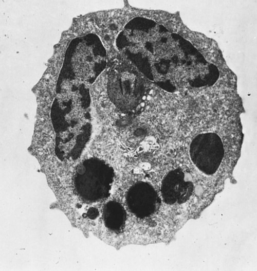
Chédiak-Higashi syndrome is an inherited disease of Hereford, Japanese black, and Brangus cattle, Aleutian mink, blue smoke Persian cats, white tigers, beige (bg/bg) mice, Orca whales, and humans. It is an autosomal recessive disease resulting from a mutation in a gene (LYST) that encodes a protein that controls lysosomal membrane fusion. The LYST gene is found on bovine chromosome 28. In Chédiak-Higashi cattle, there is a missense A : T → G : C mutation that results in replacement of a histidine with an arginine residue. The defect produces abnormally large secretory lysosomes in neutrophils, monocytes, eosinophils, and pigment cells (Figure 37-1). The enlarged neutrophil granules result from the fusion of primary and secondary granules. The leukocyte granules of affected animals are more fragile than those of normal animals, rupturing spontaneously and causing tissue damage, such as cataracts in the eye. These leukocytes have defective chemotactic responsiveness, reduced motility, and reduced intracellular killing. Cytotoxic T cells fail to excrete their granzyme-rich lysosomes.


Clinically, the syndrome is associated with multiple abnormalities. In hair, the melanosomes also fuse together, causing the dilution of coat color (sometimes only obvious in the newborn) and light-colored irises (pseudoalbinism). Other eye abnormalities include photophobia, and animals may develop cataracts. Their eyes have a red fundic light reflection rather than the normal yellow-green. Because of the neutrophil defects, affected animals may be more susceptible to respiratory infections and neonatal septicemia. Some affected breeds of cattle, such as Herefords, tend to be more susceptible to infection than others, such as Japanese black cattle. The Chédiak-Higashi gene also impairs the function of natural killer (NK) and cytotoxic T cells. As a result, affected animals may show increased susceptibility to tumors and to viruses such as the Aleutian disease virus in mink. Platelets from affected animals also contain enlarged lysosomes, and their function is abnormal. Affected animals tend to bleed abnormally after surgery and develop hematomas at injection sites. Death from acute hemorrhage is common.


Chédiak-Higashi syndrome may be diagnosed by examining a stained blood smear for the presence of grossly enlarged granules within leukocytes or by examining hair shafts for enlarged melanosomes. Treatment is symptomatic.


Pelger-Huët Anomaly


Pelger-Huët anomaly is an inherited disorder characterized by a failure of granulocyte nuclei to segment into lobes. The neutrophils therefore appear on first sight to be very immature (a left shift). The anomaly is usually detected when an animal is observed to have a persistent left shift that cannot be reconciled with its good health. Although Pelger-Huët neutrophils closely resemble band forms, their nuclear chromatin is condensed, reflecting their maturity. In humans, the anomaly is due to a mutation in the gene coding for lamin B, a nuclear membrane receptor that interacts with chromatin to determine the shape of the nucleus. Pelger-Huët anomaly has been observed in humans, Arabian horses, domestic short-hair cats, and various dog breeds such as Cocker Spaniels, Basenjis, Boston Terriers, Foxhounds, and Coonhounds. In Foxhounds and Australian Shepherds, the anomaly is inherited as an autosomal dominant trait. Pelger-Huët anomaly has a minimal effect on the health of animals. Nevertheless, fewer pups are weaned from affected dogs than from unaffected ones. In addition, Pelger-Huët neutrophils are less able to emigrate from blood vessels in vivo. This reduced mobility may be due to inflexible nuclei. B cell responses may also be impaired since normal canine B cells exposed to serum from affected dogs show depressed responses to antigens.


Canine Leukocyte Adhesion Deficiency


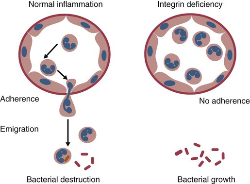
In order for neutrophils to leave inflamed blood vessels, they must first bind to vascular endothelium. This adhesion is mediated by neutrophil integrins. In the absence of these integrins, neutrophils cannot bind to endothelial cells and are unable to emigrate into tissues (Figure 37-2). Thus bacteria in tissues can grow freely without fear of attack by neutrophils. Canine leukocyte adhesion deficiency (CLAD) results from a defect in the integrin CD11b/CD18 (Mac-1). In Mac-1-deficient dogs, neutrophils cannot respond to chemoattractants, trap complement-coated bacteria (Mac-1 is a complement receptor), or bind to endothelial cells. Affected dogs suffer recurrent infections, despite the fact that their blood neutrophils are greatly elevated.


CLAD has been described in Irish Red Setters (as well as in the related Red and White Setter breed), in which it is an autosomal recessive disease. Affected animals die early in life as a result of recurrent severe bacterial infections (osteomyelitis, omphalophlebitis, gingivitis), lymphadenopathy, impaired pus formation, delayed wound healing, weight loss, and fever. Animals have a marked leukocytosis (>200,000/µL), primarily a neutrophilia and eosinophilia. Although these granulocytes look normal, functional tests reveal defects in adhesion-dependent activities, including impaired adhesion to glass or plastic surfaces or to nylon wool fibers. They cannot ingest C3b-opsonized particles. Normal canine granulocytes aggregate after activation with phorbol myristate acetate, but those of CLAD animals do not. Migration in response to chemotactic stimuli is poor. Neither CD11b nor CD18 can be detected by immunofluorescence.


The lesion results from a single missense mutation at position 107 in the β chain of the CD18 gene, which results in the replacement of a highly conserved cysteine residue (Cys36) by a serine. As a result, the mutation disrupts a disulfide bond in CD18, altering its structure and function. CD11b (the α chain) is not expressed because it must be associated with the β chain before the dimer can be expressed on the cell surface. A diagnostic test for the CLAD mutation has been developed. Thus genomic DNA is amplified by polymerase chain reaction (PCR) using primer sets for the mutated region. The PCR products may then be sequenced and the presence of the mutation determined. Matched related bone marrow allografts from normal animals have been given to CLAD dogs and effectively treated the disease.


Canine granulocytopathy syndrome was an autosomal recessive disease observed in Irish Setters. Some investigators have suggested that the disease is identical to CLAD, but because it was described before integrins were discovered, this cannot be confirmed. These animals had suppurative skin lesions, gingivitis, osteomyelitis, pododermatitis, and lymphadenopathy. Affected dogs had a pronounced leukocytosis, and their neutrophils were morphologically normal, although there was a persistent left shift. The affected animals were hypergammaglobulinemic and anemic as a result of the persistent infections. Their lymph nodes showed diffuse, suppurative, nongranulomatous lymphadenitis, which is inconsistent with a diagnosis of CLAD. Examination of the neutrophils of these dogs showed that their respiratory burst was depressed, as reflected by a decrease in glucose oxidation. Nevertheless, they were more effective than normal cells at reducing nitroblue tetrazolium, implying that O2 was produced in greater quantities than normal or, perhaps, that it was not effectively removed. Despite this, these cells were unable to kill opsonized Escherichia coli or Staphylococcus aureus, suggesting that they had a killing defect rather than an adhesion defect.


A form of canine neutrophil dysfunction related to CLAD has been reported resulting from excessive downregulation of β2-integrin. This occurs in mixed-breed dogs that present with recurrent pyogenic infections. Their neutrophils produce significantly reduced amounts of CD18 and hence β2-integrin. As a result of this reduced expression, defects occur in several adhesion-dependent neutrophil functions, including superoxide production.


Bovine Leukocyte Adhesion Deficiency


An integrin deficiency has been reported in Holstein calves. Bovine leukocyte adhesion deficiency (BLAD) is an autosomal recessive trait characterized by recurrent bacterial infections, anorexia, oral ulceration, gingivitis, periodontitis, chronic pneumonia, stunted growth, delayed wound healing, peripheral lymphadenopathy, and a persistent extreme neutrophilia. Affected calves usually die between 2 and 7 months of age. The survivors grow slowly and may develop amyloidosis. These calves have large numbers of intravascular neutrophils but very few extravascular neutrophils, even in the presence of invading bacteria.


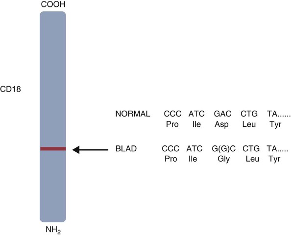
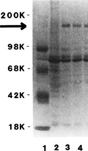
BLAD results from a point mutation in the gene coding for CD18 (Figure 37-3). As a result, an aspartic acid residue is replaced by a glycine, and functional CD18 is not produced. In the absence of this chain, complete integrins cannot be assembled. Neutrophils fail to attach to vascular endothelial cells and cannot emigrate from blood vessels. Healthy carriers have a single copy of the mutated gene and thus have abnormally low levels of CD18 (Figure 37-4). Through the use of a PCR test, the presence of the altered gene can be demonstrated. In this way it has been shown that one bull, Osborndale Ivanhoe, with thousands of registered sons and daughters, was a carrier of this gene. As a result, the defective gene was widespread and common among Holstein cattle in the United States (14% of bulls, 5.8% of cows). Fortunately, carrier animals can be rapidly detected and removed from breeding programs.


Because CD18 integrins are also expressed by T cells attracted to sites of antigen invasion, BLAD calves show poor delayed hypersensitivity responses. Their neutrophils show reduced responsiveness to chemotactic stimuli and diminished superoxide production and myeloperoxidase activity. They have increased expression of Fc receptors but decreased binding and expression of C3b and IgM on neutrophils, implying an alteration in receptor function. This is reflected by greatly reduced endocytosis and killing of S. aureus.


Canine Cyclical Neutropenia


Canine cyclical neutropenia (gray collie syndrome) is an autosomal recessive disease of Border Collies. Affected dogs have dilution of skin pigmentation, eye lesions, and regular cyclic fluctuations in leukocyte numbers. Their hair is a characteristic silver-gray color, and their nose is gray—a diagnostic feature. The loss of neutrophils occurs about every 11 to 12 days and lasts for about 3 days. It is followed by normal or elevated neutrophil counts for about 7 days. Severe neutropenia suppresses inflammation and increases susceptibility to bacterial and fungal infections. (Their neutrophils also have reduced myeloperoxidase activity, so the disease is not entirely due to a neutrophil deficiency). In humans, the disease is due to a defect in the gene coding for neutrophil elastase, an enzyme found in azurophil granules. The animals have severe enteric disease, respiratory infections, mouth infections (gingivitis), bone disease (arthralgia), and lymphadenitis and rarely live beyond 3 years. Because platelet numbers also cycle, affected dogs may also have bleeding problems, including gingival hemorrhage and epistaxis. Immunoglobulin levels rise as a result of the recurrent antigenic stimulation, but complement levels cycle in conjunction with the neutropenia. The disease begins to express itself as maternal immunity wanes. Affected puppies are weak, grow poorly, have wounds that fail to heal, and have a high mortality rate. If they are kept alive by aggressive antibiotic therapy, chronic inflammation may lead to amyloidosis.


Treatment involves the repeated use of antibiotics to control the recurrent infections. If endotoxin is administered repeatedly, it can stimulate the bone marrow and stabilize neutrophil, reticulocyte, and platelet numbers. Lithium carbonate has a similar effect. Unfortunately, both endotoxin and lithium carbonate are toxic, and the disease recurs when the treatment is discontinued.


Other Examples of Defective Neutrophil Function


An inherited defect in neutrophil bactericidal activity has been reported in Dobermans. Dogs had bronchopneumonia and chronic rhinitis that developed soon after birth and persisted despite antimicrobial therapy. Although their chemotaxis and ingestion were apparently normal, their neutrophils were unable to kill S. aureus. Since these cells showed reduced reduction of nitroblue tetrazolium and superoxide production, it was suggested that there was a defect in the respiratory burst pathway.


Young Weimaraner dogs have been described as suffering from an immunodeficiency syndrome with a wide range of clinical signs. These include recurrent fevers, diarrhea, pneumonia, pyoderma, osteomyelitis, stomatitis, and osteomyelitis. They may have defective neutrophil function, as shown by a depressed chemiluminescent response to phorbol ester, implying a defect in the respiratory burst mechanism. Their IgG levels may be significantly lower than normal and their IgM and IgA levels somewhat reduced; the other immunological parameters of these animals fall within normal ranges.


A persistent neutropenia attributable to a deficiency of granulocyte colony-stimulating factor (G-CSF) has been reported in a 3-year-old male Rottweiler. The animal had a fever due to multiple recurrent infections, especially a chronic bacterial arthritis in the presence of a persistent neutropenia. A bioassay showed that the animal was not making G-CSF. Its myeloid stem cells responded readily to additional G-CSF, suggesting that they were functionally normal. Bone marrow examination suggested that its neutrophil precursors had failed to mature.


A possible autosomal recessive neutropenia has been described in Border Collies. This disease, called trapped neutrophil syndrome, resulted in recurrent bacterial osteomyelitis and gastroenteritis. Animals presented with persistent fever and lameness due to lytic bone lesions. They had myeloid hyperplasia and dense accumulations of neutrophils in the marrow but few in the blood. The neutropenia apparently resulted from an inability of the neutrophils to escape from the bone marrow into the bloodstream, perhaps as a result of a deficiency of granulocyte-macrophage colony-stimulating factor. In humans this disease has been called myelokathexis.


Inherited Defects in the Adaptive Immune System
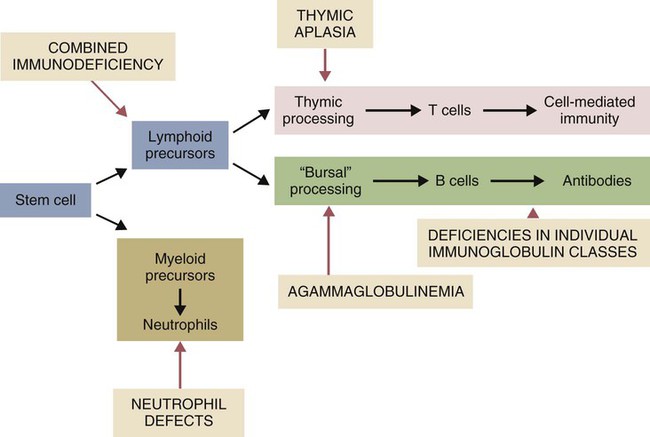

The inherited immunological defects have served to confirm the overall arrangement of the immune system, as outlined in Figure 37-5. For example, if both the cell- and antibody-mediated immune responses are defective, it may be assumed that the genetic lesion operates at a point before thymic and bursal cell processing—that is, a stem cell lesion. A defect that occurs only in thymic development is reflected in an inability to mount cell-mediated immune responses, although antibody production may be normal. Similarly, a lesion restricted to B cells is reflected by impaired antibody responses.


Recent advances in molecular genetics have enabled many new primary immunodeficiency disorders to be identified in humans. For example, at least 10 different mutations can result in severe combined immunodeficiency. Likewise mutations in many different genes can disable B cell function and result in immunoglobulin deficiencies.

< div class='tao-gold-member'>

Stay updated, free articles. Join our Telegram channel

Jul 18, 2016 | Posted by in PHARMACOLOGY, TOXICOLOGY & THERAPEUTICS | Comments Off on Primary Immunodeficiencies

Full access? Get Clinical Tree

Get Clinical Tree app for offline access