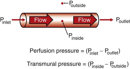
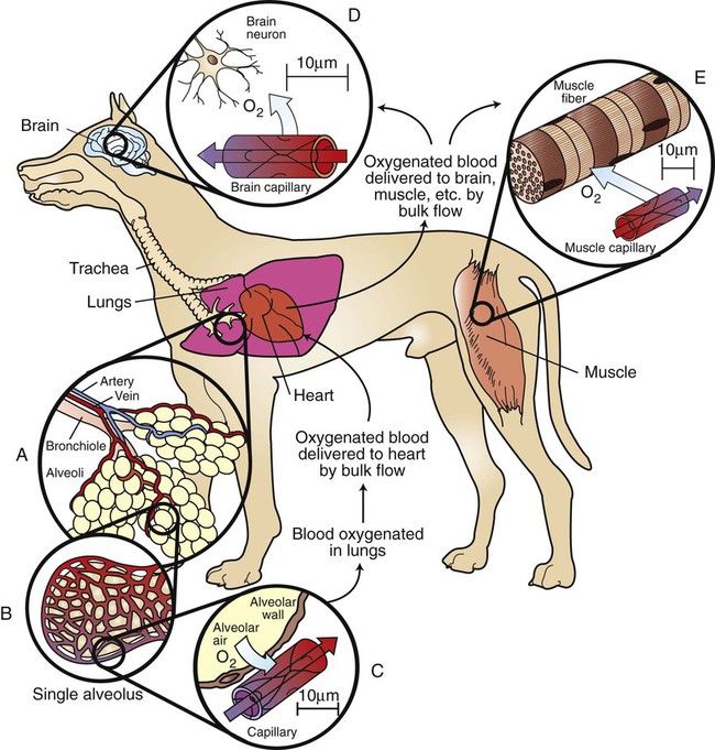
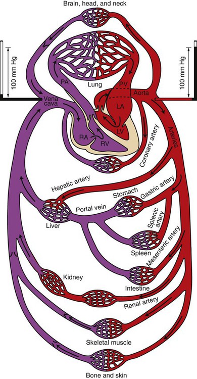
Overview of Cardiovascular Function


Overview of Cardiovascular Function


Because Normal Cardiovascular Function Is Essential for Life and Health, a Practical Understanding of Cardiovascular Function and Dysfunction Is Vital to the Veterinary Clinician


Cardiovascular physiology is the study of the function of the heart, the blood vessels, and the blood. The primary function of the cardiovascular system can be summarized in one word: transport. The bloodstream transports numerous substances that are essential for life and health, including the oxygen and nutrients required by every cell in the body. Blood also carries carbon dioxide and other metabolic waste products away from metabolically active cells and delivers them to the lungs, kidneys, or liver, where they are excreted.


To appreciate the importance of cardiovascular transport, consider what happens if the heart stops contracting and circulation ceases: unconsciousness results within about 30 seconds, and irreversible damage to the brain and other sensitive body tissues occurs within a few minutes. However, circulation does not have to stop completely for significant dysfunction to occur. For example, the loss of as little as 10% of the normal blood volume can impair exercise performance.


In each tissue of the body, normal function depends on the delivery of adequate blood flow. The higher the rate of metabolism in a tissue, the greater is the requirement for blood flow. The condition of inadequate blood flow to any tissue is called ischemia. Even transient ischemia leads to dysfunction. Persistent ischemia leads to permanent tissue damage (infarction) and eventually to cell death (necrosis).


Many veterinary students have difficulty understanding cardiovascular physiology. They tend to agree with William Harvey, the father of cardiovascular physiology, whose initial impression was that the motions of the heart and the blood were so complicated that they could be comprehended only by God. Harvey persisted, however, in a careful, deliberate study of cardiovascular function and in 1628 set forth the first proof that the heart propels blood through the blood vessels in a circulatory pattern. Before Harvey’s time, it was thought that blood flowed out of the heart into the blood vessels and then returned to the heart by backward flow in the same vessels. In other words, blood was thought to flow in a tidal manner, in much the same way that air flows through a single set of airways: first into the lungs and then back out.


We now take for granted that the cardiovascular system is a circulatory system, not a tidal system. However, the circularity of the cardiovascular system is precisely what makes it difficult to understand. It has no clear beginning or ending, and disturbances in one part of the cardiovascular system end up affecting all other parts as well. In recognition of this complexity, Chapters 18 to 26 are written with the goal of identifying the most basic and important concepts of normal cardiovascular function and explaining them in a way that best prepares the reader to understand, diagnose, and treat cardiovascular dysfunction (cardiovascular disease). The remainder of this chapter reviews the general features of the cardiovascular system. Chapters 19 to 25 discuss the various elements of the cardiovascular system in detail. Chapter 26 summarizes cardiovascular function and dysfunction by describing the overall effects of heart failure, hemorrhage, and exercise.


Cardiovascular Dysfunctions Sometimes Reflect Primary Cardiovascular Disturbances or Diseases, But More Often They Are Secondary Consequences of Noncardiovascular Disturbances or Diseases


Impairment in the transport functions of the cardiovascular system is encountered frequently in veterinary medicine. Some of these cardiovascular dysfunctions are primary, in that the fundamental disturbance or disease process affects the cardiovascular system directly. One example of primary cardiovascular dysfunction is hemorrhage (loss of blood from blood vessels). Another is myocarditis (literally, muscle-heart-inflammation), which can be caused by a toxic chemical or by a viral or bacterial infection that inflames the heart muscle and impairs the ability of the heart to pump blood.


Cardiovascular dysfunction and disease can be either congenital (present at birth) or acquired (developing after birth). Congenital cardiovascular diseases frequently involve defective heart valves, which either cannot open fully or cannot close completely. Congenital cardiac defects are common in certain breeds of dogs and horses. Although a heart that has a congenital defect or an acquired disease may be able to pump an adequate amount of blood when the animal is at rest, it usually cannot deliver the increased blood flow required by the body during exercise. When a dysfunction in the heart impairs its ability to pump the amount of blood flow normally needed by the body, the condition is called heart failure (or pump failure). The patient with heart failure classically exhibits a limited ability or willingness to exercise (exercise intolerance).


Parasites are a common cause of acquired cardiovascular dysfunction. In dogs, for example, adult heartworms (Dirofilaria immitis) lodge in the right ventricle and pulmonary artery, where they impede the flow of blood. These worms also release substances into the circulation that disrupt the body’s ability to control blood pressure and blood flow. In horses, bloodworms (Strongylus vulgaris) lodge in the mesenteric arteries and decrease the blood flow to the intestine. The resulting intestinal ischemia depresses digestive functions (motility, secretion, and absorption), and the horse exhibits signs of gastrointestinal distress (colic).


In many other disease states, cardiovascular complications develop even though the cardiovascular system is not the primary target of the disease. These secondary cardiovascular dysfunctions often become the most serious and life-threatening aspects of the disease. For example, severe burns or persistent vomiting or diarrhea leads to substantial losses of water and electrolytes (small, soluble ions in the body fluids; e.g., Na+, Cl, K+, Ca2+) . Even if the blood volume is not depleted to dangerously low levels in these conditions, the alteration in electrolyte concentrations can lead to abnormal heart rhythms (cardiac arrhythmias) and ineffective pumping of blood by the heart (heart failure). The electrolyte abnormalities in such a patient can be made even worse if incorrect fluid therapy is given. Incorrect fluid therapy can also lead to an accumulation of excess fluid in the tissues of the body; this “waterlogging” of tissues is called edema. If the excess fluid gathers in the lung tissue, the condition is called pulmonary edema. Pulmonary edema is life threatening because it slows the flow of oxygen from the pulmonary air sacs (alveoli) into the bloodstream.


Pulmonary edema is a secondary complication in many disease states. A further example is shock-lung syndrome, which results when toxic substances in the body trigger an increase in the permeability of the lung blood vessels. These “leaky” vessels allow water, electrolytes, plasma proteins, and white blood cells to leave the bloodstream and accumulate in the lung tissue and airways. The resulting pulmonary edema can lead to death.


Whereas the effects of shock-lung syndrome are most serious in the pulmonary circulation, other types of shock depress the cardiovascular system in general. Hemorrhagic shock is a generalized cardiovascular failure caused by severe blood loss. Cardiogenic shock is a cardiovascular collapse caused by heart failure. Septic shock is caused by bacterial infections in the bloodstream (bacteremia). Endotoxic shock occurs when endotoxins (fragments of bacterial cell walls) enter the bloodstream; this often occurs when the epithelial lining of the intestines becomes damaged. Epithelial damage can result from bacterial infections in the intestines or from ischemia in the intestinal walls (as with bloodworms in horses). When the intestinal epithelium breaks down, endotoxins from the intestine can enter the bloodstream. These endotoxins then cause the body to produce substances that depress the pumping ability of the heart. The resulting heart failure leads to low blood flow and ischemia in all the vital body organs. Kidney (or renal) failure, respiratory failure, central nervous system (CNS) depression, and death follow.


Anesthetic overdose is another clinical problem in which the most serious and life-threatening symptoms are the secondary cardiovascular complications. Most anesthetics depress the CNS, and the resulting abnormal neural signals to the heart and the blood vessels can depress cardiac output and lower blood pressure. Some anesthetics, particularly the barbiturates, also depress the pumping ability of the heart directly.


There are many other examples of primary and secondary cardiovascular dysfunction, but those just mentioned illustrate the importance and variety of cardiovascular dysfunctions encountered in veterinary medicine. The distinction between primary and secondary cardiovascular dysfunction is sometimes unclear, but this difficulty simply emphasizes how intimately the cardiovascular system is interconnected with all the other body systems and how dependent all the other systems are on the normal functioning of the cardiovascular system.


Substances Transported by the Cardiovascular System Include Nutrients, Waste Products, Hormones, Electrolytes, and Water


The blood transports the metabolic substrates needed by every cell of the body, including oxygen, glucose, amino acids, fatty acids, and various lipids. The blood also carries away from each cell in the body various metabolic waste products, including carbon dioxide, lactic acid, the nitrogenous wastes of protein metabolism, and heat. Although the heat produced by metabolic processes within cells is not a material waste product, its transport by the cardiovascular system to the body surface is essential, because tissues deep within the body would otherwise become overheated and dysfunctional.


Blood also transports vital chemical messengers: the hormones. Hormones are synthesized and released by cells in one organ and are carried by the bloodstream to cells in other organs, where they alter organ function. For example, insulin, which is produced by cells of the pancreas, is carried by the blood to cells throughout the body, where it promotes the cellular uptake of glucose. Inadequate insulin production (as in type 1 diabetes) results in inadequate entry of glucose into cells, whereas glucose concentrations in the blood rise to very high levels. The low intracellular glucose concentration is particularly disruptive to neural function, and the consequences can be serious (diabetic coma) or lethal. Another hormone, adrenaline (a mixture of epinephrine and norepinephrine), is released into the bloodstream by cells in the adrenal medulla during periods of stress. The epinephrine and norepinephrine circulate to various body organs, where they have effects that prepare a threatened animal for the “fight or flight” response. These effects include an increase in heart rate and cardiac contractility, dilation of skeletal muscle blood vessels, an increase in blood pressure, increased glycogenolysis, dilation of the pupils and airways, and piloerection (hair standing on end).


Finally, the blood transports water and essential electrolytes, including Na+, Cl, K+, Ca2+, H+, and HCO3. The kidneys are the organs primarily responsible for maintaining normal water and electrolyte composition in the body. The kidneys accomplish this by altering the electrolyte concentrations in blood as it flows through the kidneys. The altered blood then circulates to all other organs in the body, where it normalizes the water and electrolyte content in the extracellular fluids of each tissue.


Two Modes of Transport Are Used in the Cardiovascular System: Bulk Flow and Diffusion


Blood moves through the heart and blood vessels by bulk flow. The most important feature of bulk flow is that it is rapid over long distances. Blood that is pumped out of the heart travels quickly through the aorta and its various branches; within 10 seconds it reaches distant parts of the body, including the head and limbs. Transport requires energy, and the source of energy for bulk flow is a hydrostatic pressure difference; unless the pressure at one end of a blood vessel is higher than the pressure at the other end, flow will not occur. The difference in pressure between two points in a blood vessel is called the perfusion pressure difference or, more often, simply perfusion pressure. Perfusion literally means “through-flow,” and the perfusion pressure is the pressure difference that causes blood to flow through blood vessels. The muscular pumping action of the heart creates the perfusion pressure that constitutes the driving force for bulk blood flow through the circulation.


It is important to distinguish between perfusion pressure difference and transmural pressure difference (usually shortened to transmural pressure). Transmural means “across the wall,” and transmural pressure is the difference between the blood pressure inside a blood vessel and the fluid pressure in the tissue immediately outside the vessel (transmural pressure equals inside pressure minus outside pressure). Transmural pressure is the pressure difference that would cause blood to flow out of a vessel if a hole were poked in the vessel wall. Transmural pressure is also called distending pressure, because it corresponds to the net outward “push” on the wall of a blood vessel. Figure 18-1 emphasizes the distinction between perfusion pressure and transmural pressure.


Diffusion is the second mode of transport in the cardiovascular system. Diffusion is the primary mechanism by which dissolved substances move across the walls of blood vessels, from the bloodstream into the interstitial fluid, or vice versa. Interstitial fluid is the extracellular fluid outside capillaries. It is the fluid that bathes each cell of a tissue. Most of the movement of substances between the blood and the interstitial fluid takes place across the walls of the capillaries, the smallest blood vessels. For a substance (e.g., oxygen) to move from the bloodstream to a tissue cell, it diffuses across the wall of a capillary and into the tissue interstitial fluid, and then diffuses from the interstitial fluid into the tissue cell.


The source of energy for diffusion is a concentration difference. A substance diffuses from the bloodstream, across the wall of a capillary, and into the interstitial fluid only if the concentration of the substance is higher in the blood than in the interstitial fluid (and if the capillary wall is permeable to the substance). If the concentration of a substance is higher in the interstitial fluid than in the blood, the substance will diffuse from the interstitial fluid into the capillary blood. It is important to distinguish diffusion, in which a substance moves passively from an area of high concentration toward an area of low concentration, from active transport, in which substances are forced to move in a direction opposite to their concentration gradient. In general, substances are not transported actively across the walls of capillaries. The movement of substances between the bloodstream and the interstitial fluid occurs by passive diffusion.


Because Diffusion Is Very Slow, Every Metabolically Active Cell in the Body Must Be Close to a Capillary Carrying Blood by Bulk Flow


To understand more fully how the two types of transport (bulk flow and diffusion) are used in the cardiovascular system, consider the transport of oxygen from the outside air to a neuron in the brain. With each inspiration, fresh air containing oxygen (O2) moves by bulk flow through progressively smaller airways (trachea, bronchi, and bronchioles) and finally enters the alveolar air sacs (Figure 18-2, A). The thin walls separating alveoli contain a meshwork of capillaries (see Figure 18-2, B). Blood flowing through these alveolar capillaries passes extremely close (within 1 µm) to the air in the alveoli (see Figure 18-2, C). The blood in an alveolar capillary has just returned from the body tissues, where it gave up some of its oxygen. Therefore the concentration of oxygen in alveolar capillary blood is lower than the concentration of oxygen in alveolar air. This concentration difference causes some oxygen to diffuse from the alveolar air into the capillary blood.


A large dog has about 300 million alveoli, with a total surface area of about 130 m2 (equal to half the surface area of a tennis court). This huge surface area is laced with pulmonary capillaries. Thus, even though only a tiny amount of oxygen diffuses into each pulmonary capillary, the aggregate uptake of oxygen into the pulmonary bloodstream is substantial (typically, 125 mL O2/minute in a large, resting dog, increasing by a factor of 10 or more during strenuous exercise). In summary, both the large alveolar surface area and the proximity of alveolar air to the blood in alveolar capillaries promote efficient diffusion of oxygen; it takes less than 1 second for the blood in an alveolar capillary to become oxygenated.


As it leaves the lungs, each 100 mL of oxygenated blood normally carries 20 mL of oxygen. About 1.5% of this oxygen is carried in solution; the other 98.5% is bound to the protein hemoglobin within the erythrocytes (red blood cells). The oxygenated blood moves by bulk flow from the lungs to the heart. The heart pumps this oxygenated blood out into the aorta, and from there it is distributed via a complex system of branching arteries to all parts of the body (including the brain and skeletal muscles, as illustrated in Figure 18-2). Capillaries in the brain bring a bulk flow of oxygenated blood very close to each brain neuron (see Figure 18-2, D). Metabolic processes within the neurons consume oxygen, so the oxygen concentration inside neurons is low. The gradient of oxygen concentration between the capillary blood (high) and the neurons (low) provides the driving force for oxygen to diffuse first from the blood into the interstitial fluid and then into the neurons.


Each brain neuron must be within about 100 µm of a capillary carrying blood by bulk flow if diffusion is to deliver oxygen rapidly enough to sustain normal metabolism in the neuron. Diffusional exchange over distances up to 100 µm typically takes only 1 to 5 seconds. If the distance involved were a few millimeters, diffusion would take minutes to occur. Diffusion of oxygen a few centimeters through body fluid would take hours. Therefore, normal life processes require that every metabolically active cell of the body be within about 100 µm of a capillary carrying blood by bulk flow. If this bulk flow is interrupted for any reason, perhaps because of a thrombus (blood clot) in the artery that delivers blood to a particular region of a tissue, that region of tissue becomes ischemic. As stated earlier, ischemia leads to dysfunction; persistent, severe ischemia leads to infarction and eventually to necrosis. Cerebral infarction causes the condition commonly known as stroke.


Figure 18-2, E, shows a capillary carrying bulk flow of blood past a skeletal muscle cell (muscle fiber). Oxygen moves by diffusion from the capillary blood into the muscle interstitial fluid and then into the muscle cell, where it is consumed in the metabolic reactions that provide energy for muscle contraction. The oxygen consumption of a skeletal muscle depends on the severity of its exercise; at a maximum, oxygen consumption may reach levels 40 times greater than the resting level. Because of its tremendous metabolic capacity, muscle tissue has an especially high density of capillaries. In fact, several capillaries are typically arrayed around each skeletal muscle fiber. This arrangement provides more surface area for diffusional exchange than would be possible with a single capillary and brings the bulk flow of blood extremely close to all parts of each skeletal muscle cell.


Heart muscle, like skeletal muscle, consumes a large amount of oxygen. Oxygenated blood is carried from the aorta to the heart muscle by a network of branching coronary arteries. This blood next moves by bulk flow into coronary capillaries, which pass close by each cardiac muscle cell. If a thrombus interrupts the bulk flow of blood in a coronary artery, the heart muscle cells supplied by that artery become ischemic. Ischemia develops even if the cardiac muscle deprived of blood flow lies within a few millimeters of the left ventricular chamber, which is filled with oxygen-rich blood. Oxygen simply cannot diffuse rapidly enough from the ventricular chamber to the ischemic cells to sustain their metabolism. Ischemic cardiac muscle loses its ability to contract forcefully; also, cardiac arrhythmias may develop. Severe myocardial ischemia causes a myocardial infarction, or heart attack.


Coronary artery disease and cerebrovascular disease are encountered more often in human medicine than in veterinary medicine. In contrast, cardiac disease (dysfunction of the heart muscle or valves, as distinguished from disease of the coronary arteries) is encountered more often in veterinary medicine than in human medicine. Therefore, Chapters 19 to 26 place more emphasis on cardiac physiology than on vascular physiology.


The Pulmonary and Systemic Circulations Are Arranged In Series, But the Various Organs Within the Systemic Circulation Are Arranged in Parallel


As shown in Figure 18-3, blood is pumped from the left ventricle into the aorta. The aorta divides and subdivides to form many arteries, which deliver fresh, oxygenated blood to each organ of the body, except the lungs. The pattern of arterial branching that delivers blood of the same composition to each organ is called parallel. After blood passes through the capillaries within individual organs, it enters veins. Small veins combine to form progressively larger veins, until the entire blood flow is delivered to the right atrium by way of the venae cavae (pleural of vena cava, includes both superior vena cava and inferior vena cava). The blood vessels between the aorta and the venae cavae (including the blood vessels in all organs of the body except the lungs) are collectively called the systemic circulation. From the right atrium, blood passes into the right ventricle, which pumps it into the pulmonary artery. The pulmonary artery branches into progressively smaller arteries, which deliver blood to each alveolar (pulmonary) capillary. Blood from pulmonary capillaries is collected in pulmonary veins and brought to the left atrium. Blood then passes into the left ventricle, completing the circuit. The blood vessels of the lungs, including the pulmonary arteries and veins, constitute the pulmonary circulation. The pulmonary circulation and the heart are collectively termed the central circulation. The pulmonary circulation and the systemic circulation are arranged in series; that is, blood must pass through the pulmonary vessels between each passage through the systemic circuit.


In one pass through the systemic circulation, blood generally encounters only one capillary bed before being collected in veins and returned to the heart, although a few exceptions to this rule exist. One exception occurs in the splanchnic circulation, which supplies blood to the digestive organs. As shown in Figure 18-3, blood that leaves the gastric, splenic, or mesenteric capillaries enters the portal vein. The portal vein carries splanchnic venous blood to the liver, where the blood passes through another set of capillaries before it returns to the heart. This arrangement of two systemic capillary beds in series is called a portal system. The splanchnic portal system allows nutrients that have been absorbed from the gastrointestinal tract to be delivered directly to the liver. There the nutrients are transformed for storage or allowed to pass into the general circulation. The liver also receives some blood directly from the aorta through the hepatic artery.


The kidneys also contain a portal system. As shown in Figure 18-3, blood enters a kidney by way of a renal artery and passes through two sets of capillaries (called glomerular and tubular) before returning to the venous side of the systemic circulation. Large amounts of water, electrolytes, and other solutes are filtered out of the blood as it passes through the glomerular capillaries. Most of this filtered material is subsequently reabsorbed into the bloodstream as it flows through the peritubular capillaries. The remainder becomes urine. The kidneys use the renal portal system to adjust the amounts of water, electrolytes, and other critical solutes in the blood.


A third portal system is found in the brain and is important in the control of hormone secretion by the pituitary gland. After traversing capillaries in the hypothalamus, blood enters portal vessels that carry it to the anterior pituitary gland (adenohypophysis) and to another set of capillaries (see Figures 33-16 and 33-17). As blood traverses the hypothalamic capillaries, it picks up several signaling chemicals that control the release of pituitary hormones. When this blood reaches capillaries in the anterior pituitary gland, these substances diffuse out of the bloodstream and into the pituitary interstitial fluid, where they act on pituitary cells to increase or decrease their secretion of specific pituitary hormones. This system is called the hypothalamic-hypophyseal portal system.


To summarize, except for a few specialized portal systems, blood encounters only one capillary bed in a single pass through the systemic circulation.

< div class='tao-gold-member'>

Stay updated, free articles. Join our Telegram channel

Jul 18, 2016 | Posted by in PHARMACOLOGY, TOXICOLOGY & THERAPEUTICS | Comments Off on Overview of Cardiovascular Function

Full access? Get Clinical Tree

Get Clinical Tree app for offline access