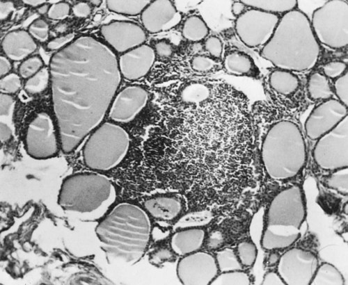
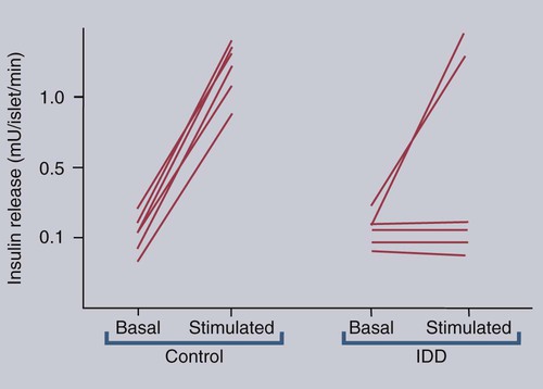
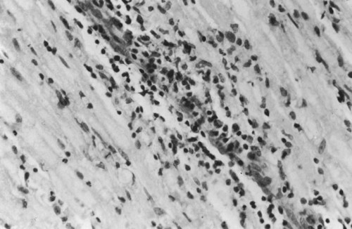
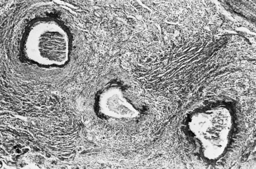
Organ-Specific Autoimmune Diseases


Organ-Specific Autoimmune Diseases


Autoimmune diseases that mainly affect a single organ or tissue presumably result from an abnormal response to a small number of self-antigens and do not necessarily reflect significant loss of control of the immune system as a whole. It is likely that all organs of the body are potentially susceptible to this form of immunological attack. Nevertheless, autoimmune diseases directed against endocrine organs, skin, blood and the nervous system, tend to be most commonly affected. Much depends on the species and breed of an animal as well as its age.


Autoimmune Endocrine Disease


Although domestic animals develop autoimmune endocrine diseases, they differ from humans insofar as these tend to present as single disorders rather than involving multiple endocrine organs. Occasionally a dog may experience two or more autoimmune endocrine disorders simultaneously (autoimmune polyglandular syndrome), but this is very uncommon.


Lymphocytic Thyroiditis


Dogs, humans, and chickens suffer from autoimmune thyroiditis as a result of the production of autoantibodies against thyroglobulin or thyroid peroxidase. These antibodies may also react with triiodothyronine (T3) or thyroxine (T4). Affected dogs may also show a delayed skin reaction to intradermally injected thyroid extract, suggesting that cell-mediated immunity contributes to the disease. Several dog breeds are predisposed to thyroiditis, and relatives of affected animals may have antithyroid antibodies although clinically normal. A familial form of hypothyroidism has been demonstrated in Beagles and Great Danes. Dogs from high-risk breeds such as Dobermans tend to develop the disease when young, whereas dogs from low-risk breeds tend to develop it when older. Unfortunately, by the time the disease is diagnosed, the dog may already have been bred. Affected thyroids are infiltrated with plasma cells and lymphocytes, and germinal center formation may occur (Figure 35-1). The invading lymphocytes probably cause epithelial cell destruction through antibody-dependent cell-mediated cytotoxicity (ADCC) and T cell cytotoxicity.


In dogs, clinical signs appear after about 75% of the thyroid is destroyed. These signs are those of hypothyroidism; that is, the animals are fat and inactive and show patchy hair loss. The most common problems are a dry, dull, coarse coat; scaling; hypotrichosis; slow hair regrowth; hyperpigmentation; myxedema; and pyoderma. Other signs include myopathy, hyperlipidemia, hypothermia, anestrus, galactorrhea, diarrhea or constipation, and polyneuropathy. Tests of thyroid function such as a radioimmunoassay for plasma T4 or T3 only confirm the existence of hypothyroidism. A thyroid-stimulating hormone (TSH) response test is more useful because it can confirm the inability of the affected thyroid to respond to TSH. (Plasma T4 levels are measured before and after injection of TSH.) In order to confirm autoimmune thyroiditis, a biopsy must show the characteristic lymphocytic infiltration. Antithyroid antibodies must be detected in serum using an enzyme-linked immunosorbent assay (ELISA), immunoblot, or an indirect fluorescent antibody test (Chapter 41). There is little correlation between antithyroid antibody titers and disease severity. Management of affected animals involves replacement therapy with sodium levothyroxine (synthetic T4). Improvement should be seen within 4 to 6 weeks. There is no cure for this disease, and the success of treatment depends on effective replacement therapy.


An autoimmune thyroiditis also occurs in the OS (obese) strain of white Leghorn chickens. The thyroids of these birds are heavily infiltrated by lymphocytes and plasma cells. Autoantibodies are directed against thyroglobulin, and affected birds are hypothyroid. These birds also make antibodies against their adrenal gland, exocrine pancreas, and proventricular cells. Neonatal thymectomy prevents the development of lesions.


Insulin-Dependent Diabetes Mellitus


In humans, insulin-dependent diabetes mellitus (IDDM) is an autoimmune disease mediated by autoantibodies against an islet cell enzyme called glutamic acid decarboxylase. Some cases of IDDM in dogs may also be immunologically mediated. The canine disease is associated with pancreatic islet atrophy and a loss of β cells. In some cases, the islets are infiltrated by lymphocytes. Experimentally, circulating mononuclear cells from diabetic dogs have suppressed insulin production by cultured mouse islet cells. Serum from IDDM dogs lysed these islet cells in the presence of complement (Figure 35-2). When dog serum was tested for antibodies against cultured β cells by immunofluorescence, 9 of 23 diabetic dogs showed strongly positive reactions, and an additional 3 showed a weak reaction. Only 1 of 15 normal dogs gave a positive response. Thus cytotoxic cells or antibodies or both may be responsible for β cell destruction in dogs. A familial predisposition to IDDM has been observed in Samoyeds. Genes that may influence the development of canine diabetes mellitus include those for the regulatory cytokines interferon-γ (IFN-γ), interleukin-12 (IL-12), IL-4, and IL-10. These candidate genes were screened for single nucleotide polymorphisms (SNPs) in multiple different dog breeds. Significant associations were observed for IL-4 in Collies, Cairn Terriers, and Schnauzers and for IL-10 in the Cavalier King Charles Spaniel. This suggests that cytokine genes that influence the Th1/Th2 balance may determine susceptibility to diabetes.


Nonobese diabetic (NOD) mice develop spontaneous IDDM associated with infiltration of the pancreatic islets by lymphocytes. Their disease resembles human type 1 diabetes. The development of diabetes in NOD mice is influenced by their microflora. Thus pathogen-free NOD mice that lack MyD88 protein (MyD88 is an adaptor molecule for innate receptors such as the toll-like receptors [TLRs]) do not develop diabetes, whereas totally germ-free MyD88− NOD mice do. If defined commensal microbes are given to these germ-free mice, their diabetes is less severe. Likewise commensal microbes from the intestine of pathogen-free MyD88− NOD mice attenuate the disease when given to germ-free mice. Somehow the interaction of the intestinal microflora with the innate immune system modifies the predisposition of these mice to develop diabetes.


Human patients with type 1 IDDM have circulating antibodies against the 67-kDa isoform of glutamic acid decarboxylase (GAD65) and/or insulinoma antigen-2 (IA-2). Some diabetic dogs also possess such autoantibodies. Thus 4 of 30 diabetic dogs had autoantibodies to GAD65, whereas 3 dogs had autoantibodies to IA-2. Two had autoantibodies to both antigens.


Diabetes mellitus is rare in cattle. Affected animals have atrophied and reduced numbers of pancreatic islets with partial or complete loss of β cells. Lymphocytes commonly infiltrate the remaining islets.


Autoimmune Neurological Disease


An autoimmune brain disease known as experimental allergic encephalomyelitis may be produced by immunizing animals with brain tissue emulsified in Freund’s complete adjuvant. After a few weeks, dogs or cats develop focal encephalitis and myelitis, possibly with paralysis. The brain lesions consist of focal vasculitis, mononuclear cell infiltration, perivascular demyelination, and axon damage. Antibodies to brain tissue can be detected in the serum of these animals, although the lesion itself is a result of a cell-mediated response.


A similar encephalitis used to occur following administration of rabies vaccines containing brain tissue to humans. For this reason, the use of adult brain tissue was stopped, and suckling mouse brain tissue obtained prior to myelination is used in the production of rabies vaccines. Postdistemper demyelinating leukoencephalopathy may also be of autoimmune origin, although the production of antimyelin antibodies appears to be common response to central nervous tissue damage, regardless of its cause.


Equine Polyneuritis


Equine polyneuritis (neuritis of the cauda equina) is an uncommon disease of horses affecting the sacral and coccygeal nerves. Affected horses show hyperesthesia followed by progressive paralysis of the tail, rectum, and bladder and localized anesthesia in the same region. The disease may also be associated with facial and trigeminal paralysis. Although sacral and lumbar involvement is usually bilateral, the cranial nerve involvement is often unilateral. A chronic granulomatous inflammation develops in the region of the extradural nerve roots. Affected nerves are thickened and discolored. They show a loss of myelinated axons; infiltration by macrophages, lymphocytes, giant cells, and plasma cells; and deposition of fibrous material in the perineurium. In severe cases the nerve trunks may be almost totally destroyed. Affected horses have circulating antibodies to a peripheral myelin protein called P2. P2 can induce experimental allergic neuritis in rodents (see later). Although equine polyneuritis may be an autoimmune disease, equine adenovirus-1 has been isolated from its lesions, so the cause is complex. Because of the severe nerve damage, immunosuppressive or antiinflammatory therapy is rarely successful. Neuritis of the cauda equina has also been reported in a dog. The dog presented with a flaccid tail and urinary incompetence. The nerve roots of its cauda equina and lumbar nerves were infiltrated by T and B cells.


Canine Polyneuritis


Canine polyneuritis or coonhound paralysis affects dogs following a bite or scratch from a raccoon. It presents as an ascending symmetrical flaccid paralysis with mild sensory impairment. The bitten limb is usually affected first, but the disease is progressive and will worsen for 10 to 12 days following the bite. In severe cases the dog may develop flaccid quadriplegia and lose the ability to swallow, bark, or breathe. The disease is, however, self-limited, and if respiration is not impaired, the prognosis is good. Dogs usually recover completely. Affected nerves show demyelination and axonal degeneration with macrophage infiltration. An acute polyneuritis similar to coonhound paralysis has also been described following vaccination of dogs with rabies or other vaccines.


Coonhound paralysis and postvaccinal polyneuritis both closely resemble Guillain-Barré syndrome in humans. This syndrome may follow upper respiratory tract infection, gastrointestinal disease, or even vaccination. It is mediated by autoantibodies against peripheral nerve glycolipids. Management of Guillain-Barré syndrome requires plasmapheresis and administration of intravenous immunoglobulins (IVIG). Veterinarians treating canine polyneuritis have traditionally administered corticosteroids, but their effectiveness is unclear.


If sciatic nerve tissue is used to immunize experimental dogs, it provokes experimental allergic neuritis. After a latent period of 6 to 14 days, the animals develop an ascending polyneuritis and gradual paralysis (Figure 35-3). The disease is due to peripheral nerve demyelination resulting from autoimmune attack.


Steroid-Responsive Meningitis-Arteritis


Steroid-responsive meningitis-arteritis (SRMA) is characterized by sterile inflammation of the meningeal arteries and cervical meningitis. Two different forms of the disease are recognized. In the acute form, affected dogs show anorexia, fever, lameness, and listlessness followed by progressive cervical rigidity; hyperesthesia along the vertebral column; generalized, cervical, or spinal pain; ataxia; seizures; and behavioral changes. The typical disease course consists of severe episodes with symptom-free remissions. The less common chronic form may develop following relapses of acute disease or inadequate treatment. Additional neurological signs consistent with lesions in the brain and spinal cord such as paresis or ataxia may develop. These dogs may have concurrent immune-mediated polyarthritis. The prognosis in young dogs is fair to good since aggressive antiinflammatory and immunosuppressive therapy using prednisolone or prednisone leads to rapid clinical improvement. Once the disease is in remission, the steroid dose should be gradually reduced to the minimum necessary to prevent relapses. Treatment may be stopped 6 months after clinical status, cerebrospinal fluid (CSF) and blood return to normal. It may not be possible to discontinue treatment completely in chronic cases, although azathioprine is effective in such cases. Large dogs, such as Boxers, Weimaraners, and Bernese Mountain Dogs, are commonly affected, although the disease has also been reported in Beagles. Dogs under 2 years of age are most commonly affected.


The CSF in acute cases of SRMA contains high IgA and CXCL8 levels and mature neutrophils. Serum IgA and acute-phase proteins (C-reactive protein and α2-macroglobulin) are also elevated. Analysis of a patient’s T cells indicates that production of IL-2 and IFN-γ are depressed, whereas Th2 production of IL-4 is enhanced and probably accounts for the increased IgA production. About 30% of these dogs have a positive lupus erythematosus (LE) cell test but no detectable antinuclear antibody activity (Chapter 36). In chronic cases the CSF contains predominantly mononuclear cells. On necropsy, the spinal meningeal arteries show fibrinoid degeneration, intimal or medial necrosis, and hyalinization and are infiltrated with lymphocytes, plasma cells, and macrophages, and a few neutrophils (Figure 35-4). Complete obliteration of the blood vessel lumina may occur, whereas rupture and thrombosis of inflamed vessels may lead to hemorrhage, compression, and infarction.


Immune-mediated vasculitis is usually associated with immune complex deposition and neutrophil infiltration in blood vessel walls. In the meningitis described earlier, however, the cellular infiltration may not contain neutrophils. In the Beagle cases, no immunoglobulin deposits were detected in the lesions, although numerous IgG-containing plasma cells were present in the leptomeninges and in the walls of affected vessels.


Necrotizing Meningoencephalitis


Three common inflammatory diseases of the canine central nervous system are recognized. They may be immunological in origin. Canine necrotizing meningoencephalitis (NME) is an inflammatory disease of unknown etiology that has been described in various toy breeds such as Pugs (young females), Maltese Terriers, Pekinese, and Chihuahuas. The necrotic lesions are multifocal and asymmetrical, restricted to the gray and white matter in the cerebra, and are accompanied by a severe meningitis. Macrophages predominate in the lesions, but both scattered T cells and dendritic cells are present in the lesions, whereas B cells are restricted to the meninges. Dogs with NME have autoantibodies to glial fibrillary acidic protein, but the significance of these is unclear. In at least one case, an affected dog had a concomitant glomerulonephritis with smooth linear basement membrane staining for IgG, suggesting the presence of anti–basement membrane antibodies.


A similar disease has been reported in Yorkshire Terriers and French Bulldogs. Called necrotizing leukoencephalitis (NLE), it is characterized by the presence of multiple necrotic foci in the white matter of the forebrain and brainstem. These foci are characterized by cavitation, necrosis, demyelination, and perivascular cuffing. The primary infiltrating cells are T cells. Some investigators consider NLE to be a variant of NME.


A third form of canine nonsuppurative encephalitis is granulomatous meningoencephalitis (GME). This is common and may account for one fourth of canine central nervous system diseases. GME is characterized by the formation of multifocal granulomas in the cerebellum and brainstem. T cells predominate in the lesions. GME may be disseminated, focal or ocular. The prognosis is poor although aggressive immunosuppression with corticosteroids may be beneficial.


Degenerative Myelopathy


Affected dogs show progressive ataxia affecting the hind limbs until they can no longer walk. Forelimb problems eventually develop, and dogs die in 6 to 12 months after disease onset. On necropsy, these dogs have a degenerative myelopathy with widespread demyelination and loss of axons in the thoracolumbar region. The cause of the disease is unknown, but some investigators believe it to be immune mediated. Affected dogs have circulating immune complexes, depressed lymphocyte mitogenic responses, and deposits of IgG and C3 in the lesions and nearby normal tissues. Boxers and Newfoundland dogs affected with inflammatory myopathies have circulating autoantibodies to sarcolemmal autoantigens. It is not clear whether these autoantibodies are a cause or effect of the myopathy. However, detection of these antibodies may be a useful diagnostic test.

< div class='tao-gold-member'>

Stay updated, free articles. Join our Telegram channel

Jul 18, 2016 | Posted by in PHARMACOLOGY, TOXICOLOGY & THERAPEUTICS | Comments Off on Organ-Specific Autoimmune Diseases

Full access? Get Clinical Tree

Get Clinical Tree app for offline access