Immune Complexes and Type III Hypersensitivity


Immune Complexes and Type III Hypersensitivity


Immune complexes formed by the combination of antibodies with antigen activate the classical complement pathway. When these immune complexes are deposited in tissues, the activated complement generates chemotactic peptides that attract neutrophils. The accumulated neutrophils may then release oxidants and enzymes, causing acute inflammation and tissue destruction. Lesions generated in this way are classified as type III or immune complex–mediated hypersensitivity reactions.


Classification of Type III Hypersensitivity Reactions


The severity and significance of type III hypersensitivity reactions depend, as might be expected, on the amount and site of deposition of immune complexes. Two major forms of reaction are recognized. One form includes local reactions that develop when immune complexes form within tissues. The second form results when large quantities of immune complexes form within the bloodstream. This can occur, for example, when an antigen is administered intravenously to an immune recipient. Immune complexes generated in the bloodstream are deposited in glomeruli in the kidney, and the development of glomerular lesions (glomerulonephritis) is characteristic of this type of hypersensitivity. If the complexes bind to blood cells, anemia, leukopenia, or thrombocytopenia may also result. Complexes may also be deposited in blood vessel walls to cause a vasculitis or in joints to cause arthritis.


It might reasonably be pointed out that the combination of an antigen with antibody always produces immune complexes. However, the occurrence of clinically significant type III hypersensitivity reactions results from the formation of excessive amounts of these immune complexes. For example, several grams of an antigen are needed to sensitize an animal, such as a rabbit, in order to produce experimental type III reactions. Minor immune complex–mediated lesions probably develop relatively frequently following an immune response to many antigens, without causing clinically significant disease.


Local Type III Hypersensitivity Reactions


If an antigen is injected subcutaneously into an animal that already has a very high level of antibodies in its bloodstream, acute inflammation will develop at the injection site within several hours. This is called an Arthus reaction after the scientist who first described it. It starts as a red, edematous swelling; eventually local hemorrhage and thrombosis occur; and if severe, it culminates in local tissue destruction.


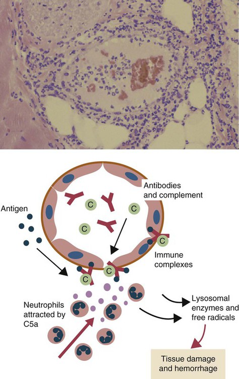
The first events observed following antigen injection are neutrophil adherence to vascular endothelium followed by their emigration into the tissues. By 6 to 8 hours, when the reaction has reached its greatest intensity, the injection site is densely infiltrated by large numbers of these cells (Figure 30-1). As the reaction progresses, destruction of blood vessel walls results in hemorrhage and edema, platelet aggregation, and thrombosis. By 8 hours, mononuclear cells appear in the lesion, and by 24 hours or later, depending on the amount of antigen injected, they become the predominant cell type. Eosinophils are not a significant feature of this type of hypersensitivity.


The fate of the injected antigen can be followed if is labeled with a fluorescent dye. The antigen first diffuses from the injection site through tissue fluid. When small blood vessels are encountered, the antigen diffuses into the vessel walls, where it encounters the circulating antibodies. Immune complexes form and are deposited between and beneath vascular endothelial cells. Complement components activated by the classical pathway will be deposited here as well.


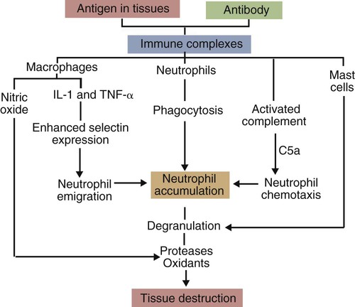
Immune complexes formed in tissues must be removed. The first step involves binding to Fc and complement receptors on cells. The most widespread of these Fc receptors is FcγRIIa expressed on macrophages. Immune complexes binding to these receptors stimulate production of nitric oxide, leukotrienes, prostaglandins, cytokines, and chemokines. Immune complexes also bind to mast cells through FcγRIII and trigger them to release their vasoactive molecules. Among the molecules released by mast cells are chemotactic factors and proteases that activate complement, cytokines, kinins, and lipid mediators. All these mediators promote inflammation by acting on vascular endothelium and stimulating neutrophil adherence and emigration.


Immune complexes activate complement and generate the chemotactic peptide C5a (Figure 30-2). Neutrophils, attracted by C5a and mast cell–derived chemokines, emigrate from the blood vessels, adhere to immune complexes, and promptly phagocytose them. Eventually the immune complexes are digested and destroyed. During this process, however, proteases and oxidants are released into the tissues. When neutrophils attempt to ingest immune complexes attached to structures such as basement membranes, they secrete their granule contents directly into the surrounding tissues. Neutrophil proteases disrupt collagen fibers and destroy ground substances, basement membranes, and elastic tissue. Normally tissues contain antiproteinases that inhibit neutrophil enzymes. However, neutrophils can subvert these inhibitors by secreting OCl. The OCl destroys the inhibitors and allows tissue destruction to proceed.


Although it has long been assumed that immunoglobulin molecules do not themselves damage antigens, recent evidence has shown that they can kill microorganisms and cause tissue damage. When provided with singlet oxygen from phagocytic neutrophils, antibodies catalyze the production of oxidants such as ozone. This ozone kills not only bacteria but also nearby cells. Biopsy specimens from Arthus reactions contain detectable amounts of ozone!


Neutrophil proteases also act on C5 to generate C5a, which promotes further neutrophil accumulation and degranulation. Other enzymes released by neutrophils make mast cells degranulate or generate kinins. As a result of all this, inflammation and destruction of blood vessel walls result in the development of the edema, vasculitis, and hemorrhage characteristic of an Arthus reaction.


Although the classical direct Arthus reaction is produced by local administration of an antigen to hyperimmunized animals, any technique that deposits immune complexes in tissues will stimulate a similar response. A reversed Arthus reaction can therefore be produced if antibodies are administered intradermally to an animal with a high level of circulating antigen. Injected, preformed immune complexes, particularly those containing a moderate excess of an antigen, will provoke a similar reaction, although, as might be anticipated, there is less involvement of blood vessel walls, and the reaction is less severe. A passive Arthus reaction can be produced by giving antibody intravenously to a nonsensitized animal, followed by an intradermal injection of an antigen, and real enthusiasts can produce a reversed passive Arthus reaction by giving antibody intradermally followed by intravenous antigen.


Although it is unusual for pure hypersensitivity reactions of only a single type to occur under natural conditions, there are some diseases in the domestic animals in which type III reactions play a major role. Experimentally, Arthus reactions are usually produced in the skin since that is the most convenient site at which to inject the antigen. However, local type III reactions can occur in many tissues, with the precise site depending on the location of the antigen.


Blue Eye


Blue eye is a condition seen in a small proportion of dogs that have been either infected or vaccinated with live canine adenovirus type 1 (see Figures 26-8 and 26-9). These animals develop an anterior uveitis leading to corneal edema and opacity. The cornea is infiltrated by neutrophils, and virus-antibody complexes are present in the lesion. Blue eye develops about 1 to 3 weeks after the onset of infection and usually resolves spontaneously as the virus is eliminated.


Hypersensitivity Pneumonitis


Type III hypersensitivity reactions may occur in the lungs when sensitized animals inhale antigens. For example, cattle housed during the winter are exposed to dust from hay. Normally, these dust particles are relatively large and are deposited in the upper respiratory tract, trapped in mucus, and eliminated. If, however, hay is stored when damp, bacterial growth and metabolism will result in heating. As a result of this warmth, thermophilic actinomycetes will grow. One of the most important of these thermophilic actinomycetes is Saccharopolyspora rectivirgula, an organism that produces large numbers of very small spores (1 µm diameter). On inhalation, these spores can penetrate as far as the alveoli. If cattle are fed moldy hay for long periods, constant inhalation of S. rectivirgula spores will result in sensitization and in the development of high-titered antibodies to S. rectivirgula antigens in serum. Eventually inhaled spore antigens will encounter antibodies within the alveolar walls, and the resulting immune complexes and complement activation will cause a pneumonia (or pneumonitis), the basis of which is a type III hypersensitivity reaction.


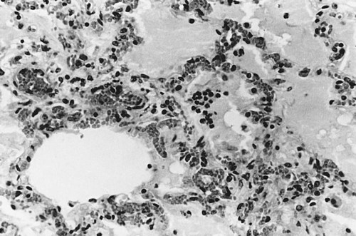
The lesions of hypersensitivity pneumonitis consist of an acute alveolitis together with vasculitis and exudation of fluid into the alveolar spaces (Figure 30-3). The alveolar septa may be thickened, and the entire lesion is infiltrated with inflammatory cells. Since many of these cells are eosinophils and lymphocytes, it is obvious that the reaction is not a pure type III reaction. Nevertheless, examination of the lungs of affected cattle by immunofluorescence demonstrates deposits of immunoglobulin, complement, and antigen. In animals inhaling small amounts of an antigen over a long period, proliferative bronchiolitis and fibrosis may be observed. Clinically, hypersensitivity pneumonitis presents as a pneumonia occurring between 5 and 10 hours after acute exposure to grossly moldy hay. The animal may have difficulty breathing and develop a severe cough. In chronically affected animals, the dyspnea may be continuous. The most effective method of managing this condition is by removing the source of the antigen. Administration of steroids may be beneficial.


A hypersensitivity pneumonitis also occurs in farmers chronically exposed to S. rectivirgula spores from moldy hay and is called farmer’s lung. Many other syndromes in humans have an identical pathogenesis and are usually named after the source of the offending antigen. Thus pigeon breeder’s lung arises following exposure to the dust from pigeon feces, mushroom grower’s disease is due to hypersensitivity to inhaled spores from actinomycetes in the soil used for growing mushrooms, and librarian’s lung results from inhalation of dusts from old books! Hay sickness is a hypersensitivity pneumonitis seen in horses in Iceland that is probably an equine equivalent of farmer’s lung.


Equine Respiratory Disease


Two forms of chronic respiratory disease occur in horses. Recurrent airway obstruction (RAO) is seen in older horses, and inflammatory airway disease (IAD) is seen in horses of any age. Both are forms of chronic bronchiolitis associated with exposure to molds and other allergens in dusty stable air.


RAO occurs most obviously in horses that inhale large amounts of organic dusts such as those generated in dusty stables. It includes obstructive pulmonary disease seen in stabled horses and summer pasture-associated obstructive pulmonary disease. RAO is defined as a severe debilitating disease characterized by coughing and an increased breathing effort due to cholinergic bronchospasm, airway hyperreactivity, and neutrophil and mucus accumulation in the airways. Characteristically horses with RAO suffer from respiratory difficulty even while at rest.


RAO is probably a hypersensitivity disease associated with an enhanced Th2 response, although there is also limited evidence for a Th1 or mixed response. Some studies suggest that RAO is simply a nonspecific response to endotoxin-like molecules. Horses with these syndromes may show positive skin reactions to intradermal inoculation of actinomycete and fungal extracts (such as Rhizopus nigricans, Candida albicans, S. rectivirgula, Aspergillus fumigatus, or Geotrichum deliquescens). Some horses react to mite extracts. There is no evidence of IgE involvement in RAO. There is evidence for a genetic predisposition.


Affected horses may respond to aerosol challenge with extracts of these organisms by developing respiratory distress. Clinical signs may resolve on removal of the moldy hay and reappear on reexposure. However, there is little correlation between skin test results and severity of disease. Affected animals usually have large numbers of neutrophils or eosinophils in their small bronchioles and high titers of antibodies to equine influenza in their bronchial secretions. The significance of the latter is unclear. High concentrations of the chemokine CXCL8 (interleukin-8 [IL-8]) are found in the bronchoalveolar washings of affected animals. Exposure of cultured equine bronchial epithelial cell cultures to hay dust or lipopolysaccharide increases IL-8, CXCL2, and IL-1β expression. It has been suggested that continuous prolonged activation of bronchoalveolar epithelial cells by dust particles and air-borne endotoxins leads to excessive production of neutrophil chemotactic chemokines. These neutrophils then cause damage by producing proteases, peroxidases, and oxidants.


Removal of clinically affected horses to air-conditioned stalls results in improvement of the disease, but this is reversed if the horses are returned to dusty stables. In some cases, RAO may persist even when horses are moved to low dust environments, probably as a result of airway remodeling.


IAD affects up to 30% of young horses in training. Although commonly linked to bacterial or viral infections, in many cases no infectious agent can be isolated. Horses with IAD show poor performance, exercise intolerance, and coughing. There is evidence of nonseptic inflammation detected by cytological evaluation of bronchiolar lavage fluid. Excessive airway mucus is apparent. Unlike RAO, the disease is not clinically apparent at rest. The pathogenesis is unknown, but like RAO, IAD is associated with inhalation of organic dusts and aeroallergens.

< div class='tao-gold-member'>

Stay updated, free articles. Join our Telegram channel

Jul 18, 2016 | Posted by in PHARMACOLOGY, TOXICOLOGY & THERAPEUTICS | Comments Off on Immune Complexes and Type III Hypersensitivity

Full access? Get Clinical Tree

Get Clinical Tree app for offline access