How Antigen-Binding Receptors Are Made


How Antigen-Binding Receptors Are Made


One of the central problems encountered in understanding adaptive immunity is to explain how lymphocytes recognize the enormous diversity of microbes that may invade the body. Given that microorganisms change rapidly, the immune system must be able to respond not only to existing organisms but also, within reason, to newly evolved organisms. The ability of the adaptive immune responses to respond specifically to an enormous number of foreign antigens implies the existence of an enormous number of different lymphocytes, each with its own specific antigen receptors. This then raises the question, How do lymphocytes generate such an enormous diversity of antigen-specific receptors?


The ability of a receptor to bind an antigen is determined by the shape of its binding site. This shape depends on the folding of its peptide chains, which is governed, in turn, by their amino acid sequences. Each amino acid in a peptide chain exerts an influence on its neighboring amino acids, which determines their relative configuration. The shape of a peptide chain therefore represents the contributions of all amino acids in the chain as the peptide assumes its most energetically favorable conformation. The folding of a protein is determined by its amino acid sequence, and that sequence is determined by the sequence of bases in the DNA coding for that protein. The diversity of antigen receptors implies either a corresponding diversity in the genes coding for these receptors or a mechanism that generates diversity from a limited pool of receptor genes. This second mechanism is now known to be the method employed by the adaptive immune system.


Receptor-Antigen Binding


When an antigen and its receptor bind, they interact through the chemical groups on the surface of the antigen and on the complementarity determining regions (CDRs) of the receptor. In classic chemical reactions, molecules are assembled through the establishment of firm, covalent bonds. These bonds can be broken only by the input of a large amount of energy, energy that is not readily available in the body. In contrast, the formation of noncovalent bonds provides a rapid and reversible way of forming complexes and permits reuse of molecules in a way that covalent bonding would not allow. However, noncovalent bonds act over short intermolecular distances and, as a result, form only when two molecules approach each other very closely. The binding of an antigen to a BCR or TCR is exclusively noncovalent, so the strongest binding occurs when the shape of the antigen and the shape of the receptor conform to each other. This requirement for a close conformational fit has been likened to the specificity of a key for its lock.


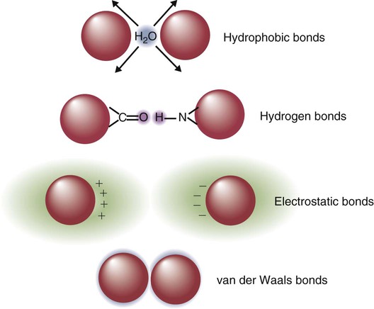
The major bonds formed between an antigen and its receptor are hydrophobic (Figure 17-1). When antigen and antibody molecules come together, they exclude water molecules from the area of contact. This frees some water molecules from constraints imposed by the proteins and is therefore energetically stable. (The bond can be likened to two wet glass microscope slides stuck together. Anyone who has tried to separate two wet glass slides can confirm the effectiveness of this type of bonding.)


A second type of binding between an antigen and its receptor is through hydrogen bonds. When a hydrogen atom covalently bound to one electronegative atom (e.g., an–OH group) approaches another electronegative atom (e.g., an O=C—group), the hydrogen is shared between the two electronegative atoms. This situation is energetically favorable and is called a hydrogen bond. The major hydrogen bonds formed in antigen-receptor interaction are O–H–O, N–H–N, and O–H–N. Hydrogen bonds are already present between proteins and water molecules in aqueous solution, so the binding of an antigen to its receptor by hydrogen bonds requires relatively little net energy change.


Electrostatic bonds formed between oppositely charged amino acids may contribute to antigen-receptor binding, but the charge on many protein groups is commonly neutralized by electrolytes in solution. As a result, the importance of electrostatic bonding is unclear.


When two atoms approach very closely, a nonspecific attractive force, called a van der Waals force, becomes operative. It occurs as a result of a minor asymmetry in the charge of an atom because of the position of its electrons. This force, although very weak, may become collectively important when two large molecules come into contact. It can therefore contribute to antigen-receptor binding.


The binding of a receptor to its antigen is therefore mediated by multiple noncovalent bonds. Each bond is relatively weak in itself, but collectively the bonds may have a significant binding strength. All these bonds act only across short distances and weaken rapidly as that distance increases. Electrostatic bond and hydrogen bond strengths are inversely proportional to the square of the distance between the interacting molecules; the van der Waals forces and hydrophobic bonds are inversely proportional to the seventh power of that distance. Thus the strongest binding between an antigen and its receptors occurs when their shapes match perfectly and multiple noncovalent bonds form. Antigens can bind to receptors when they fit less than perfectly, although the strength of this binding will be much reduced.


Antigen Receptor Genes


The information needed to make all proteins, including antigen receptors, is stored in an animal’s genome. All that is required for the production of these molecules is that the necessary genes be turned on. Once the appropriate genes are activated, they can be transcribed into RNA and translated into the appropriate receptor protein on B or T cells. It has been estimated that mammals can produce up to 1015 different antigen receptors to be expressed on B and T cells. In order to produce this enormous diversity, they use fewer than 500 genes!


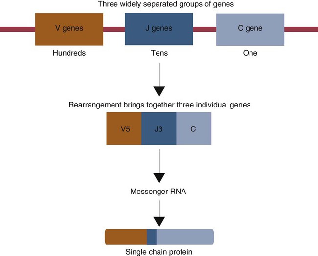
Multiple genes code for each receptor peptide chain. Several genes code for each variable region, whereas only one codes for a constant region. As a result, the single constant-region gene can be combined with any one of several different variable-region genes to make a complete receptor chain (Figure 17-2). Instead of having genes for all possible receptor chains, it is only necessary to have genes for all the variable regions and to join these to an appropriate constant-region gene as required. In addition, antigen receptor chains may be paired in different combinations to yield even greater diversity, a process called combinatorial association.


Immunoglobulin/B Cell Receptor Diversity


To make as many different antibodies as possible, it is necessary to diversify the amino acid sequences of the variable domains in both light and heavy chains. Since these amino acid sequences are determined by the nucleotide sequences in the genes coding for these variable regions, mechanisms must exist for generating this nucleotide sequence diversity. In practice, gene diversity is generated through three distinct mechanisms: gene recombination, somatic mutation, and gene conversion. All three mechanisms alter and diversify antibody gene sequences in such a way that an incredibly diverse array of antigen receptors is generated. The relative importance of each of these mechanisms differs among species, and the diversity-generating mechanisms that operate in humans and mice are not the same as those that operate in domestic mammals. The assembly of these diverse antigen receptors is carefully controlled during lymphocyte development by such factors as DNA methylation, chromatin structure, and location within the nucleus.


Gene Recombination


Gene recombination results from the random selection of one gene from each of several groups of genes followed by recombining these selected genes to generate sequence diversity. It is well seen in the genes that code for immunoglobulins.


Three gene loci code for immunoglobulin peptide chains, and each is found on a different chromosome (Figure 17-3). One locus, called IGK, codes for κ light chains; one, called IGL, codes for λ light chains; and one, called IGH, codes for heavy chains.


IGH Locus


In humans, heavy chain V regions are coded for by three genes, IGHV, IGHD, and IGHJ. The IGH locus contains about 90 different IGHV genes. Mouse IGH may have as many as 1500 different IGHV genes, but up to 40% of these are pseudogenes. The IGH locus also contains several IGHJ genes situated 3′ to the IGHV genes. Several short genes, called IGHD genes (D for diversity), are located between the IGHV and IGHJ genes (see Figure 17-3). In mice there are about 12 IGHD genes, and in humans there are at least 30. A large noncoding region separates the IGHJ genes from the IGHC genes. The IGHC genes consist of a series of constant-region genes, one for each heavy chain class and subclass, arranged in the order 5′-Cµ-Cδ-Cγ-Cε-Cα-3′ along the chromosome.


Generation of Junctional Diversity


Gene Rearrangement


The most obvious way to generate V-region diversity is to randomly select one V gene from the available pool and join it to one randomly selected J gene—a process called recombination. Since many different V and J genes are available, the number of possible combinations can be very large. For example, if there are 100 V genes and 10 J genes, then 100 × 10 = 1000 different V regions can be constructed.


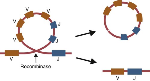
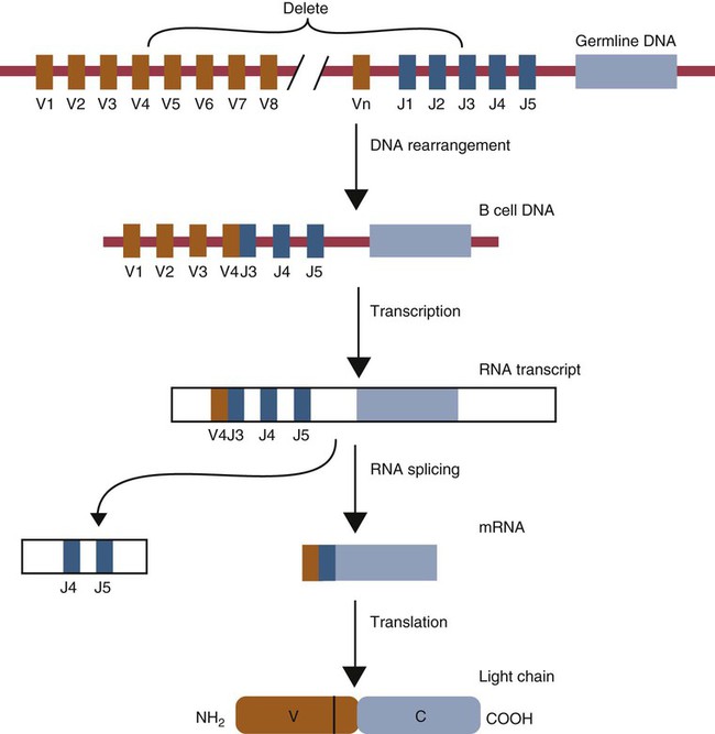
Light chain assembly requires the combination of one V, one J, and one C gene. During B cell development, the intervening genes are looped out, excised, and discarded. The V and J genes have sites at each end that guide the cutting enzymes (Figure 17-4). The looped-out genes are chopped off, and the free ends of the DNA are rejoined so that the V and J genes form a continuous sequence. Two sets of enzymes are used in this process. Recombinases cut the DNA at two points, thus excising unwanted genes. Following this, DNA repair enzymes join the two free ends to reform a continuous sequence. If these enzymes are defective, antibodies (and TCRs) cannot be made. In foals with severe combined immunodeficiency, for example, there is a defect in the DNA repair enzyme that joins the cut ends. As a result, these foals cannot make either TCRs or BCRs and thus have no functional B or T cells (Chapter 37).


Light chain gene recombination occurs in two stages. Randomly selected V and J genes are first joined to form a complete V-region gene. The joined V-J genes remain separated from the C gene until messenger RNA (mRNA) is generated. At that time the unwanted J genes are excised, and the complete V-J-C mRNA is then translated to form a light chain (Figure 17-5).


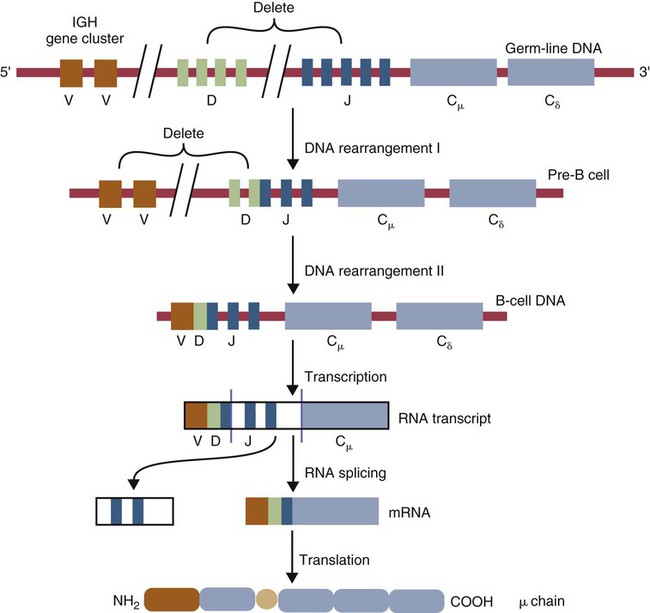
When a heavy chain V region is assembled, its construction requires the splicing together of IGHV, IGHD, and IGHJ genes (Figure 17-6). This use of three randomly selected genes enormously increases the amount of variability. For example, if a pool of 100 V, 10 J, and 10 D genes are recombined, then 100 × 10 × 10 = 10,000 different V regions can be constructed. The recombination of these genes also occurs in a specific order. Thus IGHD is first joined to IGHJ, then V genes are attached to make a complete V-region gene. After transcription, any unwanted J genes are deleted, the C gene mRNA is attached, and the completely assembled V-D-J-C mRNA is translated to form a heavy chain.


Base Insertion


In immunoglobulin heavy chain gene processing, additional nucleotides may be inserted at the V-D and D-J splice sites. Some of these nucleotides (N-nucleotides) are added randomly by an enzyme called terminal deoxynucleotidyltransferase (TdT). Up to 10 N-nucleotides may be inserted between V and D and between D and J.


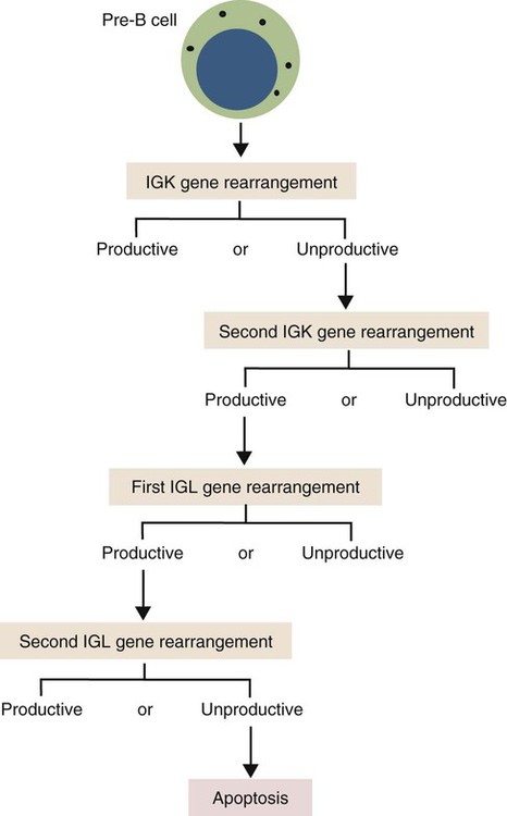
Although the random selection of genes from two or three different pools generates a large number of different combinations, not all of these combinations will produce usable antibodies. Some combinations may result in a nucleotide sequence that cannot be translated into protein. These are called nonproductive rearrangements. For example, nucleotides are read as triplets called codons, each of which codes for a specific amino acid. If the codons are to be read correctly, then the sequence must be in the correct reading frame. If nucleotides are inserted or deleted so that the codon reading frame is changed, the resulting gene may code for a totally different amino acid sequence. If this frameshift results in inappropriate splicing, translation is prematurely terminated. It is probable that nonproductive rearrangements are produced in two of three attempts during B cell development. When this happens, the B cell has several additional opportunities to produce a functional antibody. For example, immature B cells initially rearrange one of the IGK genes (Figure 17-7). If this fails to produce a functional light chain, they switch to the other IGK allele for a second attempt. If this does not work, the B cell will use one of the IGL alleles, and if this fails, the second IGL allele represents the last resort. If all these efforts fail to produce a functional light chain, the B cell cannot make a functional immunoglobulin. It will undergo apoptosis without participating in an immune response.


The sequence of events described previously has been worked out in mice and humans and may not apply to domestic mammals. One obvious difference lies in the use of κ and λ light chains. In mice, rabbits, pigs, and humans, κ chains are preferentially used (95% in mice, 90% in rabbits, 60% in pigs, 60% in humans). In the other domestic species, λ light chains predominate (98% in ruminants, 60% to 90% in horses). The reasons for these differences are unknown.


It should also be pointed out that immunoglobulin gene rearrangement is not entirely random. For example, in rabbits, mice, and humans the most 3′ IGHV genes tend to be used most often. This preferential use of certain genes results from a combination of factors, including the recombination signal sequences, the accessibility of the genes to the recombinase enzyme, sequences at the splicing sites, and way in which DNA can fold.

< div class='tao-gold-member'>

Stay updated, free articles. Join our Telegram channel

Jul 18, 2016 | Posted by in PHARMACOLOGY, TOXICOLOGY & THERAPEUTICS | Comments Off on How Antigen-Binding Receptors Are Made

Full access? Get Clinical Tree
Get Clinical Tree app for offline access