Acid-Base Homeostasis


Acid-Base Homeostasis


Key Points


Acid-Base Regulation


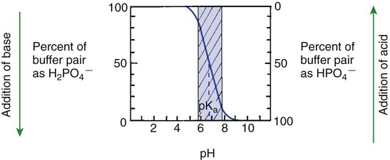
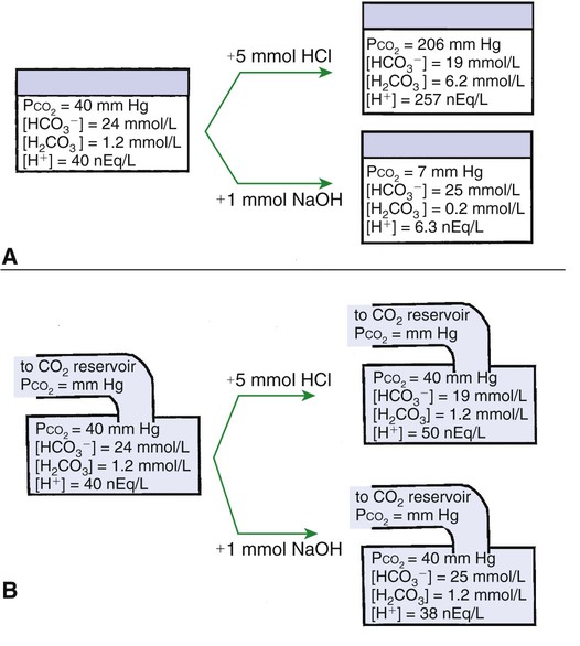
1. Relative constancy of the body’s pH is essential because metabolism requires enzymes that operate at an optimal pH.


2. Hydrogen ion concentration is measured as pH.


3. An acid can donate a hydrogen ion, and a base can accept a hydrogen ion.


4. Buffers are combinations of salts and weak acids that prevent major changes in pH.


5. Hemoglobin and bicarbonate are the most important blood buffers.


6. The first defense against a change in blood pH is provided by the blood buffers, but the lungs and kidneys must ultimately correct the hydrogen ion load.


7. Changes in ventilation can rapidly change carbon dioxide tension and therefore alter pH.


8. Metabolic production of fixed acids requires that the kidneys eliminate hydrogen ions and conserve bicarbonate.


9. Intracellular pH is regulated by buffers and ion pumps.


Acid-Base Disturbances


1. Acid-base abnormalities accompany many diseases, and the restoration of normal blood pH should be a consideration in the treatment of any disease.


2. Respiratory acidosis is caused by the accumulation of carbon dioxide, which decreases blood pH.


3. Respiratory alkalosis is caused by the loss of carbon dioxide, which increases blood pH.


4. Metabolic acidosis is caused by the accumulation of fixed acids or the loss of buffer base, which decreases blood pH.


5. Metabolic alkalosis is caused by the excessive elimination of hydrogen ions or by the intake of base, such as bicarbonate, which increases blood pH.


6. Respiratory compensations for acid-base abnormalities occur rapidly; renal compensations occur over several hours.


7. Hydrogen and potassium ions are interrelated in acid-base homeostasis.


8. The diagnosis of acid-base disturbances depends on interpretation of measurements of arterial blood pH and carbon dioxide tension, from which bicarbonate concentration and total buffer base are calculated.


9. Over the years, many terms have been used to explain acid-base balance.


Acid-Base Regulation


Relative Constancy of the Body’s pH Is Essential Because Metabolism Requires Enzymes That Operate at an Optimal pH


For optimal functioning of the cells, the ionic composition of body fluids is maintained within fairly narrow limits. Hydrogen ion (H+, proton) concentration is extremely important because it determines the acidity or alkalinity, or pH, of the body fluids. Serious deviations of pH outside the normal range disrupt cell metabolism and therefore body function. For example, the activity of the sodium-potassium (Na+-K+) pump decreases by half when pH falls by one unit, and the activity of phosphofructokinase (a key regulatory enzyme in the glycolytic pathway) decreases by 90% when pH decreases by only 0.1.


When veterinarians use the terms acidosis and alkalosis, they are comparing the pH of arterial blood with the normal value of 7.4. A pH above and below 7.4 are referred to as alkalosis and acidosis, respectively. The range of pH compatible with life is 6.85 to 7.8, but such changes are rare because buffers, the lungs, and the kidneys work in concert to regulate pH.


In a 70-kg animal, 15,000 mmole of H+ are produced daily when carbon dioxide (CO2) is added to the blood for transport from the tissues to the lungs. If the lungs eliminate CO2 as fast as it is produced in the tissues, there is no net H+ gain by the body. However, the balance between CO2 production and CO2 elimination may be disturbed during exercise or in respiratory disease, thus threatening the acid-base homeostasis of the body.


Forty mmole/day of H+ are produced during protein metabolism (sulfuric and phosphoric acids), fat metabolism (keto-acids), and the incomplete oxidation of glucose (lactic acid) and another 30 mmole/day are absorbed from the intestine. Although H+ from these sources are few compared with those produced in CO2 transport, the kidneys must eliminate them continually. In a disease state the H+ load imposed on the body is frequently increased because of an increase in tissue breakdown (catabolism) or because the kidneys fail to eliminate H+. In less common situations, such as vomiting, H+ are lost from the body.


To understand how the body regulates pH and how acid-base disorders are diagnosed, it is necessary to first review acids, bases, and buffering.


An Acid Can Donate a Hydrogen Ion, and a Base Can Accept a Hydrogen Ion


Hydrochloric acid (HCl) is a strong acid because it dissociates completely in water into H+ and Cl. Chloride ion is a base because it has the potential to accept an H+, but it is a weak base because HCl dissociates so completely in water. Carbonic acid (H2CO3), in contrast, is a weak acid because it dissociates incompletely in solution into hydrogen and bicarbonate ions. Bicarbonate (HCO3), however, is a relatively strong base that can accept an H+ and form undissociated carbonic acid. The latter reaction removes H+ from solution, and the [H+] decreases, causing the pH to rise. Bases do not have to be ions; for example, ammonia (NH3) is a base because it can accept a proton and become ammonium ion (NH4+). This reaction is of little importance in the blood, but it is important in the renal collecting duct. In addition, proteins also act as buffers by virtue of the terminal carboxyl and amino groups, which can donate (R-COOH → R-COO + H+) or accept (R-NH2 + H+ → R-NH3+) protons, respectively.


Buffers Are Combinations of Salts and Weak Acids That Prevent Major Changes in pH


Buffers “soak up” free H+ and prevent their accumulation in body fluids. By so doing, buffers prevent drastic changes in pH. Buffers are mixtures of weak acids and their salts. For example, sodium bicarbonate dissociates completely into sodium and bicarbonate ions; carbonic acid dissociates incompletely into hydrogen and bicarbonate ions. Thus, in a solution containing sodium bicarbonate and carbonic acid, there are sodium, hydrogen, and bicarbonate ions and undissociated carbonic acid. If a strong acid such as hydrochloric acid is added to the solution, the added H+ upsets the dissociation equilibrium of carbonic acid. Hydrogen ions combine with bicarbonate ion to form carbonic acid, thus reducing the [H+], that is, preventing a major change in pH.


If, in contrast, sodium hydroxide is added to the solution, the hydroxyl ions, formed by dissociation of sodium hydroxide, combine with H+ to form water. The decrease in H+ causes dissociation of more carbonic acid and liberation of H+, again preventing a large change in pH.


The dissociation of a weak acid, and therefore the [H+], base, and undissociated acid, is determined by the dissociation constant (Ka) and can be described by the law of mass action. For carbonic acid:


Ka=[H+][HCO3]/[H2CO3].


image

Taking logarithms of both sides of this equation results in:


logKa=log[H+]+log[HCO3]/[H2CO3].


image

Rearrangement of this equation yields:


log[H+]=logKa+log[HCO3]/[H2CO3].


image

However, −log[H+] is pH and −logKa is called pKa; therefore:


pH=pKa+log[HCO3]/[H2CO3].


image

This is the Henderson-Hasselbalch equation written for the bicarbonate–carbonic acid system. It can be written for any buffering system in the generic form:


pH=pKa+log[base]/[acid].


image

This equation shows that the pH of a solution is determined by the ratio of the concentration of base (the H+ acceptor) to that of undissociated acid (the H+ donor) and by the pKa of the buffering system.


Figure 52-1 shows the change in pH that results when acid is added to a phosphate buffer with a pKa of 6.8. This is a graphic presentation of the Henderson-Hasselbalch equation. Initially, as acid is added, there is a large decrease in pH. As considerably more acid is added to the solution, the pH changes little. H+ ions combine with HPO42− and form H2PO4. Finally, the pH decreases considerably. The zone over which the pH changes little as acid is added (i.e., where buffering capacity is optimal) is within ±1 pH unit of the pKa. Note that when the pH equals the pKa, 50% of the buffer has been consumed. From this buffer curve, it is obvious that an effective buffer must have a pKa within ±1 pH unit of the solution in which it operates. Thus the optimal blood buffers should have a pKa between 6.4 and 8.4. In addition, buffers must be sufficiently plentiful to be effective.


Hemoglobin and Bicarbonate Are the Most Important Blood Buffers


Hemoglobin is an important blood buffer because it is plentiful and because the imidazole residues of globin histidine have a pKa close to the blood pH. In actuality, the pKa of hemoglobin changes with the degree of oxygenation. Because deoxyhemoglobin has a pKa (7.93) closer to blood pH than does oxyhemoglobin (pKa = 6.68), deoxyhemoglobin provides more buffering capacity. When arterial blood enters the tissue capillaries, oxygen leaves hemoglobin, so the resulting deoxyhemoglobin is an excellent buffer for the H+ produced when CO2 is added to the blood.


The other blood buffer with an optimal pKa is the HPO42−/H2PO4 system, with a pKa of 6.8 (see Figure 52-1). The normally low phosphate concentration in the blood makes this buffering system quantitatively unimportant; however, it is important in the renal tubules, where phosphate becomes concentrated. Plasma proteins also provide a small amount of blood buffering.


Although a pKa of 6.1 seems to make the HCO3/H2CO3 buffer unimportant for blood buffering, this is not so for two reasons. First, there is a large amount (24 mEq/L) of HCO3 in the blood, making it readily available for buffering. Second, the kidneys can regulate the concentration of HCO3, and the lungs can regulate the concentration of H2CO3. Because the base and acid concentration can be regulated, the HCO3/H2CO3 system is said to be an open system.


Figure 52-2 shows the value of this open system in maintaining body pH. It should be noted that the concentration of H2CO3 ([H2CO3]) in solution is directly proportional to the carbon dioxide tension (PCO2); 1 molecule of H2CO3 is in equilibrium with 340 molecules of CO2. Therefore, [H2CO3] can be calculated as 0.03 × PCO2. In Figure 52-2, A and B, top panels, 5 mmol of hydrochloric acid is added to plasma. Figure 52-2, A, shows what happens if the PCO2 and thus [H2CO3] is held constant. In such a closed system, the 5 mmol of HCl reacts with 5 mmol of HCO3 to form 5 mmol of H2CO3. As a consequence of this reaction, [HCO3] decreases from 24 to 19 mmol/L, [H2CO3] increases from 1.2 to 6.2 mmol/L, and PCO2 increases from 40 to 206 mm Hg. Using the law of mass action, one can calculate that [H+] increases from 40 to 257 nEq/L or, stated in other terms, pH decreases from 7.4 to 6.5. If the system is open, however (as it is in the top panel of Figure 52-2, B) carbon dioxide evolves to the environment as fast as it is produced so that PCO2 and therefore [H2CO3] remain constant, then [H+] increases to only 50 nEq/L and the pH decreases only to 7.3. The lower panel in Figure 52-2, B, shows similar advantages to the open system when a strong base is added to the buffer system. Under most conditions, the body functions as an open system with regard to the HCO3/H2CO3 system so that pH changes are minimized. When tissues are ischemic, however, they have no connection to the lungs, so CO2 cannot be eliminated. The ischemic tissue then functions as a closed system, and pH changes within the tissue can therefore be drastic.


The HCO3/H2CO3 buffering system is of great value to clinicians because its components can be readily measured in the clinical laboratory and thereby used to diagnose acid-base disturbances. It is not necessary to measure the components of every buffering system to diagnose acid-base disturbances. If one system is known, changes in other systems can be predicted. It is standard to measure pH and PCO2 and to use the Henderson-Hasselbalch equation to derive [HCO3]. In actual practice, these calculations are now done for the clinician by computers in the blood gas machine.


For clinical use, the Henderson-Hasselbalch equation for the HCO3/H2CO3 system is written as follows:


image

Under normal conditions, the pH of arterial blood is 7.4, [HCO3] is 24 mEq/L, and the arterial carbon dioxide tension (PaCO2) is 40 mm Hg:


7.4=6.1+(log24)/(0.03×40)=6.1+log20.


image

This equation demonstrates that a normal blood pH requires an [HCO3]/[0.03 × PCO2] ratio of 20 : 1. An increase or decrease in this ratio increases or decreases pH, respectively.


The First Defense Against a Change in Blood pH Is Provided by the Blood Buffers, but the Lungs and Kidneys Must Ultimately Correct the Hydrogen Ion Load


When body pH is threatened by a change in the production or elimination of H+, the blood and tissue buffers provide the first line of defense. However, buffers only prevent drastic changes in pH; they cannot correct the problem by increasing or decreasing the elimination of H+ or by replacing lost buffering capacity. Ultimately, pH must be corrected by adjustments in ventilation or by changes in renal function. Because the lungs can alter PaCO2 and the kidneys can regulate the concentration of HCO3, the Henderson-Hasselbalch equation has been written as follows:


pH=pKa+log(renal function/ventilation)


image
< div class='tao-gold-member'>

Stay updated, free articles. Join our Telegram channel

Jul 18, 2016 | Posted by in PHARMACOLOGY, TOXICOLOGY & THERAPEUTICS | Comments Off on Acid-Base Homeostasis

Full access? Get Clinical Tree

Get Clinical Tree app for offline access