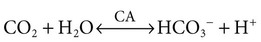
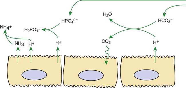
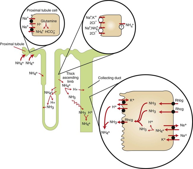
Acid-Base Balance


Acid-Base Balance


Buffers, Lungs, and Kidneys Together Maintain Acid-Base Balance


Normal blood pH is approximately 7.4; normal cellular function requires a pH close to this value. Three systems maintain acid-base homeostasis: (1) intracellular and extracellular buffers, (2) the lungs, and (3) the kidneys. The first two make rapid corrections of blood pH, whereas the kidneys more slowly control acid-base homeostasis and excrete excess hydrogen ion (H+).


Maintaining acid-base balance usually requires preventing excess acid in the body. Acid is constantly produced in the body as a byproduct of metabolism. The amount of acid produced varies depending on changes in diet, exercise, other organ functions, and in birds the phases of the egg-laying cycle. Therefore the systems that maintain acid-base homeostasis must adapt to changes in the acid load. Less often there is an excess base load that must be eliminated.


Several intracellular and extracellular buffers titrate H+ to maintain a physiological pH. These include hemoglobin and other proteins, carbonate in bone, phosphate, and bicarbonate (HCO3). These buffers rapidly normalize the pH after acute changes in the acid load, unless the buffering capacity is exceeded. In addition, during chronic metabolic acidosis, bone provides a reservoir of buffer that is mobilized to help normalize systemic pH. Excess H+ and low HCO3 in the extracellular fluid promote physicochemical as well as osteoclast-mediated dissolution of bone, releasing carbonate, which buffers H+. In chronic acidosis, this can lead to abnormally low bone mineral density.


The respiratory system also responds rapidly to maintain normal blood pH by altering the rate of removal of carbon dioxide (CO2) from the blood. The enzyme carbonic anhydrase (CA), present in red blood cells and many other cells, catalyzes the following reaction:


image

Removal of CO2 from the blood by respiration shifts this reaction to the left, and the concentration of H+ is consequently reduced (pH is raised). Thus the lung is important for stabilizing blood pH, particularly in response to rapid changes in the acid load.


The kidney is the third line of defense of acid-base balance. Although buffering and respiration are able to stabilize blood pH, the kidneys are responsible for the actual excretion of most excess H+.


Acid Excretion Is Achieved by Proton Secretion by Tubule Epithelial Cells, Buffering in the Tubule Fluid, and Bicarbonate Absorption


The kidney excretes acid efficiently by the combined effects of (1) carbonic anhydrases, which make protons and bicarbonate readily available for transport; (2) transporters that move H+ from the epithelial cells into the tubule fluid and bicarbonate into the interstitial fluid; and (3) buffers that minimize increases in H+ concentration in the tubule fluid.


The kidneys excrete acid by secretion of H+, primarily in the proximal tubule, the thick ascending limb of Henle’s loop, and the collecting duct. These segments use different mechanisms to excrete excess acid and to control blood pH precisely. The proximal tubule secretes the majority of acid, whereas the collecting duct controls net acid excretion and the final urine pH.


Most secreted H+ is transported across the apical plasma membrane by the following three transporters: (1) a sodium ion (Na+)/H+ exchanger, (2) an H+-adenosine triphosphatase (ATPase) pump, and (3) an H+,K+-ATPase pump. The Na+/H+ exchanger secretes acid by electrically neutral exchange of luminal Na+ for intracellular H+. The Na+ gradient generated by basolateral Na+,K+-ATPase drives apical Na+/H+ exchange (secondary active transport). Na+/H+ exchange is the main route of H+ secretion in the proximal tubule and the thick ascending limb.


The electrogenic H+-ATPase pump actively transports intracellular H+ across the apical plasma membrane and contributes a net positive charge to the tubule fluid. The collecting duct H+,K+-ATPase pumps, which are similar to gastric and colonic proton pumps, actively secrete acid by electrically neutral exchange of intracellular H+ for K+ in the tubule fluid. Although the H+-ATPase pump is responsible for most H+ secretion by the collecting duct, H+,K+-ATPases may equal or exceed the acid secretion rate of the H+-ATPase pump under some conditions.


Buffering of the tubule fluid is necessary for efficient acid excretion. Buffers accept secreted H+ and minimize the decrease in tubule fluid pH that would otherwise follow rapid H+ secretion by the epithelial cells. In mammals the most important buffers are bicarbonate, phosphate, and ammonia (NH3); to a lesser extent, creatinine and citrate serve as luminal buffers. In birds, urates significantly contribute to titration of secreted acid. Figure 44-1 illustrates the removal of acid by intraluminal buffers.


In the proximal tubule, HCO3 is the most important intraluminal buffer, for two main reasons. First, the concentration of HCO3 in the tubule fluid is high. Although large amounts of HCO3 are reabsorbed in the proximal tubule, roughly proportional amounts of H2O are reabsorbed, and the HCO3 concentration remains similar to that of the glomerular filtrate. Second, secreted H+ combines with luminal HCO3 to form H2O and CO2; this reaction is catalyzed by carbonic anhydrase associated with the apical plasma membrane. The CO2 crosses the plasma membrane, partly by diffusion and partly facilitated by membrane proteins serving as gas channels, such as the water channel, aquaporin-1, which has been shown to function as a CO2 channel in proximal tubule cells. Intracellular carbonic anhydrase catalyzes CO2 hydration to form H+ and HCO3, which are transported across the apical and basolateral plasma membranes respectively, resulting in acid secretion and bicarbonate reabsorption.


Filtered phosphate also buffers the tubule fluid. Secreted H+ titrates HPO42− to form H2PO4. The monovalent phosphate ion (H2PO4) is lipid insoluble and is transported only at very low rates on the apical Na+-inorganic phosphate (Pi) co-transporters. Thus the secreted protons bound to H2PO4 are retained in the tubule fluid. In birds, titration of luminal urate forms uric acid. Besides being lipid insoluble, uric acid also has a low aqueous solubility, and thus a significant portion of acid is removed as uric acid precipitates. The role of NH3 in acid excretion is discussed in the following sections.


Apical acid secretion is coordinated with basolateral bicarbonate transport. In the proximal tubule bicarbonate reabsorption is mediated primarily by the sodium bicarbonate co-transporter, NBCe1. In the collecting duct, the basolateral anion exchanger, kAE1, is a chloride/bicarbonate exchanger that is responsible for the majority of bicarbonate uptake by acid-secreting cells.


Renal Ammonia Metabolism Generates New Bicarbonate and Promotes Acid Excretion


Renal ammonia metabolism is a major component in the maintenance of acid-base balance and is illustrated in Figure 44-2. In proximal tubule cells the amino acid glutamine is metabolized to produce NH4+. This process is called ammoniagenesis. The intracellular NH4+ enters the tubule fluid through secondary active transport by substitution for H+ on the Na+/H+ exchanger. Glutamine metabolism also produces new bicarbonate anions, which are transported across the basolateral plasma membrane. Thus proximal tubule ammoniagenesis enables bicarbonate production and absorption and distal delivery of ammonia. Renal ammoniagenesis is enhanced by acidosis and is an important renal response to an increase in the acid load.


In the thick ascending limb of Henle’s loop, luminal NH4+ is reabsorbed by substitution for K+ on the apical Na+,K+,2Cl co-transporter. NH4+ reabsorption in this segment reduces the amount of ammonia species delivered to the late distal tubule and increases ammonia (NH3) in the medullary interstitium.


High NH3/NH4+ concentrations are enhanced and maintained in the medullary interstitium by a countercurrent multiplication system in the loops of Henle, similar to that described in Chapter 43. This creates a steep concentration gradient for NH3, which favors its movement into the medullary collecting duct. Until recently, the prevailing model for NH4+ excretion in the collecting duct was NH3 diffusion and trapping. The belief was that ammonia freely diffused across plasma membranes and into the luminal fluid where it bound protons, rendering it impermeant across plasma membranes and trapped in the luminal fluid. However, NH3, although not a charged molecule is a polar molecule, much like water, and it is now known that the lipid bilayer is relatively impermeable to NH3. In fact, specific ammonia transporters, the Rh glycoproteins, Rhcg and Rhbg, are present in plasma membranes of the majority of cell types in the collecting duct and facilitate transepithelial ammonia transport. These transporters are required for normal collecting duct ammonia transport and renal ammonia excretion and their abundance and subcellular distribution are regulated in accordance with physiologic conditions that increase renal ammonia excretion, such as acidosis. In addition, in the terminal inner medullary collecting duct, NH4+ is transported by substitution for K+ on the basolateral Na+,K+-ATPase.


The majority of secreted NH3 is titrated by secreted H+ to form NH4+ in the tubule luminal fluid. The formation of NH4+ from intraluminal NH3 and H+ lowers the concentration of both NH3 and H+ in the tubule fluid. This contributes to the maintenance of a favorable gradient for the transport of NH3 into the tubule fluid and reduces the electrochemical gradient for H+ that is created by active proton secretion in the collecting duct.


Finally, the comparative aspects of ammonia excretion are intriguing. Ammoniagenesis and ammonia excretion are important mechanisms controlling acid-base homeostasis in mice, rats, dogs, chickens, and humans. In these species, ammonia excretion accounts for up to 60% of net acid excretion in basal conditions and can increase to 90% of net acid excretion in models of metabolic acidosis. However, these findings cannot be applied generally to other species. Rabbits have low basal urinary ammonia excretion rates and do not increase ammonia excretion during metabolic acidosis. In one model of metabolic acidosis, domestic cats acidified the urine but apparently did not increase renal ammoniagenesis and only increased urinary ammonia excretion to a level comparable to basal excretion in mice. In humans and rats, dietary potassium restriction increases renal ammonia excretion despite concurrent development of metabolic alkalosis, but potassium restriction causes metabolic acidosis in dogs and cats and reduces renal ammonia excretion in dogs.

< div class='tao-gold-member'>

Stay updated, free articles. Join our Telegram channel

Jul 18, 2016 | Posted by in PHARMACOLOGY, TOXICOLOGY & THERAPEUTICS | Comments Off on Acid-Base Balance

Full access? Get Clinical Tree

Get Clinical Tree app for offline access