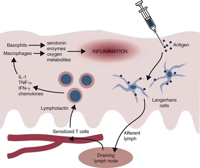
Type IV Hypersensitivity: Delayed Hypersensitivity


Type IV Hypersensitivity


Delayed Hypersensitivity


Certain antigens, when injected into the skin of sensitized animals, provoke an inflammatory response at the injection site after a delay of 12 to 24 hours. Since these “delayed” hypersensitivity reactions can only be transferred from sensitized to normal animals by lymphocytes, they must be cell mediated. Delayed hypersensitivity reactions are classified as type IV hypersensitivities and result from interactions among the injected antigen, antigen-presenting cells, and T cells. An important example of a delayed hypersensitivity reaction is the tuberculin response. This is an inflammatory response that develops in the skin of an animal infected with tuberculosis following intradermal injection of tuberculin. Delayed hypersensitivity reactions can be considered a specialized form of inflammation directed against organisms that are resistant to elimination by conventional responses.


The Tuberculin Reaction


Tuberculin is the name given to extracts of mycobacteria used to skin-test animals in order to identify those suffering from tuberculosis. Several types of tuberculin have been employed for this purpose. The most important is purified protein derivative (PPD) tuberculin, prepared by growing organisms in synthetic medium, killing them with steam, and filtering. The PPD tuberculin is precipitated from this filtrate with trichloroacetic acid, washed, and resuspended in buffer ready for use. Thus PPD tuberculin is a crude antigen mixture. Its major antigenic component is probably the heat-shock protein (HSP) 65. Many of its proteins are shared among different mycobacterial species, thus ensuring that tests that use PPD tuberculin are relatively nonspecific. It is possible to increase the specificity of the tuberculin test with a defined mycobacterial protein such as early secretory antigenic target-6 (ESAT-6). ESAT-6 is a mycobacterial protein of unknown function that is recognized strongly by T cells. However, the reactions induced by very pure proteins tend to be minimal and require greater amounts of antigen to induce a satisfactory response.


When tuberculin is injected into the skin of a normal animal, there is no apparent response. On the other hand, if it is injected into an animal infected with mycobacteria, a delayed hypersensitivity response occurs. In these animals, a red, indurated (hard) swelling develops at the injection site. The inflammation begins after 12 to 24 hours, reaches its greatest intensity by 24 to 72 hours, and may persist for several weeks before fading gradually. In very severe reactions, tissue destruction and necrosis may occur at the injection site. The lesion is infiltrated with mononuclear cells (lymphocytes, macrophages), although neutrophils are present in the first hours of the reaction (Figure 31-1).


The tuberculin reaction is mediated by T cells. When an animal is infected with Mycobacterium tuberculosis, the organisms are readily phagocytosed by macrophages. Some of this mycobacterial antigen triggers a Th1 response and generates memory cells. These memory T cells will respond to injected mycobacterial antigens such as tuberculin. Since a positive tuberculin test can be elicited many years after exposure to an antigen, some of these memory T cells must be very long lived.


When tuberculin is injected intradermally, it is taken up by Langerhans cells, which then migrate to the draining lymph node (Figure 31-2). Here they present antigen to memory T cells that respond by generating Th1 effector cells. The circulating Th1 cells recognize the antigen when they encounter it in the skin and accumulate around the antigen deposit. By 12 hours in cattle, the injection site is infiltrated with T cells. (In humans and mice, α/β T cells tend to predominate, whereas in sheep and cattle, γ/δ, WC1 T cells predominate.) There are no B cells in the lesion.


The γ/δ T cells recruit other Th1 lymphocytes and macrophages to the site. The Th1 cells secrete interferon-γ (IFN-γ), interleukin-2 (IL-2), and IL-16. The first two act on endothelial cells to increase expression of adherence molecules. IL-2 stimulates production of the chemokines CXCL8, CCL5, and XCL1, which attract and activate more T cells. IL-16 attracts CD4+ T cells. The macrophages also release serotonin and chemokines such as CXCL1 and CCL2, which attract basophils. Basophil-derived serotonin (in rodents) or histamine (in humans) causes yet more inflammation and enhances migration of mononuclear cells into the lesion. The T cell–derived chemokines CCL2 and CCL3 can induce mast cell degranulation, whereas some CD4+ T cells can activate mast cells directly through MHC class II–bound antigen.


T cell–derived chemokines cause inflammation and attract even more T cells. Most of these new T cells are not specifically sensitized to the inducing antigen. Only a very small proportion, perhaps 5%, of the lymphocytes seen in a delayed hypersensitivity reaction are specific for the antigen. Most are attracted nonspecifically by XCL1 (lymphotactin). By 60 to 72 hours, the predominant lymphocytes are α/β+, CD4+, and CD8+. Macrophages accumulate in the lesion attracted by CXCL8 and may be activated by IFN-γ. Some of the tissue damage in intense delayed hypersensitivity reactions may be due to the release of proteases and oxidants from these activated macrophages. The macrophages ingest and eventually destroy the injected antigen. This, plus the appearance of regulatory cells in the lesion, permits the tissues to return eventually to normal.


Cutaneous Basophil Hypersensitivity


Sometimes, basophils predominate in a delayed hypersensitivity reaction (Figure 31-3). This type of reaction, called cutaneous basophil hypersensitivity (CBH), can be transferred between animals with antibody, with purified B cells, or even with T cells. CBH is therefore mediated by several different mechanisms. CBH occurs in chickens in response to intradermal Rous sarcoma virus, in rabbits in response to schistosomes, and in humans with allergic contact dermatitis and renal allograft rejection. CBH reactions may contribute to the development of flea allergy dermatitis in dogs.


Tuberculin Reactions in Cattle


Because a positive tuberculin reaction occurs only in animals that have, or have had, tuberculosis, skin testing may be used to identify animals affected by this disease. Indeed, the tuberculin test has provided the basis for all tuberculosis eradication schemes that involve the detection and subsequent elimination of infected animals.


Skin testing of cattle may be performed in several ways (Table 31-1). The simplest is the single intradermal (SID) test. In this test, 0.1 mL of PPD tuberculin derived from M. tuberculosis or Mycobacterium bovis is injected into one caudal fold (the folds of skin underneath the tail), and the injection site is examined 72 to 96 hours later. A comparison is easily made between the injected and the uninjected folds, and a positive reaction consisting of a firm lump or marked discoloration at the injection site is readily detected.


In the United States, two separate tests are performed. Thus two injections of tuberculin are made—one into the mucocutaneous junction of the vulva and the other into a caudal fold; in other countries, tuberculin is normally injected into the skin on the side of the neck. The neck site is more sensitive than the caudal folds, but restraint of the animal may be more difficult, and good injection technique is critical.


The advantage of the SID test is its simplicity. Its main disadvantage is that because of cross-reactions, it cannot distinguish between tuberculosis and infection by related mycobacteria such as Mycobacterium avium and Mycobacterium avium paratuberculosis, or the Nocardia group of organisms. A second disadvantage is that some animals react positively to the test but on necropsy do not have detectable tuberculosis lesions. The reasons for this are unclear but may be false positives resulting from exposure to nonpathogenic environmental mycobacteria such as Mycobacterium phlei.


False-negative SID tests may occur in animals with advanced tuberculosis, in animals with very early infection, in animals that have calved within the preceding 4 to 6 weeks, in very old cows, and in animals tested during the preceding 1 to 10 weeks. The lack of reaction (anergy) seen in advanced cases of tuberculosis is also seen in clinical Johne’s disease and appears to be due to the presence of an IgG antibody that prevents T cells from reacting with antigen. There is also evidence for the involvement of regulatory cells in anergy. Repeated short-interval tuberculin testing leads to desensitization associated with elevated IL-10 and decreased IL-1β responses. (It does not influence IFN-γ responses.) Because of these defects in the SID, several modifications of this test have been developed. The comparative cervical test, for example, involves intradermal inoculation of both avian and bovine tuberculins. Each tuberculin is injected into the side of the neck at separate sites, and these sites are examined 72 hours later. In general, if the avian tuberculin site shows the greatest reaction, the animal is considered to be infected with M. avium or M. avium paratuberculosis. On the other hand, if the M. bovis site shows a significantly greater reaction, then it is believed that the animal is infected with M. bovis or M. tuberculosis. This test is useful when a high prevalence of avian tuberculosis or Johne’s disease is anticipated. PPD from M. bovis is more specific in cattle than M. tuberculosis, giving less cross-reaction with M. avium as well as being more appropriate for use in cattle, and is therefore preferred. In practice, the comparative test has a sensitivity of 90% (10% false negatives) and a specificity of greater than 99% (<1% false positives); however, this depends on the criteria used to read the results.


Anther modified tuberculin test is the short thermal test, in which a large volume of tuberculin solution is given subcutaneously, and the animal is examined for a rise in temperature between 4 and 8 hours later. (Presumably the tuberculin acts on T cells that then provoke the release of IL-1 and other cytokines from macrophages.) The Stormont test relies on the increased sensitivity of a test site, which occurs after a single injection; it is performed by giving two doses of tuberculin at the same injection site 7 days apart. Both tests are relatively sensitive. As a result, they may be used in postpartum cows as well as for the testing of heavily infected animals. Repeated tuberculin testing results in a period of decreased reactivity and the induction of antibodies against M. bovis antigen HSP 70.


Tuberculin Reactions in Other Animals


Tuberculin skin testing has never been a widely employed procedure in domestic animals other than cattle, so information on these is scanty. Nevertheless, it appears that the ability of different species to mount a classic tuberculin reaction varies greatly. In pigs and cats, for example, the tuberculin test is unreliable, being positive for only a short period following infection. In pigs and dogs, the best test is an SID test given in the skin behind the ear, whereas in cats, the short thermal test is probably best. In sheep and goats, the antigen is usually given in the anal fold, but the results are usually unreliable in these species as well. Horses appear to be unusually sensitive to tuberculin, and the dose used must be reduced accordingly. Nevertheless, the results obtained do not always correlate well with the disease status of the animal. In birds, good reactions may be obtained by inoculating tuberculin into the wattle or wing web.


< div class='tao-gold-member'>

Stay updated, free articles. Join our Telegram channel

Jul 18, 2016 | Posted by in PHARMACOLOGY, TOXICOLOGY & THERAPEUTICS | Comments Off on Type IV Hypersensitivity: Delayed Hypersensitivity

Full access? Get Clinical Tree

Get Clinical Tree app for offline access