Chapter 17
Video Telescopic Operating Microscope
A Recent Development in Reptile Microsurgery
The Demand for Improved Reptile Surgery
1. A huge diversity in taxa-specific anatomy, physiology, and disease
2. A heavy reliance on magnification for adequate surgical visualization and microsurgical techniques
This need for excellent visualization using a focused light source and magnification have been well-documented and have, in-part, fueled recent developments in minimally invasive endoscopic and endosurgical techniques (see endoscopy chapters).1–3 Nevertheless, we are far from being able to perform most procedures endoscopically; therefore the need for traditional, open surgical techniques in small reptiles remains of paramount importance.
Illumination and Magnification: Video Telescopic Operating Microscope
The recent development of a video telescopic operating microscope (VITOM-25, Karl Storz Veterinary Endoscopy America Inc, Goleta, Calif; www.ksvea.com) has provided another option for delivering high-quality focused illumination and magnification. The VITOM uses existing endoscopy equipment, specifically the light source, camera, and documentation hardware that is common in practices, to perform extracorporeal visualization of surgical procedures.
The VITOM 25 system is composed of a 11-cm, zero-degree rod-lens telescope that is held 25 to 60 cm above the surgical site by means of a mechanical arm that clamps to the operating table (Figure 17-1). The system provides a depth of field of 2 to 6 cm and a range of fields of view and magnifications dependent on the distance from the surgical site (Table 17-1). The articulated, mechanical arm has a single lock mechanism that controls all five joint functions, thereby facilitating easy, rapid positioning. A metal clamp holds the telescope and light guide cable in place. Illumination is controlled at the light source, while the surgeon controls zoom, focus, white balance, and photo/video documentation from the camera. The VITOM system is autoclavable and typically sterilized before placement and positioning by sterile surgical staff. The array of potential uses is vast and includes almost any situation in which microsurgery would be an advantage; however, just one example is provided here.
TABLE 17-1
Specifications of the VITOM System When Used with an Image 1 Camera, Xenon Light Source, and 26-Inch (66 cm) Monitor
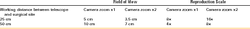
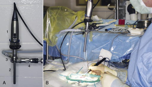
FIGURE 17-1 A, Endoscopy camera (1), video telescopic operating microscope (VITOM) telescope (2), and light-guide cable (3), held in place with the clamp and articulated arm (4). B, Intraoperative view of the VITOM system positioned above the surgical site. (Photo courtesy Dr. Stephen J. Divers, University of Georgia, Athens, Ga.)
Stay updated, free articles. Join our Telegram channel

Full access? Get Clinical Tree


