Fig. 1
Illustration of the real-time monitoring and alarm system of the IZSLER Veterinary Biobank facility
3 Sample Types and Preparation
Pre-analytical conditions are a key factor in maintaining the high quality of biospecimens. They are necessary to achieve the highest specificity of the laboratory test used for clinical diagnosis as well as for accurate reproducibility of experiments in the field of biomarker discovery. Inappropriate collection, handling, and storage of samples, as well as errors in data analysis and documentation, may all contribute to the generation of irreproducible and unreliable data. Preservation and optimization of biosample integrity to foster relevant research results and outcomes is a guiding principle of sample management.
The field of veterinarian research is rapidly evolving with new technologies and new standards. Samples are collected and stored also for a long period of time (years) before being used. For this reason, sample handling procedures should be defined and appropriated in order to guarantee a suitable use for the technology of tomorrow.
Biological samples have different storage requirements. For this reason, various approaches can be defined in order to obtain the most correct procedure.
3.1 Blood Samples
The routine use and collection of blood samples for diagnosis has provided general information on optimal methodologies and potential pitfalls.
Although procedures for collecting, processing, storing, and shipping blood components are generally standardized and well documented, several important factors need to be considered.
An important early decision in blood collection is whether to collect anticoagulated blood (consisting of plasma, buffy coat, and red blood cells) or coagulated blood (consisting of serum and red blood cell clot). Hemolysis of the specimen affects the accuracy of laboratory tests, particularly chemical and serological tests; therefore, it should be prevented by employing careful handling techniques according to optimum needle size, proper handling of the tubes, and proper pipetting techniques; however, if hemolysis is observed (pink to red tinge in sample), this information should be recorded [21]. In fact, hemolyzed samples would not be used for proteomics analysis, but destroying them may be unnecessary as any sample is worth saving, unless storage space is constrained. Annotation of all pertinent information about the samples allows the identification of potential factors that could influence outcomes. Serum and plasma specimens are of better quality for analysis if smaller-volume aliquots are initially prepared rather than larger ones that have to be thawed, handled, and refrozen, perhaps several times. Indeed, the ability to provide ready-to-use (RTU) aliquots without additional handling steps facilitates the sharing of samples and provides multiple replicates handled in an identical manner. It is also important to consider new or alternative methods and reagents that may offer longer-term stability or increased efficiency in the collection and preservation of blood [22, 23].
3.2 Cell Cultures
The ability to cryopreserve and successfully recover cell lines has been critical to the conservation of these biological samples, in particular the preservation of stem cells and the preparation of well-characterized cell banks. Indeed, the systematic storage and establishment of cryopreserved banks of cells for the stem cell research community is essential to the promotion of standardization in stem cell research and its use in clinical applications. Despite the significant potential for the use of stem cells in research and therapy, they are challenging to preserve and have been shown to be unstable after prolonged culture, often resulting in permanent alterations in their genetic background, which ultimately alters the phenotype of the culture. The working process related to cell culture isolation, amplification, control, storage, and shipment should be carried out in accordance with a quality management system such as ISO 9001:2008. Preparation and propagation of cell cultures are performed as indicated by international guidelines, and their sensitivity and reproducibility have been evaluated during interlaboratory tests. Each cell culture process provides ideal conditions for the growth of many organisms [24]; for this reason, particular attention must be placed on quality controls that should be performed on all samples with the purpose of assessing their purity and safety. These tests are carried out using microbiology, virology, serology, and molecular biology methods, as reported by the European Pharmacopoeia or other international guidelines. These assays are mainly based on in vitro tests. However, in vivo methods are used on laboratory animals, in the absence of validated in vitro systems or as indicated by international guidelines. In particular, the tests usually performed are aimed at detecting contamination from bacteria, fungi, yeasts, and mycoplasmas as well as animal and human viruses for human cell lines. Furthermore, bacterial endotoxin level, tumorigenicity, and cross contamination are investigated. Tests on cell cultures for detecting bacteria, fungi, and yeasts must be performed in an isolation unit, according to the European Pharmacopoeia.
3.3 Chlamydiaceae
Serological and molecular screening focused on the identification of chlamydial infections is routinely performed. Isolation of chlamydial species is carried out on cell culture.
Identification of the species belonging to the Chlamydiaceae is carried out using molecular tests, i.e., real-time PCR and PCR-RFLP analysis, which amplify specific genome sequences. The identification of new chlamydial species not yet classified is also performed using new real-time PCR and specific gene sequencing.
3.4 Field and Immune Sera
The field sera include samples from different animal species, mainly farmed, pets, and wildlife animals. The samples are collected by blood, sent to IZSLER to be tested in accordance with national or regional control programs or for specific serological tests. All these samples can be used as positive reference due to the presence of antibodies toward a specific pathogen, turning them very useful for retrospective serological surveys and validation of innovative serological tests. All serum samples collected and stored in the biobank are tested toward a panel of pathogens with the aim to know the presence/absence of antibodies.
Different serological techniques can be used and these are reported in specific sheets. Titers of humoral antibodies of serum samples are also registered and values are performed at preestablished intervals. The immune sera consist in polyclonal sera obtained from different animal species (rabbit, goat, mouse, rat, guinea pig) and toward several bacterial and viral antigens. They have been produced with antigens mostly purified and have a specificity panel available at the facility where they have been produced.
3.5 Hybridomas
The hybridomas that produce monoclonal antibodies are selected to be used in in-house diagnostic assays. They were generated to produce monoclonal antibodies against a wide spectrum of viruses, bacteria, and proteins, mainly of veterinary interest, and immunoglobulin isotypes of various animal species that were shown to be strategic tools for both research and diagnostic purposes. Hybridoma cultures are usually collected for freezing during the exponential growth phase and then are submitted to a double series of cloning procedure to assure the stability of the hybridoma and the clonality of the produced antibody. The monoclonal antibodies expressed by each hybridoma are controlled through a series of immunological assays aimed at identifying and characterizing their reactivity profile.
3.6 Microbiological and Parasitological Samples
Bacterial/fungal strains are identified by either phenotypic or genotypic tests. The former consists of microscopic observation and isolation in specific culture media. Identification is made through biochemical tests performed using commercial tests like the “API” or the “Vitek” systems. Genotypic tests, mainly PCR-based, are carried out to detect genes of virulence. Moreover, the 16S rDNA gene sequence analysis can be used to confirm the identity of bacterial species.
Parasites are identified using either phenotypic or genotypic tests. The former consists of microscopic observation and comparison with identification keys. Genotypic tests, mainly PCR-based, are carried out to identify genus or species.
3.7 Prototheca Algae
The identification of Prototheca algae (species and eventually subspecies) is currently carried out through microscopic examination, growth in specific culture media, and molecular assays (HMR-PCR, end point PCR, or sequencing).
3.8 Tissue Samples
Tissue samples can be stored in different ways, depending on their intended purpose. Indeed, molecular tools for tissue profiling, such as real-time PCR and expression microarrays, generally require collection of fresh frozen tissues as sources of high-quality RNA. Frozen tissue sections are made from high-quality tissues immediately snap-frozen in liquid nitrogen after being excised and identified by a pathologist. Available tissues include normal, diseased, and tumor animal tissues. The tissues excised are immediately frozen by liquid nitrogen and then stored at −80 °C. Tissue sections of 5–10 μm in thickness are mounted on positively charged glass slides. Furthermore, 0.4–0.7 g samples of standard size could be frozen in blocks into liquid nitrogen for 20–40 min after surgical excision. Otherwise, tissues could be fixed in 10 % neutral formalin for a minimum of 24 h and stored as paraffin blocks of 0.5 × 1 × 1 cm. Formalin fixation and paraffin embedding (FFPE) preserve the morphology and cellular details of tissue samples. Thus, it has become the standard preservation procedure for diagnostic surgical pathology [25]. Historically, the archived FFPE blocks have been successfully used for immunohistochemistry application. However, formalin-fixed archival samples are known to be poor materials for molecular biology applications due to the irreversible modifications caused by formalin fixation on macromolecules. In the last ten years, there has been an exponential increase in the development of molecular assays using FFPE blocks. At present, when sections from FFPE blocks are to be used for molecular extraction, focus is placed on the time of fixation in formalin in order to avoid over-fixation. The advances in the field of molecular biology techniques have attempted to overcome the issue of formalin cross-linking and have successfully extracted DNA, RNA, and proteins, although fragmented.
3.9 Viruses and Viral Pathological Materials
Cell-associated viruses that can be grown in adherent or suspension cell cultures or chorioallantoic membranes of embryonated hen’s eggs can be isolated from several types of samples. The main principle of isolating viruses is to choose the most suitable cell line and mechanically lyse infected cells and subsequently carry out several amplification passages to increase the titer in order to produce the master sample and then the working samples. Virus batches are tested for potential microbiological and viral adventitious contaminations; the tests are performed using microbiology, virology, serology, and molecular biology methods. The primary sources of potential viral contamination come from infected animal tissues used to prepare biological reagents and media and during laboratory manipulation. Virus detection and identification can be made by employing several methods, mainly based on serology tests using monoclonal antibodies and standard and real-time PCR. These assays amplify specific viral genome sequences known to be characteristics of a virus with a nucleotide sequence available in database collections. Extraneous viral contaminations can be verified through tests based on molecular biology techniques that allow the detection of viral DNA and RNA of other viruses. Mycoplasma contamination can be detected using real-time PCR methods. Bacterial contamination is determined through inoculation of nonselective culture media. In addition, electron microscopy is available for viral detection and identification using negative staining methods. Such “catchall” methods (able to detect even non-suspected/unknown viruses) benefit from “Airfuge” ultracentrifugation, increasing the sensitivity of the detection level. Indeed, immune electron microscopy methods (IEM and IEM gold) based on the use of hyperimmune sera and/or monoclonal antibodies may help viral identification and classification.
4 Storage of Biological Resources
Proper storage requires the use of cryovials and labeling systems that will withstand the intended storage conditions: vessels, labels, and bar codes or other printing systems are chosen for extended storage periods.
Samples have been deposited in freezers or other appropriate storage containers according to specific storage systems in order to preserve several parameters known to influence the condition of biospecimens.
In accordance with features, intended use and estimated length of storage, specimens may be stored at: room temperature, 4 °C, −20 °C, −80 °C, or −196 °C (vapor phase nitrogen) (Figs. 2 and 3). Temperature is a major variable in specimen management. Moreover microorganisms and viruses can be submitted to lyophilization process that allows preserving viability for a long time. These freeze-dried samples can be stored at 4 °C or at −20 °C, reducing the necessity to have freezers with lower temperature (−80 °C); for these reasons, this is a practical and efficient method for prolonged storage, less expensive, and more available in emerging countries.
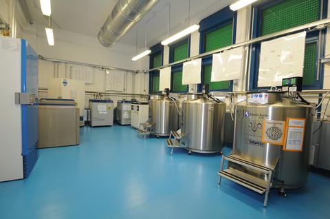
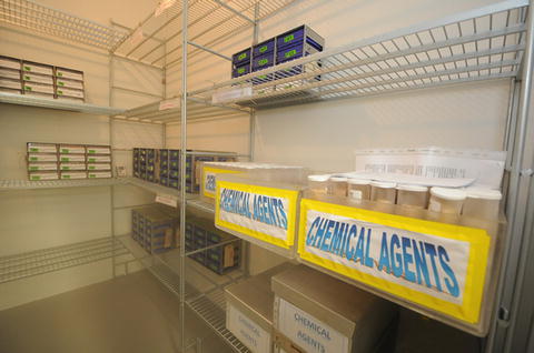
Fig. 2
Cryogenic area for preservation of biological resources
Fig. 3
Freezing area for preservation of biological resources
5 Application of Biological Resources
Two major formats of biobanks with several subtypes can be distinguished: the population-based biobank and the disease-oriented biobanks, each with distinct and complementary scientific value. The most common format is the longitudinal population-based biobank, with biological samples and data from randomly selected individuals of a general population, used as resources for future unspecified research [26–28]. The specific strength of this format is the assessment of the natural frequency of occurrence and progression of common diseases, with special emphasis on predisposing genetic variants and environmental risk factors. In contrast, in disease-oriented biobanks, which may contain tissue, isolated cells, blood, or other body fluids, specimens are collected in the context of medical diagnosis and treatment. These biosamples allow the comparison of different disease stages and/or forms of treatment at a molecular level, in order to evaluate biomarkers for the diagnosis of a disease or assessment of risk/predisposition and prognosis (research purpose), monitoring the recurrence of diseases, prediction of mortality, and response to therapy [29]. The development of precisely defined clinical data elements (CDEs) may help to ensure that clinically relevant data are collected at each time interval [12]. In veterinary medicine, the wide availability of biological samples stored in biobanks provides the basis for research, leading to a better understanding of animal disease biology and the development of new diagnostic tests that require the use of biosamples with well-defined features [13]. The European Technology Platform for Global Animal Health (ETPGAH) has identified the lack of biological material as one of the main gaps in the development of new effective tools for the control and prevention of animal diseases. Biobanks represent an important tool in improving epidemiological research dependent on the availability and quality of the biomaterials but also on the collection of associated data [6]. In particular, for retrospective studies and longitudinal designs for evaluating the course of diseases, the requirements for obtaining time-specific data are even stronger. Furthermore, biological materials are a critical resource for genetic research. Major research in genomics is being pursued to improve the efficiency of selection for healthier animals with disease resistance properties. Molecular genetic tests have been developed to select farm animals with improved traits, for example, removal of the porcine stress syndrome and selection for specific estrogen receptor alleles [30]. The sequencing of the genome to identify new genes and unique regulatory elements holds great promise in providing new information that can be used for livestock production. Currently, in vitro embryo production and embryo transfer are being the preferred means of implementing these new technologies to enhance efficiency of farm animal production. Furthermore, the possibility of developing an integrated approach of genomics and proteomics using bioinformatics is essential for obtaining complete use of the available molecular genetic information. The development of this knowledge will benefit scientists, industry, and breeders considering that the efficiency and accuracy of traditional farm animal selection schemes will be improved by the implementation of molecular data into breeding programs.
Stay updated, free articles. Join our Telegram channel

Full access? Get Clinical Tree


