Vestibular system

Chapter 8 Vestibular system


General concepts


The vestibular system (vestibulum – L = entrance) is the neural system that sets body equilibrium or balance. Equilibrium is defined as the condition in which all influences acting on a structure are cancelled out by others, resulting in a stable, balanced system. For example, if the head moves to the left, there has to be increased extensor muscle activity on the left side of the body to accommodate the shift in weight. Head movement can be primary (animal turning to look at something) or secondary due to movement of the body. The latter can be generated by the animal (e.g. sitting, locomotion) or by external forces, such as being pushed by another animal. Head movement is detected by the vestibular system, the input from which results in modification of somatic muscle activity to accommodate the new set of forces acting on the body.


Cephalisation is the concentration of the exteroceptors and neural processing centres at the leading end of the animal. Use of exteroceptors such as vision, audition, olfaction and gustation, as well as tactile receptors, (e.g. whiskers) necessitates precise control of head position and movement. Controlling head position and movement requires detailed proprioceptive input that modifies motor activity in the neck muscles. Changes in head and neck position require postural changes in axial and appendicular muscle activity to provide the correct postural support. This is reflected in increased extensor muscle activity on the side to which the head is turned. Therefore head position dictates tone in muscles through the neck, body and limbs. To maintain appropriate visual input requires that the position and movement of the eyes be linked to head position and movement.


The vestibular apparatus, located in the inner ear, houses the receptor organs for detecting head position and movement. Stimulation of the vestibular apparatus provides the major input to the vestibular nuclei in the brainstem. However, the vestibular nuclei also integrate input from the gravity and movement sensors in the inner ear with proprioceptive information from muscles and joints.


The vestibular apparatus on each side consists of five distinct organs. There are receptors in three semicircular ducts, which are sensitive to angular acceleration/head rotation, and in two otolith organs (utriclus and sacculus), which are sensitive to linear accelerations and the influence of gravity in static head positions. The receptors are housed in the inner ear, in a series of membrane-lined, fluid-filled ducts and spaces located in the bony vestibule of the petrous temporal bone.


The receptors that detect head position and movement are hair cells. These are specialised epithelial cells with fine, hair-like processes on top of them. The base of the hair cell synapses with sensory nerves that convey action potentials via the vestibular portion of cranial nerve VIII (vestibulocochlear nerve) to the vestibular nuclei. The cell bodies of these bipolar sensory nerves are spread along the fibres forming the vestibular ganglion in the petrous temporal bone. There are four pairs of vestibular nuclei, located in the brainstem, just ventral to the cerebellum. The vestibular nuclei have five main efferent connections. They give rise to UMN spinal cord tracts that reflexively stimulate ipsilateral extensor muscle activity in the limbs, trunk and neck, inhibit antagonist muscles and contralateral extensor muscles. Rostrally directed output from the vestibular nuclei affects function of the cranial nerve nuclei, III, IV, VI, causing extraocular muscle contraction for eyeball position and movement. The vestibular nuclei send information to the cerebellum and cerebrum about head proprioception for subconscious modulation of postural muscle activity, and for conscious awareness, respectively. The vestibular nuclei also connect to the vomiting centre of the brainstem reticular formation; this is the basis of motion sickness.


The basic mechanism that stimulates the hair cells is similar for the detection of either static, or changing, head position. On each hair cell are 40–80 stereocilia (long microvilli) and a single kinocilium. These processes project into an overlying gelatinous layer, which, when it moves, causes deviation of the cellular processes. This deviation induces changes in membrane potential of the hair cell and may trigger an action potential in the sensory nerve fibre that synapses with the base of the hair cell. Gravity constantly stimulates hair cells that detect static head position. However, hair cells that detect head movement will only do so during acceleration or deceleration of the head, not when the head has reached constant velocity.


Structural and functional anatomy


The vestibular system is divided into peripheral and central components (Fig. 8.1); this segregation has clinical implications. The peripheral vestibular system consists of the vestibular components of the inner ear (receptors and axons of CN VIII), while the central vestibular system comprises the vestibular nuclei and their output.


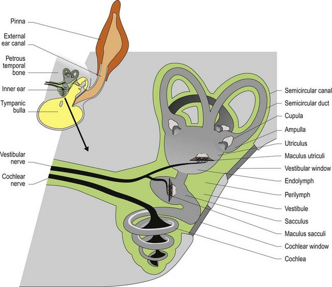
The inner ear is housed in the petrous temporal bone bilaterally. It contains a number of canals and cavities forming the bony labyrinth. On each side there are three semicircular canals, the bases of which connect to the bony vestibule. Ventral to the vestibule is the spiral cochlear canal that houses the auditory receptors (see Chapter 10). The semicircular canals arise from the vestibule, curve around and connect back to the vestibule. At one end of those connections the canal is dilated to form the ampulla. The canals and cavities are lined with membranes forming the membranous labyrinth. Perilymph is the fluid located between the membrane and the bone while endolymph fills the membranous labyrinth.


Static position, linear acceleration and deceleration of the head


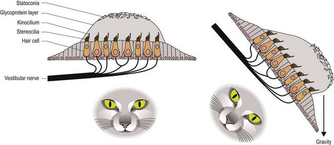
Located within the vestibule are expanded portions of the labyrinth, called the utriculus and the sacculus (Fig. 8.1). These membrane-bound portions of the vestibular system each have a region called a macula; this is where the hair cells are located. The stereocilia and kinocilium of each cell project into an overlying gelatinous matrix called the otolithic membrane. Embedded in the membrane are calcareous crystals called otoliths that, having sufficient mass, move under the effect of gravity, pulling on the otolithic membrane and causing deflection of the stereocilia and kinocilium. The macula in the utriculus is oriented in the horizontal plane, whereas in the sacculus it is oriented vertically. Thus no matter what position the head is in, gravity will be acting on one or both sets of otoliths causing deflection of the cell processes on the hair cells and stimulating the sensory nerves at their base. The stimulation is constant due to the ongoing effect of gravity on the mass of the otoliths. Linear acceleration or deceleration will also affect otolith position (Fig. 8.2).


Angular acceleration and deceleration of the head


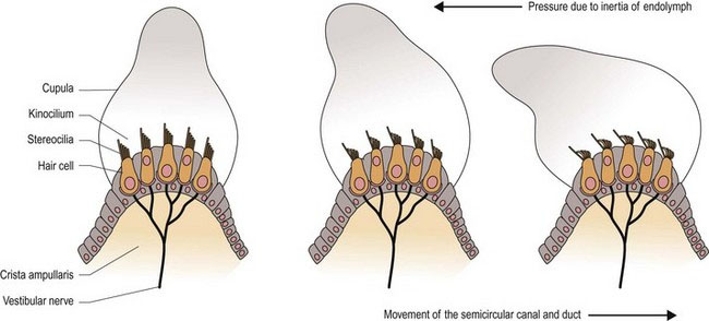
The three semicircular canals and their membranous ducts are located at right angles to each other (in the x, y and z directions) with the anterior and posterior canals being vertically oriented and the horizontal canal being laterally oriented. The semicircular ducts on each side of the head function as synergic pairs. Turning the head in one direction will cause endolymph to flow towards the ampulla on that side, but away from the ampulla in the paired duct on the opposite side. This causes stimulation of hair cells on the first side, but inhibition on the opposite side. Thus the input received by the vestibular nuclei of the brainstem is unequal. Unequal input from paired semicircular canals is interpreted as head movement. Note that if there is disease in the vestibular system on one side, then even at rest the brain will receive uneven input and thus, it is perceived that the animal’s head is moving. The uneven input from the two sides will be interpreted as head movement inducing the reflex changes in posture and eyeball position/movement (see Clinical dysfunction) that result in clinical signs of vestibular disease.


Like the semicircular canal, the semicircular duct is dilated at one end forming the ampulla. The hair cells that perceive head movement are located on a ridge of tissue (crista ampullaris) in the ampulla of the semicircular ducts. The numerous stereocilia and a single kinocilium project from the surface of the hair cells into a gelatinous matrix called the cupula; this projects into the endolymph.


As the head moves, so will the petrous temporal bone, and the associated semicircular ducts and hair cells (Fig. 8.3 and 8.4). However, due to inertia, movement of the endolymph inside the ducts lags behind. The endolymph presses on the cupula, which deflects the stereocilia towards or away from the kinocilium, resulting in stimulation or inhibition of the vestibular neurons, respectively.


Stay updated, free articles. Join our Telegram channel

Aug 26, 2016 | Posted by in INTERNAL MEDICINE | Comments Off on Vestibular system

Full access? Get Clinical Tree

Get Clinical Tree app for offline access