Chapter 1 Regional neuroanatomy
Basic systems arrangement of the nervous system
Nerve fibres are of three basic types:
1. Afferent/sensory fibres bring information into the central nervous system (CNS) from the periphery and convey that information to higher centres for processing.
2. Efferent/motor fibres take information from motor planning centres through the CNS to connect with other motor fibres that take information into the periphery.
3. Integrating fibres that may connect afferent fibres with storage centres, other processing centres or efferent fibres. They process, organise and/or store information.
Introduction to regions
Peripheral nervous system
Key points


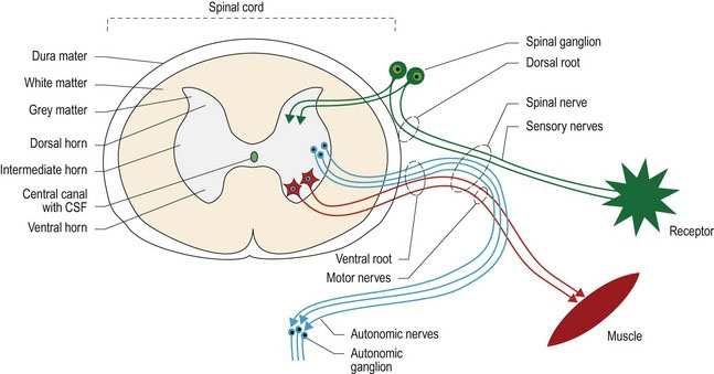
The PNS consists of the nerves and ganglia located outside the brain and the spinal cord and principally functions to connect the central nervous system (CNS) to the head, body, limbs and viscera. With respect to nomenclature, a ‘nerve(s)’ is by definition, in the PNS, making the word ‘peripheral’ (as in ‘peripheral nerve’) redundant; it is myelinated by Schwann cells. Unlike the CNS, the PNS is not protected by bone, leaving it more vulnerable to mechanical injury. Schwann cells form the insulating myelin sheaths surrounding peripheral axons, whereas in the CNS that task is performed by oligodendrocytes. The change from oligodendrocytes to Schwann cells occurs where the dura mater surrounding the spinal cord abuts the perineurium at the origin of the spinal nerves (Fig. 1.1). Afferent and efferent axons of the PNS form the spinal and cranial nerves (CNN).
Spinal nerves
Spinal nerves arise as roots from the spinal cord. A dorsal and a ventral root attach on each side of the spinal cord, and define each spinal cord segment. For example, the third cervical spinal cord segment has two dorsal roots and two ventral roots attaching to it. The dorsal roots convey primarily sensory nerve fibres into the spinal cord. Each dorsal root contains a spinal ganglion (old name ‘dorsal root ganglion’), housing the nerve cell bodies of these sensory fibres (Fig. 1.1). The ventral roots convey motor nerve fibres away from the spinal cord. Motor fibres may be somatic and innervate striated muscle, or autonomic and innervate smooth or cardiac muscle. The dorsal and ventral roots fuse at the level of the intervertebral foramen to form a spinal nerve. Distal to the intervertebral foramen, the mixed sensory and motor spinal nerve usually splits into a dorsal and ventral branch. The dorsal branch supplies the epaxial muscles and skin, while the ventral branch supplies the hypaxial muscles and skin. A third branch, carrying autonomic fibres, may also arise and pass ventrally towards the midline to supply the viscera.
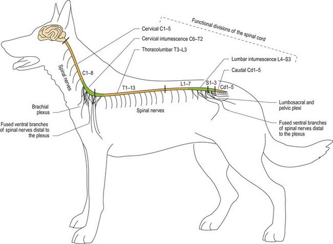
Spinal nerves may remain as single, discrete nerves all the way out into the periphery, in which case they are named for the number of the spinal cord segment from which they arise, e.g. ‘C3, ventral branch’ is the ventral division of cervical spinal nerve 3; it supplies sensory and motor innervation to the hypaxial tissue of the neck. Alternatively, in a nerve plexus, the ventral branches of two or three adjacent spinal nerves may fuse, giving rise to nerves with specific names, such as radial nerve and femoral nerve (Fig. 1.2).
A cranial nerve arises from the brain; it is known by both the number of the nerve and a specific name, e.g. trigeminal nerve or cranial nerve, CN V (Fig. 1.6A).
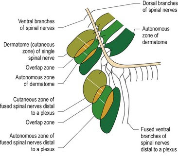
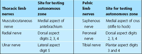
Afferent nerve fibres originate at different types of sensory receptors and most of their fibres only synapse once they reach the CNS. The sensory nerve cell bodies of spinal nerves are located in spinal ganglia sited at the level of the intervertebral foramen. Cranial nerve ganglia are located just near, or inside, the neurocranium. The area of skin innervated by a spinal nerve is called a dermatome, while the area of skin innervated by a specific named nerve, which originates from two or more spinal nerves (for example, the radial nerve) is called a cutaneous zone. Adjacent dermatomes and cutaneous zones usually overlap. The area of skin innervated purely by one nerve is called an autonomous zone (Fig. 1.3). Autonomous zones are found on the head (Figure 10.13) as well as the body and limbs (Table 1.1).
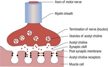
Motor neurons can be defined as upper motor neurons (UMNs) or lower motor neurons (LMNs). The UMN is confined to the CNS and its axon influences activity of LMNs. They are the ‘managers’ of the motor system (see Chapter 4). The LMNs are found in cranial nerves, originating from the brainstem, and spinal nerves from the spinal cord. They form synapses at the neuromuscular junction and innervate striated muscle, smooth or cardiac muscle. They are the ‘workers’ of the motor system. The neurochemical that connects the electrical activity of the motor nerve to the striated muscle is acetylcholine (ACh). A nerve impulse arriving at the nerve termination triggers release of ACh (Fig. 1.4). The ACh crosses the synaptic cleft, binds to the receptors on the post-synaptic membrane and may stimulate muscle membrane depolarisation and muscle contraction, depending on stimulus strength and amount of ACh released. The ACh is broken down by acetylcholine esterase and recycled back into the distal end of the LMN. For striated muscle, the motor unit is defined as an axon and the muscle fibres it innervates. Motor units range in size from 3–150 muscle fibres per axon for muscles with fine versus coarse control, respectively. Small motor units are found in extraocular muscles, whereas large motor units are found in the large, postural muscles, e.g. the quadriceps femoris muscle.


Stay updated, free articles. Join our Telegram channel

Full access? Get Clinical Tree


