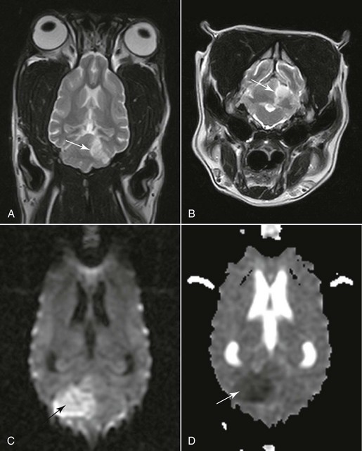
Vascular Disease of the Central Nervous System

Chapter 241


Vascular Disease of the Central Nervous System


There has been a good deal of confusion in the veterinary literature regarding the terms cerebrovascular disease, cerebrovascular accident, and stroke. Cerebrovascular disease refers to any abnormality of the brain caused by a pathologic process compromising the blood supply. Pathologic processes that may result in cerebrovascular disease include occlusion of the lumen by a thrombus or embolus, rupture of a blood vessel wall, a lesion or altered permeability of the vessel wall, and increased viscosity or other changes in the quality of the blood. This chapter considers vascular disorders of the brain (stroke) and of the spinal cord (ischemic myelopathy).


Cerebrovascular Accident (or Stroke)


Stroke or cerebrovascular accident (CVA) is the most common clinical presentation of cerebrovascular disease and is defined as a sudden onset of nonprogressive focal brain signs secondary to cerebrovascular disease. By convention these signs must persist for longer than 24 hours to qualify for the diagnosis of stroke, which usually is associated with permanent damage to the brain. If the clinical signs resolve within 24 hours, the episode is called a transient ischemic attack. From a pathologic point of view, a CVA falls into one of two broad categories: (1) ischemia with or without infarction secondary to obstructed blood vessels, and (2) hemorrhage caused by rupture of the blood vessel wall.


Ischemic Stroke


With limited stores the brain relies on a permanent supply of glucose and oxygen to maintain ionic pump function (see Chapter 228). When perfusion pressure falls to critical levels, ischemia develops, progressing to infarction if hypoperfusion persists long enough. An infarct is an area of compromised or necrotic brain parenchyma caused by a focal occlusion of one or more blood vessels. It may be caused either by vascular obstruction that develops within the affected vessels (thrombosis) or by obstruction by material that originates from another vascular bed and travels to the brain (thromboembolism).


Depending on the size of vessel involved, infarcts can be the consequence of small-vessel disease (e.g., disease of a superficial or deep perforating artery), which gives rise to a lacunar infarct, or large-vessel disease (disease of a major artery of the brain or its main branches), which gives rise to a territorial infarct. Two distinct regions can be distinguished in ischemic stroke: the core, in which ischemia is severe and infarction develops rapidly; and the penumbra, which surrounds the core and demonstrates a less severe reduction of cerebral blood flow (CBF), so that longer durations of ischemic stress can be tolerated. The relative volume of pathologic effects within these two regions changes as the infarct evolves. The factors favoring evolution of the penumbra to irreversible brain injury are multiple and complex. The time window after which the penumbra is no longer viable depends on the degree of reduction in blood flow, the region of the brain involved, and the individual. In the penumbra neurons are still viable but at risk of becoming irreversibly injured. Penumbra tissue has the potential for recovery and therefore is the target of interventional therapy in acute ischemic stroke.


Ischemic strokes have been reported infrequently in veterinary publications compared with the human medical literature. Most reports have been based on postmortem results in dogs that either died or were euthanized as a result of the severity of the ischemic stroke or the suspected underlying cause of the stroke. This retrospective reporting may affect the prevalence and type of underlying causes identified in canine CVA because only the most severe cases are likely to be reported. In a similar vein, dogs in which infarction occurred secondary to a disease with a poor prognosis also would die or be euthanized.


Suspected underlying causes identified in histopathologically confirmed cases included septic thromboemboli, atherosclerosis associated with primary hypothyroidism and with hypertriglyceridemia in miniature schnauzers, aberrant parasitic migration (Cuterebra spp.) or parasitic emboli (Dirofilaria immitis), embolic metastatic tumor cells, intravascular lymphoma, embolism with an aortic or cardiac source, and fibrocartilaginous embolism. In our study (Garosi, 2005) using magnetic resonance imaging (MRI), a concurrent medical condition was detected in just over 50% of dogs affected by brain infarcts. Hypertension was documented in 30% of dogs. In these dogs chronic kidney disease and hyperadrenocorticism were the most commonly suspected underlying causes of the hypertension. No underlying cause could be identified antemortem in nearly half of the dogs. An infarct of unknown origin is called cryptogenic. No age, sex, or breed predisposition was identified. However, cavalier King Charles spaniels and greyhounds appeared overrepresented.


Reports of ischemic strokes in cats are limited. A concurrent medical condition is found more frequently in cats than in dogs. Concurrent conditions identified include Cuterebra parasitic migration, heartworm migration, intracranial telangiectasia, cardiovascular disease, hyperthyroidism, liver disease, and neoplasia elsewhere in the body.


Hemorrhagic Stroke


In hemorrhagic stroke blood leaks from the vessel directly into the brain, forming a hematoma within the brain parenchyma, or into the subarachnoid space. The mass of clotted blood causes physical disruption of the tissue and pressure on the surrounding brain. This alters central nervous system volume-pressure relationships with the possibility of increasing intracranial pressure (ICP) and decreasing CBF. In contrast to the high incidence observed in humans, intracerebral hemorrhage resulting from spontaneous rupture of vessels is considered rare in dogs. Secondary hemorrhage in dogs has been associated with rupture of congenital vascular abnormalities, primary and secondary brain tumors, inflammatory disease of the arteries and veins, intravascular lymphoma, brain infarction (hemorrhagic infarction), or impaired coagulation. Nontraumatic subarachnoid hemorrhage has been reported in dogs but remains very rare. This is in contrast to human patients, in whom aneurysmal rupture of blood vessels is the most common underlying cause of hemorrhagic stroke. Intraparenchymal hemorrhage in cats has been associated with primary or secondary hypertension, intracranial neoplasia, cerebral amyloid angiopathy, and feline infectious peritonitis.


Clinical Presentation of Stroke


In all forms of stroke the denominative feature is the temporal profile of neurologic events. The abruptness with which the neurologic deficits develop is highly suggestive that the disorder is vascular. This is followed by an arrest and then regression of the neurologic deficit in all except fatal strokes. Worsening of edema (associated with the secondary injury phenomenon) can result in progression of neurologic signs for a short period of 24 to 72 hours. Intracranial hemorrhage can be an exception to this description, presenting with a more progressive onset over a very short period of time. Clinical signs usually regress after 24 to 72 hours; this is attributable to diminution of the mass effect caused by hemorrhage with subsequent reorganization or to edema resorption. Neurologic deficits usually refer to a focal anatomic diagnosis and depend on the neurolocalization of the vascular insult (telencephalon, thalamus, midbrain, pons, medulla, cerebellum). Infarction of an individual brain region is associated with specific clinical signs that reflect the loss of function of that specific region. With hemorrhagic stroke the total clinical picture is different because the hemorrhage usually involves the territory of more than one artery, and pressure effects cause secondary signs. Neurologic signs are related largely to increasing ICP, which gives rise to nonspecific signs of forebrain or brainstem disease. Fundus examination is important and may reveal findings such as tortuous vessels (suggestive of systemic hypertension), hemorrhage (suggestive of coagulopathy or systemic hypertension), or papilledema (suggestive of elevated ICP).


Imaging studies of the brain (computed tomography [CT], conventional and functional MRI) are necessary to confirm the suspicion of stroke, define the vascular territory involved and the extent of the lesion, and distinguish between ischemic and hemorrhagic stroke (Figure 241-1). Imaging studies also are necessary to rule out other causes of neurologic deficit such as tumor, head trauma, and encephalitis. (See References and Suggested Reading for more details.)


Ancillary diagnostic tests in ischemic stroke should focus on evaluating the animal for systemic hypertension (and underlying causes; see Chapter 169), endocrine disease (hyperadrenocorticism, hypothyroidism, hyperthyroidism, diabetes mellitus), kidney disease (especially protein-losing nephropathy), heart disease (a much greater risk factor in cats with cardiomyopathy), and metastatic disease. In cases of ischemic stroke, D-dimer assays and antithrombin III evaluation should be included routinely in the screening tests to identify thromboembolic disease as a possible cause of ischemic stroke. A D-dimer assay is considered to be a more useful test for detecting thromboembolic disease in dogs than traditional tests currently in use (platelet count and clotting times). In cases of hemorrhagic stroke, diagnostic tests should be targeted at screening the animal for a coagulation disorder (and underlying causes), hypertension (and underlying causes), and metastatic disease (particularly hemangiosarcoma).


Treatment and Prognosis of Stroke


Once the diagnosis of a stroke has been made, any potential underlying disease should be identified and treated accordingly. Generally treatment of these patients aims to provide supportive care, maintain adequate tissue oxygenation, and manage neurologic and nonneurologic complications. Nursing management of a recumbent dog is vital to the success of more specific therapies. Such management includes prevention of decubital ulceration, aspiration pneumonia, and urine scald, in addition to physical therapy and provision of enteral nutrition. More specific therapies are aimed at preventing further neurologic deterioration.


Treatment of Ischemic Stroke


Treatment of an ischemic stroke revolves around three principles: monitoring and correcting basic physiologic variables (e.g., oxygen level, fluid balance, blood pressure, body temperature), inhibiting the biochemical and metabolic cascades subsequent to ischemia to prevent neuronal death (the concept of neuroprotection), and restoring or improving CBF by thrombolytic therapy if a thrombus is present. The potentially salvageable portion of the ischemic zone (ischemic penumbra) is the presumed therapeutic target for both thrombolytic and neuroprotective stroke therapy. The time period during which injury may be reversible is called the therapeutic window. It is estimated that this “window of opportunity” is approximately 6 hours before irreversible neurologic damage occurs. Fortunately, the vast majority of ischemic stroke patients have no major difficulty maintaining the airway, breathing efforts, or circulatory competence early in the clinical course.


There is some controversy surrounding the management of hypertension in the setting of an ongoing acute ischemic stroke. Although hypertension is a potential risk factor for a CVA, it also can develop as a physiologic response to a stroke to ensure adequate cerebral perfusion pressure (CPP) in the penumbra of the infarct. Elevated blood pressure can persist for up to 72 hours after the onset of injury. Maintenance of systemic arterial blood pressure within the physiologic range is essential, and aggressive lowering of blood pressure should be avoided during the acute stages unless the patient is at a high risk of end-stage organ damage (systolic blood pressure remains >180 mm Hg). In such cases hypertension often can be controlled with an angiotensin-converting enzyme inhibitor such as enalapril (0.25 to 0.5 mg/kg twice daily PO) or benazepril (0.25 to 0.5 mg/kg twice daily PO) with or without a calcium channel blocker such as amlodipine (0.1 to 0.25 mg/kg once daily PO). Amlodipine is more effective in severe hypertension.


There is no evidence that glucocorticoid treatment provides any beneficial neuroprotection in stroke. Not only is there lack of proven benefit in veterinary stroke patients, the use of glucocorticoids may increase the risk of gastrointestinal complications and infection. Treatment strategies considered for ischemic stroke in humans that use other neuroprotective agents (N-methyl-d-aspartate antagonists, calcium channel blockers, sodium channel modulators) or antiplatelet and thrombolytic therapy remain to be evaluated clinically in dogs. Although these neuroprotective agents have resulted in a dramatic decrease in the size of stroke lesion in experimental animal models, they either have failed to prove their efficacy in clinical trials or are awaiting further investigation.


At the time of this writing, there are no definitive data in humans or animals to confirm a significant improvement in clinical outcome in patients with acute ischemic stroke treated with unfractionated heparin as anticoagulant therapy. Despite conflicting results regarding the efficacy of intravenous recombinant tissue plasminogen activator, it sometimes is used in human ischemic stroke patients if it can be given within the first 3 hours. This critical time window makes the use of thrombolytic treatment largely unrealistic in veterinary practice. Furthermore, this type of treatment carries a significant risk of intracranial hemorrhage following treatment. Antiplatelet therapy with low-dose aspirin (0.5 mg/kg once daily PO) or clopidogrel (2 to 4 mg/kg once daily PO) can be used prophylactically to prevent clot formation when a cardiac embolic source has been proven.

< div class='tao-gold-member'>

Stay updated, free articles. Join our Telegram channel

Jul 18, 2016 | Posted by in PHARMACOLOGY, TOXICOLOGY & THERAPEUTICS | Comments Off on Vascular Disease of the Central Nervous System

Full access? Get Clinical Tree

Get Clinical Tree app for offline access