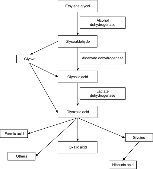
Automotive Toxins

Chapter 36


Automotive Toxins


Various compounds used in vehicle maintenance and stored around the home have known toxic properties. An incomplete list of such chemicals includes ethylene glycol (EG), propylene glycol (PG), diethylene glycol (DEG), petroleum products, and methanol. EG is the most common component of antifreeze and unfortunately is the most common automotive product associated with poisoning in small animals. PG has been substituted for EG in antifreeze brands that are advertised as “safe” or “nontoxic,” and although PG is not without adverse effects, it is much less toxic than EG.


Ethylene Glycol


EG, or 1,2-dihydroxyethane, is a colorless, sweet-tasting liquid with a density of 1.113, a high boiling point (197.2° C [326° F]), a low freezing point (−12.3° C [9.86° F]), and is miscible with water and alcohol. Less common sources of EG include deicer, hydraulic brake and transmission fluids, additives in motor oils, paints, inks, wood stains and polishes, photographic solutions, and industrial solvents.


EG is one of the most common causes of fatal poisoning in small animals, perhaps because it is readily available in most households, is toxic in low doses, and is palatable. Toxicosis is most common in the late autumn or early spring, when radiators have been drained and open containers may be available to pets. Dogs occasionally chew through closed containers. Denatonium benzoate, a bittering agent, has been added to antifreeze to make it less palatable. The states of Arizona, California, Georgia, Illinois, Maine, Maryland, Massachusetts, New Jersey, New Mexico, Oregon, Tennessee, Utah, Vermont, Virginia, Washington, and West Virginia require addition of bittering agents to commercial antifreeze at the time of this writing.


Toxicity and Toxicokinetics


The minimum lethal dose for EG in dogs is about 6.6 ml/kg. Cats are more sensitive, with a minimum lethal dose around 1.4 ml/kg. Dogs are more frequently affected than cats, although cats are likely to become intoxicated through grooming activity after dermal contamination. Intact animals are more frequently affected.


EG is absorbed rapidly from the gastrointestinal tract, particularly on an empty stomach. Peak plasma concentrations occur within 3 hours of ingestion. Metabolism begins within hours of ingestion and occurs predominantly in the liver, with minor renal and gastric metabolism. The metabolic pathway of EG is illustrated in Figure 36-1. The metabolism of EG to glycoaldehyde and then glycolic acid to glyoxylic acid are both rate-limiting steps. Oxalic acid is the most important final metabolite of EG. The plasma half-life of EG is approximately 3 hours, and elimination is almost complete within 24 hours. EG and its metabolites are eliminated in the urine.


Clinical Signs


EG toxicosis is described as having three sometimes overlapping stages, although early stages are often missed. The first stage, usually within 30 minutes of exposure, can last for 2 to 12 hours. Some animals vomit. Polyuria and polydipsia are described in dogs, and cats are frequently polyuric. Apparent “inebriation” presents as ataxia and hyporeflexia. Dogs often have a period of apparent recovery from this stage, but cats typically do not.


The second stage usually occurs 8 to 24 hours after exposure and is related to metabolic acidosis. Clinical signs include central nervous system (CNS) depression, changes in heart rate, hypothermia, muscle fasciculations, and sometimes coma. Cats often lose coordination in the pelvic limbs.


Animals that survive the first two stages enter the third stage, acute renal failure, which can begin from less than 1 to 3 days after EG ingestion. Animals progress through oliguria to anuria. Signs of uremia include oral ulcerations, salivation, vomiting, anorexia, and seizures. Palpation of cats often reveals large, painful kidneys.


Serum chemistries reveal an increased osmolal gap about an hour after EG ingestion, which usually declines within the first 18 hours. The anion gap increases in a few hours and can remain elevated for 48 hours. Hypocalcemia is reported due to calcium oxalate crystal formation. Phosphorus can be elevated early, as a result of the phosphate additives in antifreeze, and again as a consequence of renal failure. Other findings associated with renal failure include elevated serum urea nitrogen and creatinine, hyperkalemia, isosthenuria with low urine pH, hematuria, proteinuria, glycosuria, and granular and cellular casts. Calcium oxalate crystals are evident in urine within 8 hours of exposure, but are not a specific finding considering that crystalluria is identified in many healthy animals.


Diagnosis


Speed is of the essence in the diagnosis (and management) of EG toxicosis. The prognosis decreases precipitously the longer treatment is delayed. EG concentrations begin to decline within 6 hours of exposure and are often undetectable in blood or urine by the time the animal presents to the veterinarian, further complicating the diagnosis.


Various analytic tests have been used to confirm EG exposure. Commercial test kits are available, but some of these kits cross-react with PG, glycerol, sorbitol, ethanol, and other compounds that can be present in pet foods and pharmaceuticals; thus samples for testing should be collected before initiating treatment. Current test kits are able to detect EG at concentrations above 0.6 mg/dl, but due to rapid EG metabolism false-negative results can occur in samples collected more than 12 hours after ingestion. Gas chromatography (GC) is frequently used to assay blood, urine, or kidney tissue for EG at diagnostic laboratories, although results are delayed by sample shipping and processing and false-negative results are still possible if the EG has been completely metabolized.


The findings of increased anionic and osmolal gaps along with the appropriate history and presentation are highly suggestive of EG toxicoses, but are not always seen. Calcium oxalate monohydrate crystals are commonly found in the urine. Fluorescent dye, which is sometimes added to antifreeze, is visible in the urine, in the vomitus, or around the oral cavity under Wood’s lamp. However, other components of urine and some plastic containers also fluoresce.


The “halo effect,” an ultrasound finding related to increased echogenicity of the renal cortex and medulla and decreased echogenicity at the corticomedullary junction and central medulla, is supportive (although not pathognomonic) for the diagnosis of EG toxicosis. This change occurs near the onset of anuria.


Postmortem findings are frequently used to confirm the diagnosis of EG toxicosis. Typical findings include gastric hemorrhage, pulmonary edema, and pale firm kidneys. Histologic changes include degeneration and necrosis of proximal convoluted tubular epithelium with intraluminal birefringent crystals. Chronicity is indicated by evidence of tubular regeneration, interstitial fibrosis, and glomerular atrophy and synechia. Oxalate crystals are sometimes found in other tissues, including the liver and CNS.


Management


The prognosis for EG toxicosis depends on the time of presentation. Animals treated early (stage 1) have an excellent prognosis, but the prognosis is poor for animals that present in renal failure (stage 3). Due to the rapid absorption of EG, gastrointestinal decontamination is unlikely to be beneficial.


Antidotes that act by inhibiting metabolism of EG by alcohol dehydrogenase include fomepizole and ethanol. Antidotal treatment should be started as early as possible for best effect but can still be attempted if the animal is presented to the clinician up to 32 hours postexposure.


Fomepizole


The preferred antidote is fomepizole (4-methylpyrazole). This product is used in dogs at an initial dose of 20 mg/kg IV, then 15 mg/kg IV at 12 and 24 hours, and then 5 mg/kg IV at 36 hours. Cats have recovered after being treated with the much higher doses of fomepizole of 125 mg/kg IV initially, then 31.25 mg/kg every 12 hours for three more doses. The dosing regimen can be extended if the dog or cat ingested a large amount of EG. Fomepizole causes less CNS depression than ethanol. Concurrent use of fomepizole and ethanol produces severe CNS depression due to ethanol toxicosis and is therefore contraindicated. The prognosis is good if fomepizole therapy is started within 8 to 12 hours of EG ingestion for dogs or within 3 hours for cats. Ethanol can be used if fomepizole is not immediately available. Fomepizole dosage must be adjusted if hemodialysis or peritoneal dialysis is used to treat renal failure.

< div class='tao-gold-member'>

Stay updated, free articles. Join our Telegram channel

Jul 18, 2016 | Posted by in PHARMACOLOGY, TOXICOLOGY & THERAPEUTICS | Comments Off on Automotive Toxins

Full access? Get Clinical Tree

Get Clinical Tree app for offline access