32 Uveal tract – introduction
The uveal tract makes up the middle layer of the globe, and consists of the iris, ciliary body and choroid. It is usually pigmented and is the vascular layer of the globe. The anterior uvea includes the iris and ciliary body while the choroid comprises the posterior uveal tract.
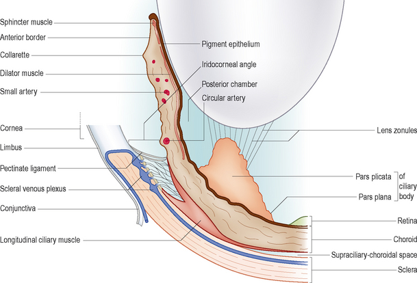
The iris is the visible coloured layer seen within the eye. It is made up of an anterior layer of stroma and iris sphincter muscle, and a posterior layer made up of an epithelial layer (which is pigmented) and dilator muscle (Figure 32.1). The colour of the iris depends on both the number of melanocytes present within the stroma and the thickness of the anterior layer itself. In addition to muscle fibres and melanocytes, the stroma contains numerous blood vessels, nerve endings, fibroblasts and collagen fibres. The main blood vessels entering the iris are the long ciliary arteries (temporal and nasal) which together form the major arterial circle visible in most animals as an undulating ring within the anterior iris stroma. The pupil is the central hole in the iris. Normally the iris rests lightly against the anterior surface of the lens. Movement of the iris muscles controls the quantity of light entering the posterior segment through the pupil. The sphincter muscle circles the iris in the dog while in the cat the fibres also criss-cross above and below the pupil allowing it to constrict to a narrow vertical slit. Other species such as horses, sheep and reptiles have different arrangements of this muscle which contribute to pupil shape. Constriction of the pupil is mediated via parasympathetic nerve fibres travelling in the oculomotor nerve (cranial nerve III). The dilator muscle is innervated by sympathetic fibres (which run with the ophthalmic branch of the trigeminal nerve (cranial nerve V)). The dilator muscle radiates out from the pupil margin towards the iris periphery in a similar pattern to bicycle spokes. In addition to regulating the amount of light entering through the pupil, altering its size also affects depth of focus and reduces optical aberrations.
Stay updated, free articles. Join our Telegram channel

Full access? Get Clinical Tree


