Proliferative (eosinophilic) keratoconjunctivitis

27 Proliferative (eosinophilic) keratoconjunctivitis


CLINICAL EXAMINATION


General clinical examination is usually normal. On ophthalmic examination menace responses are unaffected and pupillary light reflexes normal. Schirmer tear test readings can be normal, elevated or slightly reduced. Abnormalities are confined to the cornea and conjunctiva with intraocular contents being normal.


A pale pink mass, similar to granulation tissue, is seen extending across the cornea from the limbus. This is usually either at the temporolateral or ventronasal quadrant (Figure 27.1). The conjunctiva adjacent to the lesion is hyperaemic but no chemosis is usually present. A collection of white coloured tissue – almost globules or blobs – can be present on the surface of the pink mass, or adjacent to it. Some whitish discharge can accompany the signs. This looks similar to cottage cheese in consistency. There is normally corneal vascularization. Sometimes the nictitans membrane can be affected – particularly the bulbar side – so this should be examined. Ulceration is not usually a feature of the condition but care must be taken in interpreting fluorescein tests – the excess dye must be thoroughly flushed with sterile saline solution. Uptake of the green dye is quite possible on the pink fleshy tissue itself, often in tiny pinpoint patches, but not actually on the cornea – the epithelium usually remains intact.


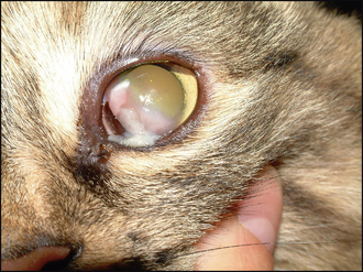
If the condition is quite advanced, the pink tissue can extend all the way across the cornea and in these cases vision will be affected. The palpebral surface of the eyelids can be involved. Although normally initially unilateral, both eyes can be affected (especially in advanced or untreated cases) but usually to different degrees – the owner might have only been aware of one eye being abnormal but on careful ophthalmic examination it is discovered that the other eye has more subtle lesions (Figure 27.2).


CASE WORK-UP


A full ophthalmic examination is obviously required to rule out other causes for the corneal infiltration – an infected ulcer or corneal foreign body for example. The white ‘cottage cheese’ discharge adherent to the granulation-type lesion is often considered pathognomonic but nonetheless further diagnostic tests are recommended to reach a definitive diagnosis. Cytology is the most appropriate test.


Under topical anaesthesia samples are collected from the lesion. This can be done either using a Kimura spatula (or more commonly the blunt end of a scalpel blade) or with a cytobrush. The collected material is gently pressed or rolled onto a microscope slide and allowed to air dry. Staining with Diff-Quik will allow rapid in-house examination of the sample although professional interpretation should also be considered. It is usual to see a collection of epithelial cells, mast cells, neutrophils, lymphocytes and, of course, eosinophils. However, the last are not necessarily abundant, and for this reason the condition can be called proliferative keratoconjunctivitis instead of eosinophilic! If eosinophils are present, it is usual to also find eosinophilic granules within the debris of the cytology samples as well as disrupted cells. Bacterial components are not normally a feature. However, if they are noted, a swab for bacterial culture and sensitivity should be taken.


In addition to cytology, a full haematology screen can be considered – a few cases do show a circulating eosinophilia.


Swabs can also be submitted for feline herpes virus (FHV-1) polymerase chain reaction (PCR). This is particularly important in cases where a history of upper respiratory infection or previous corneal disease (including ulceration) is present. Since the treatment of eosinophilic keratoconjunctivitis usually requires topical steroids, which can allow FHV-1 recrudescence, identification of this pathogen is particularly important.


Stay updated, free articles. Join our Telegram channel

Sep 10, 2016 | Posted by in SMALL ANIMAL | Comments Off on Proliferative (eosinophilic) keratoconjunctivitis

Full access? Get Clinical Tree

Get Clinical Tree app for offline access