38 Leishmaniasis
CLINICAL EXAMINATION
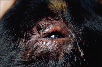
Ophthalmic examination can be similarly variable. In some cases the eyes will be normal on initial examination (but ocular changes can develop later in the course of the disease, even after treatment has been instigated). Some patients will have thickened, inflamed eyelids – in either a diffuse or a nodular pattern (Figure 38.1). Some will have marked chemosis and conjunctivitis, with or without corneal involvement. Nodular swellings at the limbus might be noted. Corneal ulceration and vascularization are occasionally present. Anterior uveitis is common, with miosis, aqueous flare and iridal swelling (Figure 38.2). Such patients will be in pain and can be blind. If the posterior segment is visible then signs of chorioretinitis might be present, but the anterior segment is more frequently the area of the uveal tract which is most inflamed. Occasionally orbital cellulitis will be present and the patient will have a painful exophthalmos. Schirmer tear test readings should be performed – some dogs will have keratoconjunctivitis sicca. Clearly fluorescein testing is mandatory. If tonometry is available, intraocular pressures should be assessed – secondary glaucoma can be a feature. Most cases are bilateral but not necessarily symmetrical.
Stay updated, free articles. Join our Telegram channel

Full access? Get Clinical Tree


