12 Medial canthal pocket syndrome INITIAL PRESENTATION Discharge PRESENTING SIGNS Medial canthal pocket syndrome (MCPS) is a very common, but rarely diagnosed, conjunctival condition. Typically it presents as a chronic or recurrent conjunctivitis, manifested by a thick mucoid or mucopurulent discharge and moderate conjunctival hyperaemia. Owners complain that the discharge is most noticeable in the morning but can recur later in the day, especially after sleeping, and that it is thick and tenacious. The dog rarely shows any signs of discomfort, despite the ‘red eye’, although occasionally can rub the eyes on waking – presumably to dislodge the discharge. The most frequently presented breeds include the Dobermann, standard poodle, Afghan hound, rough collie and lurchers. Other breeds with broad heads and deep set eyes such as the Weimaraner, golden and Labrador retriever, great Dane and Rottweiler can be affected. CASE HISTORY Typically the patient will be a young (6–18 months old), clinically well dog of dolichocephalic conformation. A chronic mucoid or mucopurulent discharge will be reported, centred medially, which does not seem to bother the dog unless it becomes copious. The patient normally improves on topical antibiotic medication, although the discharge is quick to reform as soon as the course of treatment is finished. Often the patient undergoes several treatment periods before careful examination leads to the diagnosis. CLINICAL EXAMINATION General clinical examination is normal. On ophthalmic examination all neurological tests are normal and the eyes are open and comfortable. Schirmer tear test readings are normal. The most obvious feature is a discharge at the medial canthus and some localized conjunctival hyperaemia (Figure 12.1). The bulbar conjunctiva is normally only very slightly hyperaemic whereas the ventral palpebral and nictitans conjunctiva can appear very inflamed. The discharge is bilateral but not necessarily totally symmetrical. Some discharge is seen within the ventral fornix, in a pocket formed by the gap between the eyeball and the lids (due to the relative enophthalmos inherent in the breed conformation). Follicles can be noted within the conjunctiva close to the discharge on cleaning it away. No corneal pathology is normally encountered but a mild ventral entropion is sometimes seen. Intraocular examination is unremarkable. 
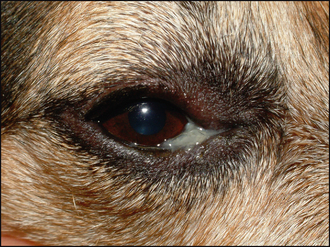
Figure 12.1 Typical grey–white mucoid accumulation at the medial canthus in a collie/German shepherd cross (XGSD). DIFFERENTIAL DIAGNOSES • Conformational – medial canthal pocket syndrome • Primary bacterial conjunctivitis • Entropion • Ectropion • Dacryocystitis • Keratoconjunctivitis sicca • Conjunctival foreign body • Allergic conjunctivitis CASE WORK-UP A thorough ophthalmic examination is essential, and normally this will lead to the diagnosis of MCPS. Careful evaluation of the globe – lid positioning, together with an appreciation of the exact location of the discharge – will help. If the discharge appears purely mucoid in nature then sample collection is not warranted; however, if any purulent component is noted then a swab should be sent for culture and sensitivity testing. Cytology samples are not usually required. It is sensible to flush the nasolacrimal ducts – normally this can be done under topical anaesthesia or light sedation – since a dacryocystitis will be one main differential (although the discharge is likely to be purulent, some discomfort is likely and the distribution is more commonly unilateral than bilateral in the case of dacryocystitis). NURSING ASPECTS Regular bathing and cleaning away of the discharge is the mainstay for this condition. Thus owners should be shown how to do this and demonstrate their proficiency to enable good management of these affected patients. EPIDEMIOLOGY Breeds affected with medial canthal pocket syndrome include: • Afghan hound Only gold members can continue reading. Log In or Register to continue You may also needComplicated entropionChronic superficial keratitisVitiligo in two dogsScratchingGeneralized progressive retinal atrophyHydrocephalusDeep corneal ulcerPrimary canine glaucoma Share this:Click to share on Twitter (Opens in new window)Click to share on Facebook (Opens in new window)Click to share on Google+ (Opens in new window) Related Tags: Saunders Solutions in Veterinary Practice Small Animal Ophthalmo Sep 10, 2016 | Posted by admin in SMALL ANIMAL | Comments Off on Medial canthal pocket syndrome