Chapter 6
Update on Fungal Infections in Reptiles
Fungi are ubiquitous on the skin of reptiles1; thus the mere isolation of a fungus from a lesion is clinically irrelevant if unsupported by compatible histopathologic findings. Morphologic characteristics of fungal elements in tissue sections are critical in assigning causality to isolates or dismissing them as contaminants. The literature is replete with cases of reptile mycoses in which various fungi were incriminated but for which causality was never established or even substantiated. Fungal identification was subjectively ventured based on hyphal or yeast morphologic features in histologic sections when material was unavailable for culture. Only when we possess a sound understanding of which fungi have in the past posed a threat to reptiles can we assign the “emerging” epithet to a fungal agent. For this to happen, we need to strictly consider cases in which identification and causality of an isolate were firmly or reasonably ascertained. When the existing literature pertaining to fungal disease in reptiles is objectively reviewed, only a few fungi have emerged as genuine repeat offenders. Common environmental organisms such as Purpureocillium lilacinum (formerly Paecilomyces lilacinus), Fusarium solani and other fusaria, Metarhizium anisopliae, Beauveria bassiana, various aspergilla, and yeast such as Candida or Cryptococcus can and will cause disease in reptiles under the right circumstances.2,3 Pigmented fungi very rarely cause disease in squamates but, in contrast, are well-represented among agents of fungal disease in chelonians, especially tortoises and box turtles (Terrapene spp.). Infection with these opportunistic fungi is usually a result of overwhelming exposure, immune compromise, and stress from inadequate or substandard captive conditions, or any combination thereof. In wild chelonians and crocodilians, stressful climatic events such as cold spells or cold-stunning in sea turtles often precede systemic or pulmonary mycoses with otherwise fairly innocuous fungi. Similarly, in captive specimens, thermal extremes from mechanical breakdowns or other causes of environmental failure can subsequently lead to mycosis. Antifungal prophylaxis is routine in sea turtles recovering from cold spells, and it may be wise to do the same for other captive reptiles that have experienced accidental cooling events.
Of course, fungi other than those already listed can infect reptiles, and none among those is more of a threat than the fungus currently referred to as the “Chrysosporium anamorph of Nannizziopsis vriesii” (CANV), which we now know is an established cause of contagious, deep, and often fatal dermatomycosis in a variety of lizards and snakes and in crocodiles.2,3 The CANV might not be a truly emerging reptile pathogen but rather one that has gone undiagnosed, misdiagnosed, or unrecognized for decades. Nevertheless, this fungus is the single most important fungal pathogen of reptiles, and research has demonstrated it may act as a primary pathogen.4 More than 30 CANV isolates collected from various sources over the last 25 years have been catalogued, and all but one were retrieved from reptile lesions. Most of these were initially misidentified, often as Trichophyton, Geotrichum, Malbranchea, or as a distinct Chrysosporium species. Repeated attempts at mating isolates to yield a teleomorph have been unsuccessful, and although all reptile CANV isolates are morphologically indistinguishable from the true anamorphic stage of Nannizziopsis vriesii, they do differ slightly when examined at the molecular level (Hambleton S, Sigler L, and Paré JA: unpublished data). They are now known to represent an assembly of closely related fungi, many of which remain to be named (Hambleton S, Sigler L and Paré JA: unpublished data). Chrysosporium ophiodiicola and Chrysosporium guarroi, recently described agents of dermatomycosis in a Black Ratsnake (Elaphe obsoleta)5 and a Green Iguana (Iguana iguana),6 respectively, are renamed fungi that would have previously fallen under the CANV group umbrella. CANV infection is contagious and typically occurs in captive reptiles as outbreaks of severe, progressive necrogranulomatous dermatitis that eventually disseminates. This fungus consistently expands its list of susceptible hosts. Recent additions include Broad-headed Snakes (Hoplocephalus bungaroides),7 Brown Anoles (Anolis sagrei),8 coastal Bearded Dragons (Pogona barbata),9 Leopard Geckos (Eublepharius macularius),10 and even Tuataras (Sphenodon punctatus).11 Of substantial concern is the recent identification of the CANV as a cause of severe disease in wild snakes, which carries significant conservation implications. The CANV, more precisely C. ophiodiicola, was strongly incriminated as the cause of disfiguring facial lesions in free-ranging Eastern Massasauga Rattlesnakes (Sistrurus catenatus catenatus)12,13 in Illinois. Four snakes collected from the wild in southern Illinois over a period of 3 years as part of a health survey were compromised by deep, severe, very aggressive granulomatous infection resulting in effacement of normal facial and cranial anatomic structures.12 The head of affected snakes becomes distorted from the infection, and the disease was dubbed facial disfiguration syndrome (FDS). The clinicopathologic picture was peculiarly yet strikingly consistent among affected snakes but was also identical to that of a Western Massasauga Rattlesnake (Sistrurus catenatus tergeminus) with extensive and invasive granulomatous fungal infection reported in 1979.14 In the latter case, the description of the hyphae in tissue sections was not consistent with the presumptive diagnosis of phycomycosis, an obsolete term for zygomycosis, and this snake may well have been a misidentified case of C. ophiodiicola facial mycosis or FDS. Extensive dermatomycosis, very suggestive of FDS, has recently been observed in wild Timber Rattlesnakes (Crotalus horridus) in New England13,15 (Figure 6-1). Necrogranulomatous lesions may not be limited to the face and may extend to the rest of the body. Snakes are typically found with lesions as they emerge from hibernacula in the spring, but some sick snakes are also seen basking outside dens in winter months (Condon J: Pers. com.,) (Figure 6-2). Infection progressively hinders feeding, and snakes become emaciated before they succumb. This disease might have substantially impacted northeastern Timber Rattlesnake populations. Investigations as to the causative agent are being conducted, but available data are also pointing to C. ophiodiicola. A wild Black Ratsnake and one of several Copperheads (Agkistrodon contortrix) with severe and ultimately fatal granulomatous skin disease in New Jersey were confirmed with C. ophiodiicola (Schantz K: unpublished data,) in the last 2 years. This spreading epidemic of fungal disease chiefly affects wild crotalid snakes but might have spilled over to sympatric Ring-necked Snakes (Diadophis punctatus), Black Racers (Coluber constrictor), Gartersnakes (Thamnophis sp.), and Watersnakes (Nerodia sp.) (Figure 6-3), all of which were seen with facial lesions consistent with CANV dermatitis. C. ophiodiicola is known from Europe and Australia and probably occurs worldwide (Sigler L and Paré JA: unpublished data), yet much remains to be known about this fungus and its impact on free-ranging animals. The ecologic niche of C. ophiodiicola and other CANV isolates remains unclear, but it has been implicated in the past as causing disease mostly in recently caught or newly imported terrestrial and aquatic reptiles. Treatment may be protracted, as lesions are often advanced by the time sick snakes are first observed. C. ophiodiicola does not grow or grows very poorly at 35°C, and this limited thermotolerance may partially account for some affected snakes with less severe lesions healing after emergence from the hibernaculum. Simple exposure to sunlight and warmer days would impede fungal growth and therefore disease progression, allowing animals to progressively improve with each shedding thereafter (Figure 6-4). In cases in which lesions are too extensive or deep, surgical debridement with topical care and long-term antifungal therapy are needed. Adjunct fluid therapy, as well as nutritional and thermal support, is usually initially indicated to stabilize the patient beforehand or concurrently with systemic medications and assist in recovery. Voriconazole, terbinafine, and itraconazole are all valid drug options, on the basis of prior CANV in vitro susceptibility studies.16,17 Secondary bacterial infection may also need to be addressed.
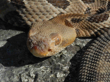
FIGURE 6-1 Wild juvenile Timber Rattlesnake (Crotalus horridus) with early Chrysosporium ophiodiicola infection, confirmed at the National Wildlife Health Center, Madison, Wisc. The rostrum is swollen, especially on the left side. Nasal, internasal, and loreal scutes are hyperkeratotic and necrotic, and the nares are effaced. (Photo courtesy of J. Condon.)
Stay updated, free articles. Join our Telegram channel

Full access? Get Clinical Tree


