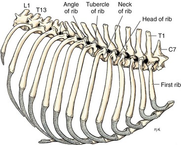
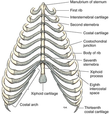
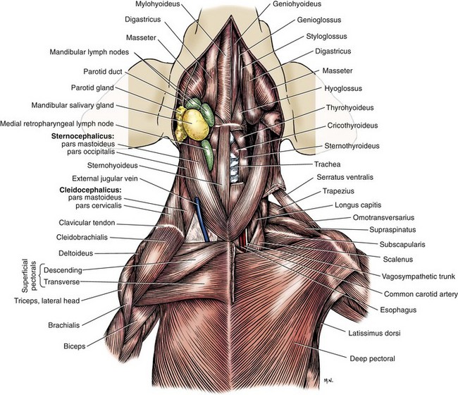
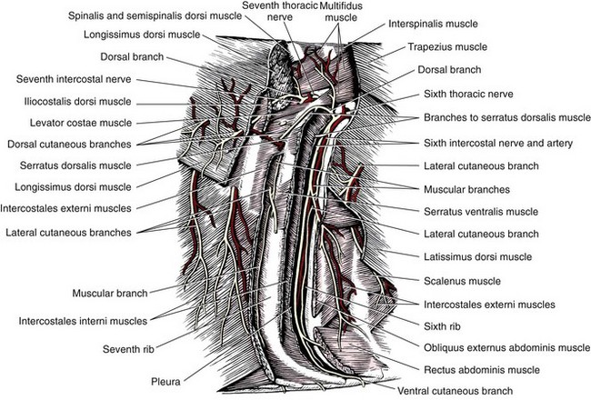
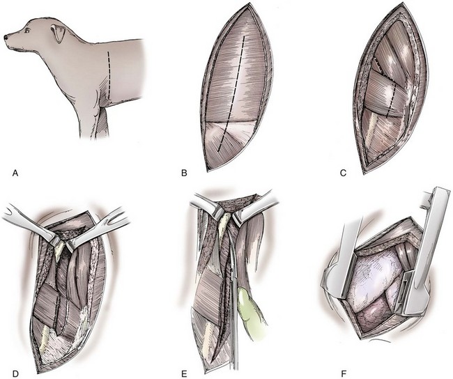
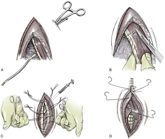
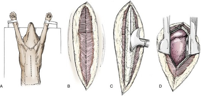
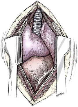
Thoracic Wall

Chapter 104


Thoracic Wall


Access to the thoracic cavity is required for treatment of a variety of conditions in dogs and cats and ranges from simple tube thoracostomy to complex approaches involving a combination of sternotomy, thoracotomy, rib pivot, and celiotomy. In the majority of cases, however, the thorax is approached in one of three ways: intercostal thoracotomy (or thoracostomy), median sternotomy, or transdiaphragmatic incision. Because the thoracic cavity is deep and narrow on a transverse plane in cats and many breeds of dog, intercostal thoracotomy provides good access for the majority of clinical procedures. Sternotomy, however, is required for major surgery of the cranial mediastinum and for access to both sides of the thorax.


Anatomy


The complex musculoskeletal structure of the thorax facilitates complete resection of tumors with adequate margins. Natural tissue planes allow separation of muscle bellies to reduce postoperative morbidity, and anatomic landmarks allow intraoperative delivery of local anesthesia.12


Boundaries of the Thoracic Cavity


The cranial extent of the thoracic cavity is the first rib. The hiatus between the ribs, sternum, and the spinal column is known as the thoracic inlet, through which travel the esophagus; trachea; great arteries and veins; and nerves such as the phrenic, vagus, and recurrent laryngeal. The right cranial lung lobes project slightly more cranially than the left. It is possible for the right lung lobes to project into the caudal neck at certain phases of ventilation, particularly in brachycephalic patients with chronic upper airway obstruction.


The caudal extent of the thoracic cavity is marked by the diaphragm, consisting of two muscular crura that arise by tendons from the fourth lumbar vertebra and have muscular attachments to the lumbar vertebrae, ribs, and sternum. The diaphragmatic crurae are joined by a central tendinous portion. Nerves, vessels, and the esophagus enter or leave the thorax through the esophageal and aortic hiatii and the caval foramen.12


Skeleton


Dogs and cats have 13 thoracic vertebrae, 13 ribs, and 9 sternebrae (Figures 104-1 and 104-2). Ribs one to nine articulate with the sternebrae via cartilaginous extensions from the costochondral junctions. The sternebrae are connected to one another by fibrocartilage. The cranialmost sternebra, the manubrium, provides the point of attachment for the sternocephalicus muscle. The caudalmost sternebra, the xiphoid, is positioned dorsal to the linea alba and ventral to the falciform ligament. The xiphoid does not have any direct muscular attachments other than the diaphragm; the fascial connection between the paired rectus abdominus muscles (linea alba) merges with the fascia of the deep pectoral muscle ventral to the xiphoid.12


Muscular Anatomy


Muscle groups (Figures 104-3 and 104-4) associated with the thorax include the intrinsic and extrinsic muscles of respiration, muscles of the abdominal wall, and locomotor musculature.12 The locomotor muscles attach the forelimb to the trunk. Most significant for thoracic surgeons are the latissimus dorsi, the serratus ventralis thoracis group, and the superficial and deep pectoral muscles. The latissimus dorsi muscle originates from the lumbodorsal fascia and thoracolumbar vertebrae and converges cranioventrally to insert on the proximal humerus. It draws the scapula and hence the forelimb caudally. The latissimus dorsi muscle may either be divided or elevated to gain access to the intercostal muscles for intercostal thoracotomy. Limb function does not seem to be unduly impaired if this muscle is separated or relocated. Elevation of the muscle has been suggested to cause less postoperative pain but can also reduce surgical access or necessitate more active retraction to maintain surgical exposure. The thoracolumbar attachments of the latissimus dorsi muscle can be incised and the muscle rotated ventrally to close large defects in the thoracic wall.


The complex serratus ventralis muscle possesses a number of bellies. Its thoracic segments arise from the caudal edge of the first seven or eight ribs and insert onto the medial serrate surface of the scapula. The serratus ventralis may either be sectioned or the bellies divided, with the belly traversing the proposed intercostal incision site elevated from the appropriate rib(s). Separating, rather than incising, the muscle bellies has no impact on exposure and facilitates creation of a leakproof seal during closure.


The scalenus muscle traverses the ventral thoracic wall from the caudal neck to the midthorax. An obvious division between its muscular and tendinous portions is visible at the fifth rib, making it a useful landmark.


The pectoral muscles travel bilaterally between the sternum and the medial aspect of the humerus. The dorsal edge of the deep pectoral muscle is often incised when a lateral intercostal incision is performed. The left and right pectoral muscles must be separated when performing a median sternotomy. Branches of the internal thoracic artery and vein perforate between the right and left deep pectoral muscles in association with each sternebra and should be avoided or ligated as the sternum is exposed for sternotomy.


The external and internal intercostal muscles connect the caudal edge of each rib to the cranial edge of the rib behind it. They are incised midway between the ribs to avoid damage to the intercostal artery and to ensure sufficient tissue remains if separate closure of the intercostal muscles is required.


The transverse thoracic muscle arises on the pleural surface of the thoracic wall and runs laterally from the sternum to blend with the endothoracic fascia at the level of the costochondral junctions. The transverse thoracic muscle is of little clinical significance except as a landmark for the internal thoracic artery that travels dorsal to it. Division of the transverse thoracic muscle, when required, should be undertaken carefully to avoid inadvertent sectioning of this large artery.


Nerves and Blood Vessels


Intercostal nerves (Figure 104-5) arise as ventral branches of the thoracic spinal nerves and pass ventrally along the caudal edge of each rib in association with the intercostal arteries and veins. There are 12 intercostal arteries on each side of the thorax. The first three or four are branches of the thoracic vertebral artery, and the remainder are branches of the aorta. Dorsal branches of the intercostal arteries descend in the intercostal spaces along the caudal borders of ribs 1 through 12. They give off branches dorsally that penetrate the epaxial muscles and ventrally that perforate the intercostal and adjacent muscles to supply cutaneous structures, including the thoracic mammary glands. Ventrally, the intercostal branches anastomose with ventral intercostal branches of the internal thoracic artery. The intercostal arteries can be avoided by incising the intercostal muscles midway between ribs in the dorsal two thirds of the intercostal space; however, small anastomotic branches are often cut ventrally during intercostal thoracotomy.


The internal thoracic arteries arise from the left and right subclavian arteries, respectively, and travel on either side of the cranial mediastinum until they travel dorsad to the transverse thoracic muscles. They provide branches to the paired sternal lymph nodes. The left and right external jugular veins and brachial veins join to form paired brachiocephalic trunks that fuse in the cranial mediastinum to form the cranial vena cava. The brachiocephalic vessels and cranial vena cava are separated from the cranial sternum by flimsy connective tissue only; care should be taken not to damage them inadvertently during median sternotomy because they collapse after retraction of the sternotomy and therefore may be confused with a plane of connective tissue.


Orientation of the heart and other viscera within the thorax is important for determining the most appropriate surgical approach for various conditions (see Table 105-1). Left intercostal thoracotomy is always used for approaches to the left side of the heart (Figure 104-6). The right atrium, venae cavae, and azygous vein are most accessible via a right lateral thoracotomy, although the cavae can also be identified and tourniquets placed from a ventral and left approach. Median sternotomy provides good visualization of the cranial vena cava from its origin as the brachiocephalic veins to where it enters the right atrium. The right ventricle arcs around the cranioventral border of the heart, and the right ventricular outflow tract and pulmonary artery are best accessed from a left intercostal or a ventral approach. The anterior free wall of the left ventricle is accessible through a left lateral thoracotomy, but the left ventricular apex is most accessible via a transdiaphragmatic approach, which is thus the best approach for insertion of epicardial pacemaker leads.


In animals with normal embryologic development, the aortic arch arises from the fibrous skeleton of the heart and travels craniodorsally on the left side of the trachea and esophagus. Surgery of the trachea and esophagus is most appropriately undertaken from a right intercostal approach. The reverse is true for animals with persistent right aortic arch because the esophagus and ligamentum arteriosum are best visualized via a left intercostal approach.


Physiology and Pathophysiology


A number of features of thoracic physiology are important in regards to diseases and surgery of the thoracic wall. These include the integrity of the pleural space, the ability to maintain negative intrathoracic pressure, the stability of the thoracic wall, the ability of the thorax to expand and contract in response to muscle action and lung volume, and the mechanisms involved in thoracic and pleural pain and their impact on efficiency of ventilation.


The thoracic wall is made of soft tissue and bony structures. The fibroelastic components of the soft tissue are passive, and the muscular elements are active. These passive and active elements work on the rigid bony ribs to produce a “bucket handle” motion, allowing expansion and contraction of the thoracic cavity. The passive elements of the thoracic wall produce either an inward or an outward elastic recoil, depending on the thoracic volume. When the passive elastic structures of the thoracic wall are relaxed (i.e., neither stretched outward nor inward), the volume of the thorax is known as V0. When the thoracic volume is less than V0, a net outward passive recoil of the thoracic wall is created, thus tending to expand the thoracic wall. When volumes are greater than V0, a net inward passive recoil of the thoracic wall is created, thus tending to collapse the thoracic wall.


Because the thoracic wall and lungs are functionally linked by negative pleural pressures, total pulmonary compliance is a function of the additive compliance of the thoracic wall and lungs. Abnormalities in total pulmonary compliance may result from changes in either lung or thoracic wall compliance.33 Alterations in thoracic volume after procedures such as thoracic wall resection or advancement of the diaphragm or spontaneous diseases such as pectus excavatum or large thoracic wall tumors have an impact on ventilation and tidal volume. Likewise, changes in lung volume after lung lobe resection or secondary to pathologic conditions such as restrictive pleuritis, chronic emphysema, or pleural effusion have an impact on thoracic volume, limited by the compliance of the thoracic wall.


Surgical Approaches to the Thorax


Intercostal Thoracotomy


The patient is positioned in right or left lateral recumbency, and the lateral thorax is clipped and prepped from dorsal to ventral midline. The elbow fold and caudal aspect of the upper foreleg is also clipped and prepped. The leg is secured in an extended position to avoid limb movement with spasms of extrinsic musculature. Positioning a rolled-up drape or sandbag beneath the thorax, just caudal to the scapula, spreads the upper ribs apart and elevates the heart, bringing the intrathoracic viscera closer to the surgical wound.


For fourth and fifth intercostal thoracotomy, the skin is incised 2 cm caudal to the scapula from the caudodorsal angle of the scapula to just below the costochondral junction (Figure 104-7). Subcutaneous tissue is incised, and the latissimus dorsi muscle is divided with scissors or electrocautery. Alternatively, the latissimus dorsi may be elevated and displaced by incising its ventral fascial attachments and retracting it dorsally. Neuromuscular paralysis assists by reducing muscle spasm during incision and allowing the ribs to be retracted farther.


After the latissimus dorsi muscle has been incised, the surgeon can count the ribs by inserting a finger deep to the muscle and sliding it cranially until the first rib is located. Ribs are counted caudally to identify the correct intercostal space. The proposed site of intercostal incision can be confirmed by visualizing the caudal extent of the muscular portion of the scalenus muscle (usually to the fifth rib).


The serratus ventralis muscle is elevated from the rib caudal to the proposed intercostal incision and retracted cranially. Muscle bellies associated with each rib are separated with sharp dissection or cautery, taking note that a branch of the intercostal artery supplying each belly of serratus ventralis and hemorrhage will have to be controlled. External and internal intercostal muscles are incised while an assistant retracts the bellies of serratus ventralis. If possible, the pleura is left intact during intercostal incision; visualization of the thoracic cavity and lungs through the intact pleura may help avoid damage if there are adhesions between the lungs and pleura. The translucent pleura is punctured carefully with a hemostat or dissecting forceps to avoid damage to the underlying lung. The intercostal incision is extended dorsally to the point where the ribs angle medially and ventrally to a point just below the costochondral junction.


Continuation of the incision dorsal to these landmarks results in damage to epaxial musculature and potential damage to intercostal arteries. Ventral continuation of the incision should be done very cautiously and only after confirming the location of the internal thoracic artery by palpation. The internal thoracic artery will be avoided if the incision does not extend beyond the lateral aspect of the transverse thoracic muscle.


Finochietto retractors are inserted, and the ribs retracted until connective tissue at the dorsal end of the incision becomes tight. Limiting retraction to this point will reduce the risk of rib fracture. Rib retraction provides better access to the thoracic cavity craniad to the incision than caudad; hence, the site chosen for incision should be located caudally, rather than cranially, if the surgeon is unsure of the exact interspace to enter.


After the intrathoracic procedure has been completed, the pleural cavity is lavaged and inspected for ongoing hemorrhage or air leakage. The sandbag or drape is removed from beneath the patient’s chest to facilitate rib apposition. Three to four cruciate sutures, or four to six encircling sutures, of 2-0 to 1 polydioxanone suture are placed around the ribs cranial and caudal to the thoracotomy (Figure 104-8). The assistant places traction of one or more of the sutures while the remaining sutures are tied. The aim of the pericostal sutures is to reduce tension on the soft tissue repair rather than immobilize the two ribs completely. The sutures should therefore be tied securely but not overly tightly. Ribs may be fractured during suture tightening or after surgery if pericostal sutures are too tight. By its nature, placement of circumcostal sutures will entrap intercostal nerves. Transcostal sutures have been used in a small case series; results suggest that animals experience less postoperative pain compared with conventional techniques.41


In larger patients, intercostal muscles may be repaired with a continuous suture pattern of 2-0 or 3-0 polydioxanone. This is rarely necessary in small dogs and cats. Bellies of the serratus ventralis muscle bellies are reapposed in a continuous suture layer that may be extended ventrally to include the scalenus muscle and pectoral muscle. If transacted, the latissimus dorsi muscle is sutured separately in a continuous pattern. The subcutis and skin are closed routinely.


Rib Resection Thoracotomy


Rib resection is usually only performed when wide access to the thoracic cavity (particularly cranially) is required, as part of en bloc excision of a thoracic wall tumor, or for removal of large masses that do not fit through a single intercostal space. In these latter cases, median sternotomy should be considered as an alternative. The rib to be removed is isolated from soft tissue attachments, and its intercostal artery is ligated at the dorsal and ventralmost extent of the proposed resection. The rib is sectioned with bone cutters and removed. Closure may be complicated if intercostal musculature is also excised, in which case establishment of a leakproof seal may be dependent on closure of more superficial layers of muscle and subcutis. Pericostal sutures may be passed around the rib cranial and caudal to the defect; care should be taken not to tie them too tightly so as to avoid undue stress on the ribs. The remainder of the incision is closed routinely.


Median Sternotomy


Median sternotomy is the approach of choice for bilateral exploration of the thoracic cavity, wide exposure of cranial mediastinal masses (especially those with involvement of the cranial vena cava), access to the right ventricle, and in patients that may require cranial abdominal exploration. Access to the dorsal mediastinum is limited with this approach; however, lung lobectomy may be performed through median sternotomy with minimal difficulty as long as adhesions to the dorsal mediastinum are not present. Sternotomy may be used for surgery of the cranial intrathoracic trachea; the large vessels must be retracted for tracheal access through this approach. Median sternotomy is not ideal for thoracic duct ligation, exploration of the tracheal bifurcation and tracheobronchial lymph nodes, or surgery of the esophagus or caudal vena cava (unless in association with celiotomy and diaphragmatic incision).


Postoperative morbidity of this approach is reduced by ensuring that the sternebrae are sectioned longitudinally without being broken. This permits stable closure and uncomplicated healing in most cases. Instability of the sternotomy causes severe postoperative pain and prolonged recovery and is probably why some surgeons avoid this approach. Sternotomy may be partial or complete, depending on the purpose of the surgery. Leaving either the manubrium or xiphoid sternum intact facilitates stable closure of the incision but substantially restricts access to the cranial or caudal thorax, respectively. Nevertheless, partial sternotomy should be performed initially; sternotomy is completed if greater access is required. Each sternebra should be sectioned completely along its length into the cartilaginous connection with the next sternebra to maximize retraction and avoid fracture.


The patient is positioned in dorsal recumbency. The front legs may be tied caudally along the sides of the thoracic wall to facilitate dissection of the cranial portion of the sternum and caudal cervical region. This results in more traction on the caudal cervical and pectoral muscles; leg ties should therefore be loosened as the wound is being closed.


A ventral midline skin incision is made, and the subcutaneous tissue is incised. The pectoral muscles are separated down the midline (Figure 104-9). Careful electrocautery reduces the amount of bleeding encountered from perforating branches of the internal thoracic artery and vein associated with each sternebra. Hemorrhage from the veins, particularly, can be difficult to control. Air embolism is common during sternotomy, and small air bubbles are often seen within the internal thoracic veins after the thorax is opened. The pectoral muscles are separated from their attachments to the ventral surface of the sternum; extending the dissection laterally can result in hemorrhage and is best avoided. After the midline and a small portion of each sternebra to either side have been exposed, a combination of palpation and visualization is used to determine the appropriate line for the sternotomy. Deviation from the midline should be scrupulously avoided. Scoring the sternebrae with a scalpel or electrocautery before osteotomy may be helpful to reduce straying to one side or the other.


The sternebrae are sectioned using a reciprocating saw, osteotome, special sternal saw, sternal splitter, or bone cutters. In smaller patients, the reciprocating saw causes less compression of their compliant chest and thus permits a more accurate incision to be made. The sternotomy is commenced at the cranial or caudal end, depending on the objective for the surgery. After the first one or two sternebrae have been sectioned, a Gelpi retractor is inserted into sternal cartilage between the split edges, and the sternum is retracted. This stabilizes the sternum and produces some tension to facilitate extended incision along the midline. It also allows the connective tissue of the ventral thorax to be perforated, thus establishing a pneumothorax to ensure that the heart, lungs, or great blood vessels are not damaged during the incision. The incision may then be easily continued into the caudal neck (Figure 104-10) or cranial abdomen (Figure 104-11) if necessary. Alternatively, partial-thickness osteotomies are performed. Osteotomy is completed with a mallet and osteotome that is positioned parallel to the longitudinal axis of the sternum.


< div class='tao-gold-member'>

Stay updated, free articles. Join our Telegram channel

Jul 18, 2016 | Posted by in PHARMACOLOGY, TOXICOLOGY & THERAPEUTICS | Comments Off on Thoracic Wall

Full access? Get Clinical Tree

Get Clinical Tree app for offline access