Ruth-Anne Richter
Therapeutic Shoeing for Tendon and Ligament Injury
Substantial advances have been made in the treatment of horses with tendon or ligament injury during the past several years, not the least of which has been the increasing use of regenerative therapies. Similarly, the understanding of limb biomechanics and farriery has also advanced, which has improved treatment success rates and returned horses to athletic performance with fewer recurrences after soft tissue injury.
The goal of treatment for soft tissue injuries in the horse should be a return to previous level of performance. Although this may not be possible in all cases, it often can be achieved with a team approach and with due diligence applied for achieving a positive result. To accomplish the ultimate return of the horse to its former level of athletic ability, several goals must be achieved: (1) reduce acute pain and inflammation, (2) optimize repair of the injured tendon or ligament, (3) reduce the adverse biomechanical forces acting on the injured structure, and (4) use rehabilitation to return the horse to athletic performance.
A chief component of successful outcome is reduction of adverse biomechanical forces. This can be done through therapeutic shoeing, achieving appropriate foot balance, and altering the ground reaction forces to reduce stress on the injured structure. This necessitates not only an accurate diagnosis and knowledge of limb anatomy and biomechanics but also an understanding of the potential sequelae to use of specific shoe types. This chapter explores some of the basic principles in shoeing horses with common tendon and ligament injuries of the distal limb.
The therapeutic shoes discussed herein are not designed to be used all of the time, or for prevention of injury. When applied as an adjunct to treatment of a specific injury, these shoes increase biomechanical stresses on other supporting structures of the limb, which can lead to failure of those structures. The shoes are meant for use while an injury is healing and during a portion of the rehabilitation period, in general for less than 1 year. The ultimate goal is to return the horse to use of a plain shoe when the injury has completely healed. This requires follow-up appointments with the veterinarian and farrier so that the shoe can be adjusted as the injury responds to treatment. Often during this time the foot will change for the better. When the injury has healed, it is incumbent on both the farrier and veterinarian to ensure that the foot remains well balanced and does not fall back into the previous configuration that could again increase stress on the formerly injured structure.
Injuries of the Superficial Digital Flexor Tendon and Suspensory Ligament
Both the superficial digital flexor tendon (SDFT) and proximal check ligament contribute to limiting hyperextension of the fetlock and carpal joints during motion. During the stance phase of the stride, tension is applied to the SDFT during extension of the fetlock, and the tendon thus carries the load of the limb at that point in the stride. The load is shared with the suspensory ligament (SL) at the point of maximal hyperextension of the fetlock. For this reason, horses with injuries that affect the SDFT and SL can be shod in a similar manner. These structures are the main energy-storing structures during locomotion and are subjected to higher levels of strain than the deep digital flexor tendon (DDFT) and the common digital extensor tendon in the forelimb, which results in the SDFT and SL having a higher injury rate than the other structures.
Recent data reveal that heel elevation can be detrimental by increasing maximal strain on the SDFT and SL during extension of the fetlock. In horses at a walk, the various shoe types do not significantly affect strain on these structures, but at the trot, heel elevation increases strain on a normal SDFT and adds load to an injured SDFT. Conversely, elevation of the toe dynamically reduces the strain on these two structures and may thus be protective. Shoes that biomechanically reproduce this beneficial effect can be used during the treatment and rehabilitation phases in horses with desmitis of the body of the SL and tendinitis of the SDFT.
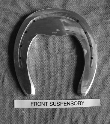
Shoes that have a wider toe (which effectively elevates the horse’s toe) and beveled branches and heels that sink slightly into the ground reduce the ground reaction force at the heel and increase it at the toe (Figure 189-1). This reduces the stresses on the injured SDFT or SL and thereby reduces extension of the fetlock joint while extending the distal interphalangeal joint.
Injuries of the Deep Digital Flexor Tendon and Its Accessory Ligament
Studies have confirmed that elevation of the heel reduces strain on the DDFT. This elevation induces extension of the fetlock joint and increases flexion of the interphalangeal joints. Although heel elevation reduces maximal strain on the DDFT at the walk and trot, toe elevation increases strain (as in horses with low heel- long toe conformation). At high loads, the DDFT limits hyperextension of the carpus and fetlock joints (counteracting the effect of the SDFT) and facilitates flexion of the proximal interphalangeal joint during weight bearing. Importantly, the DDFT stabilizes the distal interphalangeal joint by orienting P2 pressure dorsally on the articular surface of P3. At full weight bearing, the DDFT also closely contacts the distal border of the distal sesamoid (navicular) bone. Abnormal changes in the angle of insertion of the DDFT lead to uneven local stress distribution and contribute to failure of the DDFT insertion as well as the structures in the navicular region. By acting as a lever, the distal scutum facilitates foot rotation and heel takeoff at the end of the stance phase. Thus at the end of stance, muscle, DDFT, and the accessory ligaments of the DDFT induce fetlock elevation, and the DDFT causes distal interphalangeal joint extension. The maximally stretched accessory ligaments of the DDFT also contribute to distal interphalangeal joint stabilization, and limb hyperextension can therefore contribute to injury of these accessory ligaments. These structures, in combination with the branches of the SDFT, are also responsible for stability of the proximal interphalangeal joint during weight bearing.
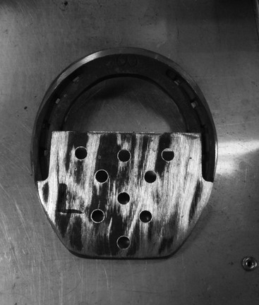
This biomechanical information can be used in the treatment of horses with injury of the DDFT, accessory ligaments of the DDFT, and associated structures. Horses with injury to the DDFT, its accessory ligaments, and the small ligaments associated with the navicular area, including the distal sesamoidean impar ligament, should be shod with functional heel elevation. Shoes with a wide heel plate create an effective “elevation” by the same principle as the wide toe does in the treatment of SDFT and SL injuries. The wide heel plate also distributes weight across a larger area of the palmar region of the foot, including the frog, rather than concentrating it centrally at the frog (as occurs with a heart bar shoe). However, straight or heart bar shoes are also useful for horses with injuries to the same structures. In the author’s experience, horses with insertional tendinopathy of the DDFT and desmitis of the distal sesamoidean impar ligament do not always tolerate a heart bar shoe and prefer the wide heel plate with dental impression material applied under the plate (Figures 189-2 and 189-3). With time, and as the lameness improves, the width of the heel plate is narrowed to the point where it becomes a straight bar shoe. From this point on, the horse can be transitioned back into a normal shoe.

Stay updated, free articles. Join our Telegram channel

Full access? Get Clinical Tree


