R. Reid Hanson, Valeria Albanese
Small Intestine Colic
Lesions causing colic in the small intestine are classified as strangulating or nonstrangulating. Strangulating lesions occlude the small intestinal lumen and its vascular supply, leading to tissue necrosis (e.g., strangulating lipoma), whereas nonstrangulating obstructions do not affect the vascular supply. Nonstrangulating lesions are further classified as obstructive or inflammatory in origin. Obstructive lesions lead to mechanical impairment of intestinal transit (e.g., ileal impactions), whereas inflammatory lesions, such as anterior enteritis, lead to ileus because of the negative effect of inflammatory mediators on the action of the enteric nervous system and intestinal musculature.
Horses with small intestinal disease show signs of moderate to severe pain. The heart rate is typically high, often 48 beats/minute or higher, depending on the degree of pain, dehydration, and endotoxemia. The respiratory rate can be normal to high. Gross distension of the abdomen may be observed, and gastrointestinal sounds can be decreased in frequency or normal. The mucous membranes vary in color, appearance, and refill time, depending on the type of lesion and the duration of disease. The volume of gastric reflux can vary; however, some volume of gastric fluid can usually be retrieved. If the lesion is located in the distal part of the small intestine and the duration of colic is short, gastric reflux may not be present.
Distended loops of small intestine are usually detected on transrectal examination. Large colon content is usually dry because of the absence of small intestinal outflow.
Transabdominal ultrasonography reveals small intestinal distension, even if it was not palpated transrectally. Ultrasound can be easily performed in the field with the same linear rectal probe as is used for reproductive examinations. Normally, contractile small intestine is difficult to see, whereas multiple, round loops of small bowel in cross section, greater than 4 cm in diameter, with thick walls, is pathognomonic for small intestinal disease. Multiple, motile, polygonal loops, less than 3 cm in diameter, are usually not a cause for concern and may simply be the effects of sedatives or other drugs (e.g., butylscopolamine).
Signs of endotoxemia, including prolonged capillary refill time, heart rate greater than 60 beats/minute, bright pink mucous membranes, and a toxic gum line, are suggestive of a strangulating lesion or anterior enteritis. Horses with strangulating lesions usually have a normal rectal temperature and complete blood count (CBC) values, with green-brown gastric reflux that can vary in volume. Abdominal pain is usually just as pronounced after gastric decompression because the pain is caused chiefly by vascular occlusion and intestinal strangulation.
Horses with anterior enteritis can be febrile, have an abnormal CBC (frequently leukopenia or leukocytosis, with left shift), and have a high fibrinogen concentration. Gastric reflux is copious, brown to red-tinged in color, and foul smelling. Abdominal pain is mostly from gastric distension and improves after decompression.
Horses with nonstrangulating obstruction of the small intestine are much less compromised systemically. Pain is usually more moderate, but endotoxemia is uncommon. Affected horses are mildly dehydrated; mucous membranes are normal in color, and capillary refill time is normal to slightly prolonged.
Peritoneal fluid analysis can be useful in determining the nature of a small intestinal lesion. As a consequence of strangulation, protein and red blood cells migrate across the bowel wall into the peritoneal fluid, followed by white blood cells. The change in peritoneal fluid color, from straw colored to red tinged, is the earliest and most sensitive indicator of a strangulating lesion, followed by increased protein concentration (normal value, <2 g/dL). These changes occur long before the white cell count increases. Although peritoneal fluid white cell counts between 5000 and 10,000 cells/µL are reported as normal in adult horses, most clinically normal horses have less than 1000 white cells/µL. In foals, peritoneal fluid white cell counts higher than 1500 cells/µL should be considered abnormal.
Strangulating Lesions of the Small Intestine
Etiology
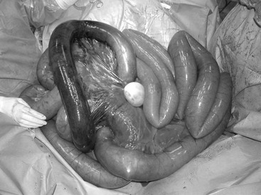
Several causes of strangulation of small intestine are recognized. Lipomas, composed of adipose tissue, are round-shaped neoplasias of the gastrointestinal tract, usually associated with the mesentery. Although benign in character, they increase in size as the horse ages, and their connection to the mesentery assumes the shape of a long, thin stalk that can wrap around a segment or loop of small intestine. This disease is infrequent in young horses but common in horses older than 15 years, especially geldings and ponies (Figure 75-1).
The epiploic foramen is the opening to the omental sac. It is usually slit-like, with a 2-inch (5-cm) opening in the cranial aspect of the right dorsal quadrant of the abdomen. Its borders are the gastropancreatic fold, the hepatoduodenal ligament caudally, the portal vein ventrally, the caudate lobe of the liver cranially, and the caudal vena cava dorsally. Small intestine can become entrapped in the foramen, in a right-to-left or left-to-right direction. Cribbing is a recognized risk factor for this condition.
Diaphragmatic hernias can be a diagnostic challenge. Affected horses usually have a history of trauma, which is thought to cause the tear in the diaphragm. Signs of colic may vary in intensity and duration as long as the intestine is not permanently entrapped in the tear. Some degree of respiratory distress may be seen when the entrapped colon becomes distended within the thorax. Signs of acute abdominal pain may begin as entrapment progresses, and emergency surgical intervention is typically required.
Scrotal or inguinal hernias arise in stallions when a segment of intestine slides through the internal inguinal ring into the inguinal canal and scrotum. This condition typically develops immediately after work. Affected horses have a unilaterally enlarged, cold scrotum, which is usually painful to touch. Transrectal examination will reveal small intestine within the inguinal ring on the affected side. Ultrasound of the scrotum will confirm the presence of distended small intestine. Other, less common causes of small intestinal strangulation include mesenteric rents, mesodiverticular bands, gastrosplenic entrapment, and small intestinal volvulus.
Pathophysiology
In most cases, initial occlusion of the mesenteric veins and partially maintained arterial blood supply induce a hemorrhagic lesion in the affected portion of intestine, which results in congestion, mucosal degeneration, and ultimately necrosis. Muscular and serosal layers are affected thereafter. The rest of the bowel may also become injured secondary to distension and may suffer reperfusion injury after correction of the strangulation. Endotoxemia is common because of increased permeability of the injured intestinal mucosa. Distal jejunum and ileum are the most commonly affected portions.
Clinical Findings and Clinical Pathology
Clinical signs parallel the degree of intestinal damage from the vascular occlusion, which results in progressive cardiovascular compromise associated with endotoxemia. Abdominal pain is moderate to severe, and heart rate is high (>60 beats/minute); these are the effects of pain and endotoxemia. Prolonged skin tenting and enophthalmos are indicative of severe dehydration. Reflux is generally obtained, and gastric decompression does not alleviate pain. Gross abdominal distension may be present in advanced cases. Hypocalcemia, hyponatremia, hypokalemia, metabolic acidosis, high anion gap value, and hyperlactatemia are common. Blood glucose concentration can be high. Liver enzyme values are generally high, usually because of gastrointestinal stasis as opposed to true hepatic compromise. Muscle enzymes can be high secondary to prolonged recumbency, repeated rolling, and transportation to a referral facility.
Diagnostic Testing
Small intestinal distension is apparent on transrectal examination and on ultrasound. The small intestinal walls appear progressively edematous and thick. Abdominocentesis yields a serosanguineous fluid with high protein concentration (>2 g/dL) and, with progression of time, high nucleated cell count (>5000 cells/µL). Peritoneal lactate is typically higher than peripheral blood lactate.
Treatment Options and Outcome
Because surgical correction is the treatment of choice, horses should promptly be shipped to a surgical facility. Xylazine (0.2 to 1 mg/kg, IV or IM) or detomidine (0.01 to 0.02 mg/kg, IV or IM) can be used alone or combined with butorphanol (0.01 to 0.02 mg/kg, IV or IM) to control pain and facilitate safe completion of the physical examination. Flunixin meglumine (1.1 mg/kg, IV) will help manage pain and reduce the effects of the endotoxin-induced inflammatory cascade. Restoration of an acceptable cardiovascular status will be necessary before anesthesia and should be initiated and maintained during transport if possible. Hypertonic saline (1 to 2 L, IV, for a 500-kg horse) and colloids (hetastarch, 8 to 10 mL/kg) can be administered initially but should immediately be followed by isotonic fluids to maintain intravascular volume.
Early surgical intervention helps reduce the need for intestinal resection and is the most important factor in determining postoperative survival. Unfortunately, most horses require resection of the affected portion of the small intestine and anastomosis as a consequence of the quick demise of the intestine and delayed diagnosis and surgical intervention. Common postsurgical complications include ileus, adhesions, and death. Postoperative ileus is defined by absence of progressive small intestinal motility, which results in continuous and copious volumes of gastric reflux. The duration of ileus is completely unpredictable: some horses reflux for weeks and require parenteral nutrition and repeated gastric decompression. Fluid therapy must account for maintenance and losses, but care should be taken to avoid hyperhydration, which can result in an increased volume of nasogastric reflux. Drugs used to treat postoperative ileus include lidocaine (1.3 mg/kg, IV, administered over 15 minutes and followed by a constant rate infusion of 0.05 mg/kg/minute). Other drugs used to treat postoperative ileus include metoclopramide (0.04 mg/kg/hour), cisapride (0.1 mg/kg IM), bethanechol (0.025 mg/kg, SC), erythromycin (0.5 mg/kg, IV), and neostigmine (0.02 mg/kg, IV).
Small intestinal adhesions may be minimal enough to cause only nonspecific, mild signs of abdominal discomfort, or severe enough to cause small intestinal obstruction or strangulation. Clinical signs are usually not apparent before 1 to 2 weeks after surgery. Lifelong dietary adjustment to a low-bulk diet may suffice in some cases, but surgical breakdown of the adhesions is needed if signs of obstruction or strangulation do not subside. Young horses are at a higher risk for forming adhesions, and chances of recurrence are also higher. Short- and long-term survival rates after small intestinal resection are approximately 75% and 65%, respectively. Factors that influence survival include duration of the lesion before surgical correction, extent of endotoxemia and systemic compromise, degree of distension and inflammation in the remainder of the small intestine, duration of surgery, surgeon’s experience, and type of anastomosis performed. Horses that undergo jejunojejunostomy have a better prognosis than those that undergo jejunocecostomy.
Stay updated, free articles. Join our Telegram channel

Full access? Get Clinical Tree


