Nicole du Toit
Donkey Dental Disease
In recent years, awareness of the importance of dental disease in donkeys has increased. A large proportion of donkeys are kept as pets, and the significance of dental disease, as has been identified in ridden horses, has often been overlooked. Furthermore, a prevailing perception is that donkeys require less maintenance and are able to survive longer without domestic intervention. However, the very fact that donkeys have greater longevity than horses places greater importance on the provision of proper dental care in these animals.
As in horses, routine dental care should start at an early age so that any developmental dental disease can be managed throughout the life of a donkey without progression of dental disease. However, it is more common for a veterinarian to be presented with an older donkey with signs of advanced dental disease. In nonridden animals, clinical signs of dental disease will be observed as problems with mastication, such as quidding, reluctance to eat specific types of feed such as stalky forage, or food pouching in the cheeks. In some cases, weight loss may be the first indication of dental disease.
Dental Examination and Instrumentation
A complete clinical examination, conducted before the dental examination, will facilitate the clinician’s obtaining a comprehensive view of the donkey’s well-being. This should include assessment of the body condition score to determine whether there has been significant weight loss and enable objective reevaluation after dental treatment. Furthermore, a donkey that is acutely anorexic or that has had a significant sudden decrease in dietary intake should be evaluated for hyperlipemia. Obtaining a blood sample to determine the triglyceride concentration in these cases is very important because some donkeys may have hypertriglyceridemia without clinical signs. In some cases, when extensive dental treatment is performed, donkeys may have an initial period of oral discomfort, and as a consequence dietary intake may be temporarily decreased. The period of decreased feed intake, together with the stress associated with handling and dental treatment, may put these donkeys at a high risk for developing hyperlipemia after dental treatment, and preventative treatment may have to be implemented.
Oral cavity examination of donkeys should be performed in the same consistent manner as it is in horses (see the fifth edition of Current Therapy in Equine Medicine, Chapter 3.1); it also may be slightly more challenging in small donkeys, as it is in smaller horses. Well-handled donkeys may be amenable to oral cavity examination and dental treatment without sedation, but routine sedation techniques can be used in donkeys that require chemical restraint for examination and treatment. The most commonly used sedative protocol is a combination of intravenously administered detomidine hydrochloride (0.02 to 0.04 mg/kg) and butorphanol tartrate (0.01 to 0.02 mg/kg). The drug dosages are generally slightly higher than what would be used in a horse of the same weight.
Depending on the size of the donkey, a standard horse or pony full-mouth speculum is needed for the examination. A standard set of hand floats can be used to perform a routine dental float in a donkey, but the use of hand floats with thin heads will facilitate easier rasping of the teeth, particularly in the smaller animals. Motorized equipment is particularly useful when more intricate targeted floating is required. However, the veterinarian should be careful to ensure that heat production during the procedure is minimized to avoid thermal damage to the teeth.
Incisor Disorders
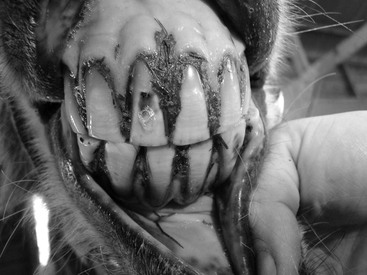
Diseases of the incisors are usually not clinically significant or advanced when seen by a veterinarian because incisors are easily visible and owners quickly detect problems involving them. In younger donkeys, fractures of the incisors may be the most common problem, and they result from the playful nature of younger animals. Retention of the deciduous incisors may also be observed occasionally. It is important that these are extracted as soon as possible because they may interfere with eruption of the permanent incisors and cause displacements. The deciduous incisors usually lie labial to the permanent incisors, but radiographs may be needed to ensure that the correct teeth are extracted. In older donkeys, the development of senile diastema (Figure 62-1) is common but usually only causes a mild gingivitis. This can be treated by asking owners to brush the incisors daily, taking care to remove the impacted feed, or by widening the diastema space so that food no longer becomes impacted. Odontoclastic tooth resorption and hypercementosis has also been observed in some older donkeys and should be ruled out radiographically in examination of incisors with gingivitis and periodontal disease. Extraction of the affected incisors is the only successful treatment for this disease.
< div class='tao-gold-member'>
Stay updated, free articles. Join our Telegram channel

Full access? Get Clinical Tree


