The Oropharynx and Tonsils

CHAPTER 8 The Oropharynx and Tonsils


Cytology is a useful, rapid screening test for lesions in the oropharynx, including masses, ulcers, draining tracts, plaques, and enlarged tonsils. It can be performed alone or in conjunction with biopsy and/or sampling for bacterial and fungal testing. Sedation or anesthesia can be necessary for complete examination of the oropharynx and to obtain adequate, representative samples. For mass lesions and plaques, aspiration of the deeper layers of the lesion to avoid any superficial secondary inflammation is usually most rewarding. If a mass lesion is nonexfoliative, scraping the lesion may yield adequate numbers of cells; however, excisional biopsy is usually necessary for a definitive diagnosis and prognosis. For flat lesions including ulcers, biopsy of the entire lesion or at least of the edge of a lesion, so that the early, primary abnormalities can be evaluated, is usually necessary to obtain an adequate number of representative cells for evaluation. However, impression smears or scrapings of ulcerative lesions can often yield cells or organisms that are distinctive and can be identified as the primary cause of the lesion rather than as secondary opportunists, and eliminate the need for biopsy. Because the oropharynx and tonsils are highly vascular, care must be taken to avoid hemodilution of the sample at the time of collection.


NORMAL FINDINGS


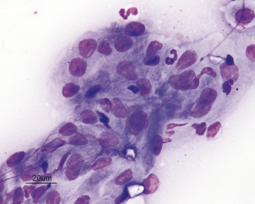
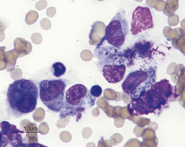
In order to correctly identify abnormal criteria, recognition of normal findings is essential. The oropharynx and tonsils are covered by mature squamous epithelial cells (Figure 8-1). These are large, flat, and round to slightly angular. They have abundant pale cytoplasm and small round nuclei that exhibit condensed chromatin. Nucleoli are not visible and some cells are anuclear. The presence of occasional intermediate squamous cells with slightly larger, less condensed nuclei is normal (Figure 8-2).


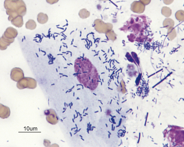
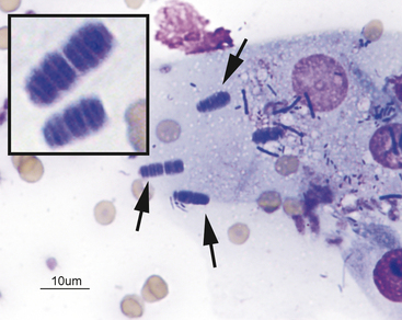
The normal squamous cells frequently exhibit a mixed bacterial population adhered to their surfaces (Figure 8-3). These bacteria are also usually present in the background between cells. The normal flora includes aerobic and anaerobic bacterial rods and cocci. Observation of spirochetes is considered normal. Yeast are never considered to be normal. One bacterium, Simonsiella spp., has a characteristic palisading appearance and is a normal inhabitant of the oropharynx (Figure 8-4). It should never be mistaken for a pathogen. If the bacterial population is dominated by only one type of bacteria, that would be considered abnormal.


The normal appearance of smears made from the tonsils is typical of other lymphoid organs. Usually, greater than 80% of the lymphoid cells are small and appear mature. The remaining lymphoid cells are intermediate-sized lymphocytes and occasional lymphoblasts. Plasma cells, neutrophils, macrophages, eosinophils, and squamous cells from the epithelial surface can be rarely observed. Occasional granules of iron pigment can be normal.


THE OROPHARYNX


Nonneoplastic Lesions


Inflammation


Acute (neutrophilic) inflammation is characterized by a predominance of neutrophils. They can be degenerate or nondegenerate. Neutrophils are most frequently degenerate if bacterial endotoxins are present. Macrophages, occasional lymphocytes, plasma cells, fibrocytes and eosinophils can also be present in low numbers.


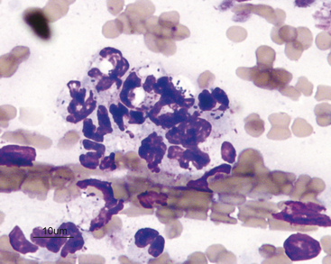
Infectious agents can be observed; however, their absence from a sample does not rule out the possibility of an infectious etiology. If the inflammatory lesion is caused by a primary bacterial infection or complicated by secondary bacterial infection, a homogenous population of bacteria is often seen and many will be phagocytized within neutrophils (Figure 8-5).


If the inflammation is superficial, secondary overgrowth of oropharyngeal bacterial flora is common. Secondary opportunistic bacterial inflammation can also be observed in association with primary, noninflammatory lesions. The presence of a heterogeneous population of bacteria that are extracellular or adhered to epithelial cells suggests overgrowth of flora.


If the lesion is granulomatous, such as from a foreign body or yeast/fungal infection, a more evenly mixed population of neutrophils, macrophages, lymphocytes, and plasma cells is observed, with variable numbers of fibrocytes and fibroblasts that are indicative of physiologic fibroplasia (Figure 8-6). In some areas of the country, histoplasmosis in cats can present with oral lesions as the predominant physical examination finding. In these cases, a diagnosis can be made based on identification of organisms from proliferative oral lesions.


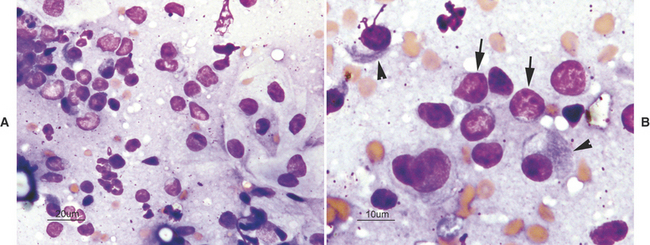
An inflammatory infiltrate characterized by a predominance of mature lymphocytes and plasma cells with scattered other inflammatory cells is seen in samples from cats with chronic gingivitis/stomatitis (i.e., lymphocytic-plasmacytic gingivitis/stomatitis). The characteristic inflammatory cells are typically admixed with normal or dysplastic epithelial cells (Figure 8-7).


Stay updated, free articles. Join our Telegram channel

Aug 31, 2016 | Posted by in GENERAL | Comments Off on The Oropharynx and Tonsils

Full access? Get Clinical Tree

Get Clinical Tree app for offline access