CHAPTER 10 The External Ear Canal
CAUSES OF OTITIS
Otitis externa is commonly encountered in veterinary practice. It may be unilateral, bilateral, acute, or chronic. Otitis externa in some form is estimated to affect 15% of dogs and 4% of cats presented for veterinary care.1 These animals generally show one or more of the following signs: head shaking, pain, pruritus, erythema, swelling, malodor, otic discharge, crusting, alopecia, excoriation, and pyotraumatic dermatitis.1–3 Conformation, habits, skin diseases, organisms, tumors, and trauma are some of the factors thought to initiate, predispose, or perpetuate otitis externa (Boxes 10-1 and 10-2). Each is briefly addressed subsequently.1–4
Conformation
A long, relatively narrow ear canal tends to trap moisture, foreign debris, and glandular secretions. Excessive hair in the external ear canal inhibits ventilation and increases retention of cerumen. Also, dogs with pendulous ears have decreased ventilation, which increases humidity within the ear canal. Such a moist environment can destroy the protective barrier of the epidermis and allow opportunistic infection.1,3,4
Habits
Dogs housed outside and hunting dogs are more likely to have foreign bodies (such as grass awns, dirt, or twigs) lodge in the ear canal.1,3 Dogs that swim or are frequently bathed may develop otitis externa, since frequent wetting of the ear canal may stimulate ceruminous gland activity resulting in overproduction of secretions. Moisture trapped in the ear canal may also affect the protective function of the epidermis.2,4
Skin Diseases
Generalized skin disease may affect the pinna and external ear canal epithelium (e.g., atopy, food allergy, contact hypersensitivity, autoimmune disease) or cause overproduction of cerumen (e.g., seborrhea, endocrinopathies). As many as 50% to 83% of atopic dogs reportedly have signs of otitis. In a few of these cases, otitis may be the only clinical sign.1,3–5
Organisms
Bacteria, yeasts, and/or parasites are commonly involved in otic disorders. Small numbers of bacteria and Malassezia yeast are present in the ear canals of many clinically normal dogs and cats. These organisms can secondarily overgrow and infect the ear canal when predisposing conditions permit. Ear mites are one of the major primary causes of otitis externa in cats with a much lower prevalence in dogs.1–6
Tumors
Any cutaneous neoplasm can occur in the ear canal and may predispose the animal to otitis externa.1,5,7,8
DIAGNOSIS
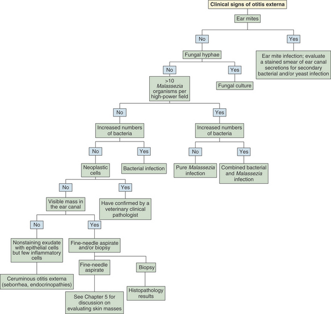
Most cases of acute otitis externa can be readily managed using the information gained from a thorough history, physical examination, otoscopic examination, and cytologic evaluation of ear canal secretions. Careful evaluation of the patient’s skin is also important because ear disease is often associated with generalized skin disease. Foreign bodies, tissue masses, abnormal ear canal conformation, mites, ticks, and other abnormalities can be detected during otoscopic examination, and any microorganisms present can be identified cytologically. Serial cytologic evaluation is also useful to monitor the response to therapy.1,3,4,6,9 The algorithm in Figure 10-1 can be used to aid in the evaluation of ear canal secretions. Additional diagnostic tests (such as culture, thyroid evaluation, intradermal skin testing, and biopsy) are generally needed to determine the underlying cause of chronic or recurrent otitis externa.1,3,6
COLLECTION AND STAINING OF SAMPLES
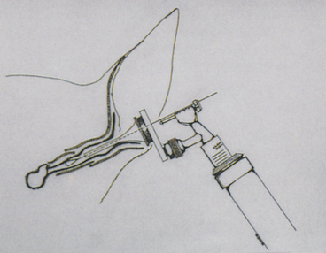
Samples of ear canal secretions for cytologic evaluation are best collected with cotton-tipped swabs. Anesthesia may be required to obtain samples from uncooperative animals or animals with painful ears. Cytology samples are preferably obtained after performing otoscopic examination to evaluate the tympanic membrane, because the swab may compress debris in the horizontal canal and obscure the tympanum. Collection of cytology samples should be performed before any cleaning agents or medications are placed in the ear. Samples may be collected by passing a cotton-tipped swab through the cone of an otoscope (Figure 10-2). The use of a separate, clean otoscope cone for each ear is recommended to prevent cross-contamination. Sterile cones are required to obtain samples for culture. Passing a cotton-tipped swab through the cone of an otoscope allows the operator to visualize the horizontal ear canal and be sure the specimen is collected from that area. Collecting ear swabs from the horizontal ear canal is desirable because most bacterial ear infections begin in this area. Another method is to carefully pass a swab into the ear canal without the aid of an otoscope collecting samples from the junction of the vertical and horizontal ear canals. Further blind advancement of the swab into the horizontal ear canal is not recommended because injury could occur if the animal moves unexpectedly. Always collect samples from both ears because animals that appear to have unilateral otitis may also have mild, less apparent disease in the other ear.1,3,6
Stay updated, free articles. Join our Telegram channel

Full access? Get Clinical Tree


