The Endocrine System


The Endocrine System


Key Points


General concepts


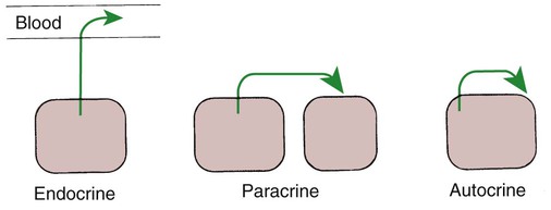
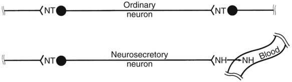
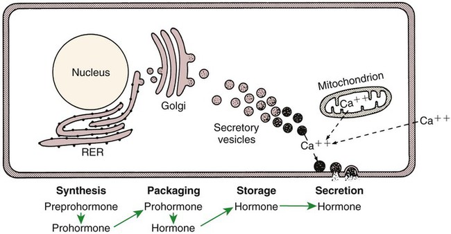
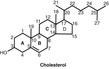
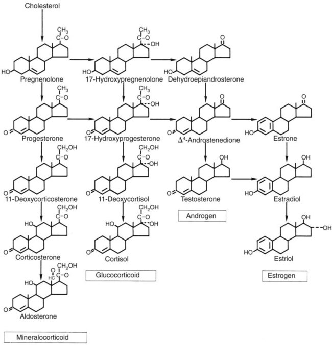
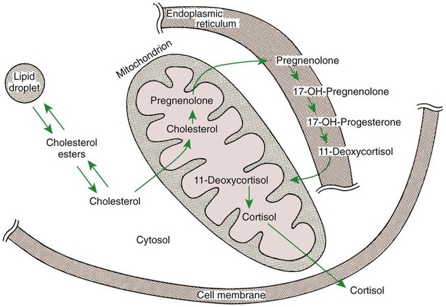
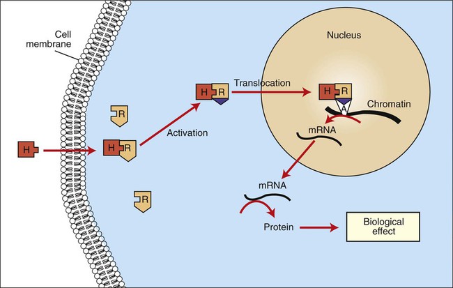
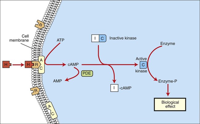
1. Hormones are chemicals produced by specific tissues that are transported by the vascular system to affect other tissues at low concentrations.


2. The endocrine and nervous systems are integrated in their control of physiological processes.


Synthesis of hormones


1. Protein hormones are initially synthesized as preprohormones and then cleaved in the rough endoplasmic reticulum to form prohormones and in the Golgi apparatus to form the active hormones, which are stored in granules before being released by exocytosis.


2. Steroids are synthesized from cholesterol, which is synthesized by the liver; steroids are not stored but are released as they are synthesized.


Transport of hormones in the blood


1. Protein hormones are hydrophilic and carried in the plasma in dissolved form.


2. Steroids and thyroid hormones are lipophilic and carried in plasma in association with both specific and nonspecific binding proteins; the amount of unbound, active hormone is relatively small.


Hormone-cell interaction


1. Protein hormones have specific receptors on target tissue plasma membranes, whereas steroids have specific receptors within the cytoplasm or nucleus.


Postreceptor cell responses


1. Steroids interact directly with the cell nucleus through the formation of a complex with its cytosolic receptor, whereas protein hormones need a messenger because they cannot enter the cell.


Metabolism of hormones


1. Steroid hormones are metabolized by conjugation with sulfates and glucuronides, which makes steroids water soluble.


Feedback control mechanisms


1. The most important feedback control for hormones is the negative-feedback system, in which increased hormone concentrations result in less production of the hormone, usually through an interaction with the hypothalamus or pituitary gland.


2. Endocrine secretory patterns can be influenced by factors such as sleep or light and can produce circadian rhythms.


The hypothalamus


1. The hypothalamus coordinates the activity of the pituitary gland through the secretion of peptides and amines.


The pituitary gland


1. The neurohypophysis has cell bodies that originate in the hypothalamus, with cell endings that secrete oxytocin and vasopressin.


2. Oxytocin and vasopressin are synthesized in cell bodies within the hypothalamus and are carried by axon flow to the posterior lobe, where they are released.


3. The main effects of oxytocin are on the contraction of smooth muscle (mammary gland and uterus); the effects of vasopressin are primarily on the conservation of water (antidiuresis) and secondarily on blood pressure.


4. Plasma osmolality controls the secretion of vasopressin.


5. The anterior pituitary produces growth hormone, prolactin, thyroid-stimulating hormone, follicle-stimulating hormone, luteinizing hormone, and corticotropin.


6. Adenohypophyseal activity is controlled by hypothalamic releasing hormones, which are released into the portal system, which in turn connects the median eminence of the hypothalamus and the anterior pituitary gland.


General Concepts


Hormones Are Chemicals Produced by Specific Tissues That Are Transported by the Vascular System to Affect Other Tissues at Low Concentrations


The endocrine system has evolved to allow physiological processes to be coordinated and regulated. The system uses chemical messengers called hormones. Hormones have traditionally been defined as “chemicals that are produced by specific endocrine organs, are transported by the vascular system, and are able to affect distant target organs in low concentration.” Although this definition is useful from a veterinary medical point of view, it should be recognized that some substances, such as prostaglandins and somatomedins, are produced by many other tissues and are still considered hormones.


Other types of control systems use chemical substances that are not transported in the vascular system to influence distant cell activity. These systems serve as means of local integration among or between cells, as follows:


• Paracrine effectors, in which the messenger diffuses through the interstitial fluids, usually to influence adjacent cells; if the messenger acts on the cell of its origin, the substance is called an autocrine effector (Figure 33-1).


• Neurotransmitters, which affect communication between neurons, or between neurons and target cells; the substances are limited in the distance traveled and the area of the cell influenced (Figure 33-2).


• Exocrine effectors, such as hormones produced by the pancreas, are released into the gastrointestinal tract.


The Endocrine and Nervous Systems Are Integrated in Their Control of Physiological Processes


The endocrine system interacts with the other main regulatory system, the nervous system, which coordinates activities that require rapid control. An example of the close interaction of the two systems is the reflex in which suckling causes the release of milk. Suckling initiates the transmission of nerve impulses from the mammary gland to the hypothalamus (by way of the spinal tract). Neurosecretory neurons within the supraoptic and paraventricular nuclei are stimulated to synthesize oxytocin. Oxytocin is transported along axons of these nerves and is released from nerve endings in the posterior pituitary into the blood vascular system. Oxytocin is then carried to the mammary gland, where it causes contraction of myoepithelial cells. These cells surround the smallest unit of milk-secreting cells, called an alveolus. This results in the movement of milk into the large cisternae adjacent to the teat and subsequently into the teat.


The interaction between the nervous and endocrine systems can be even more direct. For example, endocrine cells of the adrenal medulla are directly controlled by preganglionic neurons of the adrenal medulla, and the medullary hormones are released immediately in response to stressful stimuli. The endocrine and nervous systems also share transmitters; substances such as epinephrine, dopamine, histamine, and somatostatin are found in both endocrine and neural tissues.


The endocrine system is involved in control of physiological functions, including metabolism, growth, and reproduction. Metabolism can be divided into two forms: energy and mineral. The hormones that control energy metabolism include insulin, glucagon, cortisol, epinephrine, thyroid hormone, and growth hormone. The hormones that control mineral metabolism include parathyroid hormone, calcitonin, angiotensin, and renin. The hormones that control growth include growth hormone, thyroid hormone, insulin, estrogen and androgen (both reproductive hormones), and a large number of growth factors. The hormones that control reproduction include estrogen, androgen, progesterone, luteinizing hormone (LH), follicle-stimulating hormone (FSH), prolactin (PRL), and oxytocin.


One of the important characteristics of the endocrine system is the amplification of the signal. The action of one steroid molecule to activate a gene can result in the formation of many messenger ribonucleic acid (mRNA) molecules, and each of these can induce the formation of many enzyme molecules. Also, one protein molecule can influence the formation of many cyclic adenosine 3′,5′-monophosphate (cAMP) molecules, and each of these can activate many enzymes. Amplification is the basis for the sensitivity of the endocrine system, which allows small amounts of hormones in plasma (10−11 to 10−12 mol) to produce significant biological effects. Hormone action also influences rates of existing enzyme reactions, but not the initiation of new reactions. This implies that there are certain basal levels of enzyme activities even in the absence of hormones. Hormone action is relatively slow and prolonged, with the effects of hormones lasting minutes to days. This contrasts with the nervous system, in which the response is rapid and short (milliseconds to seconds).


Synthesis of Hormones


Protein Hormones Are Initially Synthesized as Preprohormones and Then Cleaved in the Rough Endoplasmic Reticulum to Form Prohormones and in the Golgi Apparatus to Form the Active Hormones, Which Are Stored in Granules Before Being Released by Exocytosis


The major classes of hormones include proteins (e.g., growth hormone, insulin, corticotropin [previously called adrenocorticotropic hormone, or ACTH]); peptides (e.g., oxytocin and vasopressin); amines (e.g., dopamine, melatonin, epinephrine); and steroids (e.g., cortisol, progesterone, vitamin D). The protein and peptide hormones are initially synthesized on ribosomes as larger precursor proteins, which are referred to as preprohormones (Figure 33-3). Synthesis of protein hormones begins in ribosomes, with the “pre” portion immediately attaching to the rough endoplasmic reticulum (RER), which pulls the ribosomes into close apposition with the RER. During synthesis, the preprohormone is secreted into the interior of the RER. The presence of a peptidase within the wall of the RER allows the “pre” portion of the molecule to be rapidly removed and the prohormone to leave the RER in vesicles that have been pinched off from the RER. These vesicles then move to the Golgi apparatus, where they coalesce with Golgi membranes to form secretory granules. The prohormone is cleaved during this process, so most of the hormone is in its final form within the Golgi apparatus, although some prohormone can also be found.


Protein hormones are stored in granules within the gland until needed for release. Although some of the hormone is secreted on a continuous basis, most is secreted through the process of exocytosis of granules in response to a specific signal. The process of exocytosis requires adenosine triphosphate (ATP) and calcium (Ca2+). Increased cytoplasmic calcium results from intracellular release of Ca2+ from mitochondria, or endoplasmic reticulum, or from the influx of extracellular Ca2+.


Steroids Are Synthesized from Cholesterol, Which Is Synthesized by the Liver; Steroids Are Not Stored but Are Released as They Are Synthesized


Steroids represent a class of hormones that, unlike protein hormones, are lipophilic. In general, they belong to one of two categories: adrenocortical hormones (glucocorticoids, mineralocorticoids) and sex hormones (estrogens, progesterone, androgens). They have a common four-ring, 17-carbon skeleton that is derived from cholesterol (Figure 33-4). Although the steroids can be synthesized de novo within the cell from the two-carbon molecule acetate, the majority of steroids are formed from cholesterol, which is synthesized by the liver (Figure 33-5). Low-density lipoproteins (LDLs) enter steroid-producing cells through interaction with a membrane receptor. Cholesterol is released through the degradation of LDLs by lysosomal enzymes. Cholesterol is either used immediately for steroid synthesis or stored in granules in an ester form within the cell. The first step in the synthesis of all steroid hormones from cholesterol involves cleavage of the side chain of cholesterol to form pregnenolone; this step occurs within the mitochondrion. Subsequent modifications of the steroid molecule may occur within the mitochondrion or may involve movement to other compartments of the cell (Figure 33-6). The control of movement of steroids among cell compartments during the synthesis process is not well understood.


The type of steroid hormone that is eventually synthesized depends on the presence of specific enzymes within the particular cell. For example, only cells of the adrenal cortex contain enzymes (hydroxylases) that result in hydroxylation of the eleventh and twenty-first carbon molecules, a process that is essential for the production of glucocorticoids and mineralocorticoids. The pattern for sex steroid biosynthesis is for pregnenolone to be modified in a sequence that involves progesterone, androgens, and finally estrogens. Cells that synthesize androgens (e.g., Leydig cells of the testis) have the enzymes required for the formation of pregnenolone and progesterone, as well as for the modification of progesterone to androgen, but lack the enzymes necessary to modify androgens into estrogens. Although the sex steroid–forming cells do not have enzymes that allow the formation of adrenocortical hormones, the adrenal cortex contains the enzyme systems necessary for the formation of both adrenocortical hormones and sex hormones, although the former are emphasized. As a result, the adrenal cortex normally produces small amounts of sex steroids and produces larger amounts in certain pathophysiological conditions.


There is no provision for the storage of steroid hormones within the cell; they are secreted immediately after formation by simple diffusion across the cell membrane because of their lipophilic structure. Thus, synthesis and secretion of steroid hormones occur in a tightly coupled manner, whereby the rate of hormone secretion is controlled by the rate of synthesis. The only storage form of steroids within these cells involves that of the precursor molecule, cholesterol, as an ester.


Transport of Hormones in the Blood


Protein Hormones Are Hydrophilic and Carried in the Plasma in Dissolved Form


This chapter focuses mainly on hormones that are transported to target tissues in the vascular system. The means by which hormones are transported in the blood varies according to the solubility of the hormone. Protein and peptide hormones are hydrophilic and are carried in the plasma in dissolved form. The protein hormones may circulate in monomeric (single-unit) or polymeric (multiple-unit) form (e.g., insulin). Hormones that have subunits can appear in the circulation in subunit form, although this reduces the biological potency of the molecule.


Steroids and Thyroid Hormones Are Lipophilic and Carried in Plasma in Association with Both Specific and Nonspecific Binding Proteins; the Amount of Unbound, Active Hormone Is Relatively Small


The transport of steroid and thyroid hormones is more complicated than that of protein hormones, because the steroid and thyroid hormones are lipophilic and thus have limited solubility in aqueous solutions. These hormones are transported in the blood through association with various types of proteins. Some of the proteins that bind steroids have a high affinity for a particular steroid; for example, a globulin, transcortin, has a high affinity for cortisol and corticosterone but also serves as an important transport vehicle for progesterone, even though it has a lower affinity for this hormone. The carrier proteins that have high affinities have low capacity because of their low plasma concentration. In contrast, the general class of plasma proteins called albumins have low affinities for steroid hormones but have a high capacity for steroid transport because of their high concentration in plasma.


A hormone must be in the free, or unbound, form before it can penetrate a target cell and elicit biological activity. This is accomplished by the establishment of equilibrium between bound and free hormone levels in the plasma. The free form usually represents only about 1% of the total amount of hormone in the plasma (up to 10% of cortisol may be in the free form). The system is responsive to use of the free form, and the free form is replenished quickly by dissociation of bound hormone from the protein. The total amount of the hormone is usually measured, with the exception of thyroid hormone, for which attempts are usually made to estimate the amounts of bound and free. As indicated for steroid hormones, synthesis and release are tightly linked, and because metabolic clearance rates are usually constant, concentrations of steroids in plasma are usually a good reflection of the secretion rate. Under certain physiological conditions, such as pregnancy in humans, metabolism of estrogens can change because of the increased production of estrogen-binding proteins.


Hormone-Cell Interaction


Protein Hormones Have Specific Receptors on Target Tissue Plasma Membranes, Whereas Steroids Have Specific Receptors Within the Cytoplasm or Nucleus


A central question in endocrinology is how hormones and target cells of a particular tissue interact in a specific manner. The problem seems almost overwhelming for steroids because they are lipid soluble and able to permeate all cells of the body. The solution is that target cells have receptors that are specific for a particular hormone. For steroids, the receptors are located in the cytoplasm or nucleus of the target cells, whereas receptors for protein and peptide hormones are located on the plasma membrane of the cell. In addition to specificity, receptors have a high affinity for their respective hormone. These characteristics of the receptor allow hormones to be in low concentration in the blood but effective in producing significant tissue response.


The greater the affinity of the receptor for the hormone, the longer is the biological response. Termination of the action of a hormone usually requires dissociation of the hormone from the receptor. This occurs most often as a result of a decrease in plasma concentrations of the hormone; the binding of receptor and hormone is noncovalent, and declining hormone concentrations favor a chemical equilibrium of dissociation over association. Termination of hormone action can also result from internalization of the receptor-hormone complex through the process of endocytosis. The hormone is degraded by lysosomal enzymes, whereas the receptor, protected because of its association with the vesicle membrane, can be recycled to the plasma membrane.


Receptors are present on cells in much greater numbers than required for the elicitation of a biological response. Occupancy by a hormone of considerably less than 50% of the receptors usually elicits a maximal biological response. Even so, changes in receptor numbers that affect the sensitivity of the cell, although not its maximal responsiveness, can occur. Changes in receptor number affect the probability that an interaction will occur between receptor and hormone. Receptor synthesis can be stimulated by a hormone that is different from the hormone that interacts with the receptor. For example, predominant gonadotropin receptors on ovarian granulosa cells change from FSH to LH receptors late in the ovarian follicle phase because of the influence of FSH. This allows the control of the ovarian follicle to pass from FSH to LH, which facilitates ovulation and the formation of a corpus luteum. Conversely, receptor numbers can decrease in conjunction with continued interaction of receptor and hormone. This often occurs when an agonist that has great affinity for the receptor is administered or when amounts of hormone are pathologically elevated. The receptor numbers are downregulated in this situation. The end result is that the animal becomes resistant to continued therapy with the hormone in question.


Postreceptor Cell Responses


Steroids Interact Directly with the Cell Nucleus Through the Formation of a Complex with Its Cytosolic Receptor, Whereas Protein Hormones Need a Messenger Because They Cannot Enter the Cell


The events that follow binding of the hormone and receptor depend on whether a steroid, protein, or peptide hormone is involved. With steroids, the hormone is able to interact within the cell because of its ability to penetrate the lipoprotein plasma membrane (Figure 33-7). The interaction of receptor and steroid hormone results in activation of the subsequent complex translocation to the nucleus, where it interacts with specific sites on the chromatin. The result is the production of mRNA, which, when translocated to the ribosomes, directs synthesis of proteins that produce the desired biological result.


Protein or peptide hormones require an intermediary to act in their behalf because they are not able to penetrate the plasma membrane of the cell; the intermediary substance is known as a second messenger (Figure 33-8). The best-documented second messenger is cAMP, which is produced by the activation of an enzyme, adenyl cyclase, through interaction of the hormone and receptor in the plasma membrane. The activation of adenyl cyclase and the production of cAMP result in the phosphorylation of protein kinases, which are responsible for the biological response. Other second messengers include cytosolic calcium and its associated phosphodiesterase, calmodulin, as well as inositol triphosphate (IP3) and diacylglycerol, both of which are products of phosphatidylinositol metabolism. An important action of IP3 is the stimulation of intracellular calcium release. One important response to diacylglycerol is the activation of phospholipase A and the formation of arachidonic acid, which leads to formation of members of the prostaglandin family of molecules. The biological response to a protein or peptide hormone–receptor interaction is more rapid than that to steroids; preexisting enzymes are activated, whereas the biological response to steroid requires the synthesis of enzyme protein.


< div class='tao-gold-member'>

Stay updated, free articles. Join our Telegram channel

Jul 18, 2016 | Posted by in PHARMACOLOGY, TOXICOLOGY & THERAPEUTICS | Comments Off on The Endocrine System

Full access? Get Clinical Tree

Get Clinical Tree app for offline access