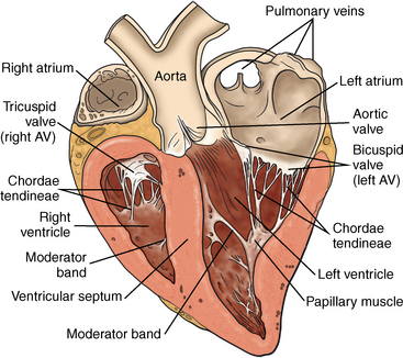
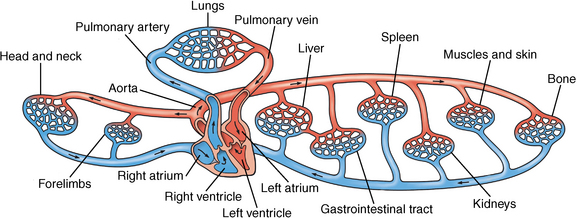
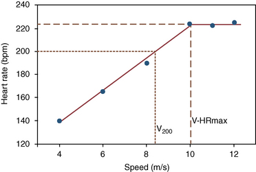
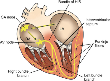
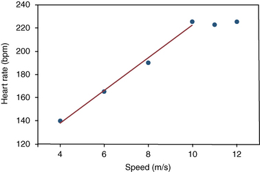
CHAPTER 11 The heart’s role is to pump sufficient blood to maintain blood pressure and oxygen flow to tissues. The anatomy of the equine heart is similar to that found in other mammals (Figure 11-1). The layout of the heart and circulatory system is illustrated in Figure 11-2. The thick-walled left ventricle pumps a stroke volume of blood into the aorta with each contraction. This establishes a wave of accelerated blood flow through the systemic arteries. Blood flow in arteries is continuous because relaxation of the elastic arterial walls compresses the blood in the vessel, forcing it away from the heart toward tissues. FIGURE 11-1 Cross-section of the equine heart showing the cardiac chambers and valves and direction of blood flow. FIGURE 11-2 The cardiovascular system illustrating the blood flow to and from the main body systems. The heart mass in Thoroughbred horses averages about 4 to 5 kilograms (kg), or 1% of body weight. Trained horses have slightly higher relative heart masses (1.1%) compared with untrained horses (0.94%), which seems to suggest that training causes hypertrophy of cardiac muscle (Evans, 2007; Kubo et al., 1974; Marr and Burton, 2010). These changes appear to occur soon after the onset of training, and this is highlighted by the finding that there is no difference in heart mass between horses in training for 2 months and those in training for 19 months. Heart mass to body weight ratio is also a function of breed. Racing horses have a relative heart mass of 0.86, compared with 0.76 in Arabian horses and 0.62 in draft horses (Kline and Foreman, 1991). The cardiac cycle is the sequence of events occurring in the heart during every contraction (systole) and relaxation (diastole). The sequence and timing of blood pressures and valvular events in the equine heart and major arteries at rest are illustrated in Figure 11-3. The cyclic nature of cardiac activity depends on normal conduction of electrical impulses from the sinoatrial (SA) node, or pacemaker, through the atrial and ventricular myocardium. The conduction of impulses in the equine heart is illustrated in Figure 11-4. The diffuse distribution of Purkinje fibers through the left ventricular wall enables rapid depolarization and development of muscular tension. However, right ventricular contraction slightly precedes left ventricular contraction. This is so because pulmonary arterial pressure is lower than aortic pressure (Holmes, 1982; Poole and Erickson, 2004). Abnormal blood flow through the heart can cause murmurs and possibly poor performance. Further discussion on this can be found in publications on equine cardiology (e.g., Marr and Burton, 2010). Heart rate in the resting horse depends mainly on the degree of relaxation of the individual horse. In relaxed horses, resting heart rate is usually in the range 25 to 40 beats per minute (beats/min). At night, when horses are relaxed or sleeping, heart rates tend toward the lower end of this range. Sudden excitement, fear, or anticipation of exercise can elevate heart rate rapidly to over 100 beats/min. Rapid heart rate changes in the range of 20 to 110 beats/min in resting horses can be explained entirely by alterations in parasympathetic nerve activity (Evans, 2007; Hamlin et al., 1972). It has been suggested that resting heart rate is lower in fit horses than in unfit horses (Evans, 2007; Littlejohn, 1987; Marr and Burton, 2010). However, resting heart rate in the horse generally does not decrease after training, as in human athletes (Bayly et al., 1983; Marr and Burton, 2010; Milne et al., 1977; Poole and Erickson, 2004; Skarda et al., 1976; Thomas et al., 1983). Use of heart rate measurements to monitor fitness is, therefore, restricted to measurements during or after exercise. Heart rate measurements during exercise in athletic horses have been used to describe the intensity of work, to measure fitness, and to study the effects of training and detraining. There are several suitable commercial heart rate meters designed for use in exercising horses (Courouce et al. 2002; Evans, 2007; Evans and Rose, 1986; Foreman and Rabin, 1984; Marr and Burton, 2010; Physic-Sheard et al., 1987). Heart rate during exercise also can be monitored electrocardiographically or digitally. The electrocardiogram (ECG) can be obtained by wiring the horse directly to the recorder, recording the ECG digitally for examination at a later time, or by radio telemetry (Evans, 2007; Marr and Burton, 2010; Patterson, 1996; Young, 2004). Commercial heart rate meters usually employ two or three electrodes incorporated into a belt or placed on the chest beneath the saddle. Such heart rate meters enable exercise testing to be performed under racetrack conditions, facilitating assessment of the response to exercise and prescription of specific exercise loads during exercise. Care must be taken to ensure that there is no faulty contact of the surface electrodes with the horse’s skin. However, under ideal conditions, the repeatability of heart rate measurements obtained during treadmill exercise is very high (Evans and Rose, 1988a; Seeherman and Morris, 1990; Evans, 2007; Young, 2004; Marr and Burton, 2010). At the onset of exercise, heart rate quickly increases and reaches a steady state in 2 to 3 minutes. This increase is associated with increased sympathetic nerve activity, catecholamine release, or both (Evans, 2007; Marr and Burton, 2010; Poole and Erickson, 2004; Young, 2004). Steady-state heart rate remains constant during submaximal workloads (Engelhardt, 1977; Evans 2007; Marr and Burton, 2010; Young, 2004). An overshooting of heart rate to values above the submaximal steady-state heart rate may occur at the commencement of exercise (Persson, 1967; Persson and Lydin, 1973; Young 2004). Mean time taken to reach maximal heart rates after onset of exercise in Thoroughbreds was 22 seconds (Krzywanek et al., 1970; Young 2004). In Standardbreds trotting at speeds of 12 to 12.5 meters per second (m/s), heart rates were not maximal until at least 700 m had been run (Courouce et al., 2002; Lindholm and Saltin, 1974). The kinetics of heart rate at the commencement of exercise without prior warmup is also dependent on the intensity of exercise (Evans 2007; Poole and Erickson, 2004). In six Standardbred horses, the typical overshoot was found at the start of exercise at 50% of A linear relationship between heart rate and submaximal work effort has been observed in horses trotting, galloping, and swimming (Ehrlein, Hornicke, Engelhardt, 1973; Lindholm and Saltin, 1974; Maier-Bock and Ehrlein, 1978; Marr and Burton, 2010; Persson, 1967; Persson and Lydin, 1973; Poole and Erickson, 2004; Senta, Smetzer, Smith, 1970; Thomas, Fregin, Gerber, 1980; Thomas and Fregin, 1981; Young 2004;). Many factors influence the position of the regression line of heart rate on work speed. These include gait, (Littlejohn et al., 1977), length of the exercise track (Ehrlein et al., 1973), and treadmill slope (Sexton and Erickson, 1990), and the position is also influenced by the presence of a breathing mask (Persson, 1983). The heart rate at a specific working velocity can vary markedly between individual horses, but if standardized treadmill exercise tests are used, the relationship between heart rate and work is very precise and reproducible for individual horses at heart rates between 120 and 210 beats/min (Courouce et al., 2002; Ehrlein et al., 1973; Evans, 2007; Evans and Rose, 1988a; Marr and Burton, 2010; Poole and Erickson, 2004; Young 2004). This relationship is usually defined by the use of treadmill tests that involve increasing the speed or, less commonly, treadmill angle and measuring heart rate at the completion of 1 to 2 minutes of exercise at each speed. After a suitable warmup, for example, 3 minutes of trotting, heart rate is stable after 1 minute of further exercise at higher speeds. Figure 11-6 illustrates heart rates recorded during the last 15 seconds at each speed in a fit Thoroughbred racehorse during a treadmill exercise test. More recently, information relating heart rate responses in field conditions have been obtained by the use of onboard heart rate monitors with speed of exercise determined by GPS (global positioning system) tracking (Evans, 2007). The heart rate–workload relationship is also affected by disease states because horses with recurrent airway obstruction (RAO) have submaximal exercise heart rates that are significantly higher than those found in normal horses (Littlejohn et al., 1977; Littlejohn et al., 1983; Young, 2004). Heart rate during standardized submaximal exercise is also higher in horses with cardiac disease such as atrial fibrillation (Evans, 2007; Young, 2004). The time to fatigue during treadmill exercise is dependent on the intensity of exercise. At heart rates of 170 to 180 beats/min, seven partially trained Thoroughbreds were able to exercise for an average of 24 minutes, whereas at a heart rate of 208 beats/min, equivalent to 100% Heart rates during swimming vary greatly between horses and are usually in the range 130 to 180 beats/min (Murakami et al., 1976; Patterson, 1996; Poole and Erickson, 2004; Thomas Fregin and Gerber, 1980; Young 2004). The mean highest heart rate recorded in nine horses during show jumping was 191 ± 3 beats/min (Art et al., 1990; Marr and Burton, 2010; Young, 2004). During prolonged strenuous submaximal exercise at a constant work rate, a gradual increase in heart rate, or cardiovascular drift, can occur. For example, during 30 minutes of constant load exercise, mean heart rate increased from 154 to 173 beats/min. This drift was accompanied by increases in minute ventilation and cardiac output, while stroke volume was unchanged (Evans, 2007; Poole and Erickson, 2004; Thomas and Fregin, 1990). In another study, horses exercising at 55% to 60% of individual maximal heart rate (HRmax) for 60 minutes had minimal changes in heart rate (Hinchcliff et al., 1990). Heart rate during prolonged exercise in horses probably depends on the intensity of exercise, environmental conditions, and possibly fitness (Evans, 2007; Young, 2004). A loss of linearity of the heart rate on velocity regression line is typical at high work speeds (see Figure 11-6). HRmax is defined as the highest heart rate measured in an incremental-speed treadmill test, which results in a plateau of heart rate. If a plateau is not demonstrated, the highest heart rate recorded in an incremental-speed treadmill exercise test is referred to as the peak heart rate. Alternatively, HRmax can be measured in horses after 1 minute of maximal exercise after suitable warmup (Courouce et al., 2002; Evans, 2007). HRmax can vary considerably between horses. Maximal heart rates recorded during racing in 19 Thoroughbreds averaged 223 beats/min, with a range of 204 to 241 beats/min (Evans, 2007; Krzywanek et al., 1970; Patterson, 1996; Poole and Erickson, 2004; Young, 2004). High individual variability in heart rates while racing also have been recorded in Standardbreds, ranging from 210 to 238 beats/min, with a mean of 221 beats/min (Åsheim et al., 1970; Courouce et al., 2002; Evans, 2007). In humans, HRmax declines with age (Åstrand and Rodahl, 1977). There is no predictable relationship between age and HRmax in horses. Mean peak heart rate in eight yearling Thoroughbreds was approximately 240 beats/min, compared with 220 to 230 beats/min in 2- to 4-year-old horses (Rose et al., 1990). Likewise, yearling, 2-year-old, and adult Thoroughbreds had similar means (229 to 231 beats/min) and ranges (215 to 254 beats/min) of peak heart rates during an incremental treadmill exercise test (Seeherman and Morris, 1991). Evans (2007) and Courouce et al. (2002) reported similar values for horses working in the field. The individual HRmax is a highly repeatable measurement in individual horses (Courouce et al., 2002; Evans, 2007; Evans and Rose, 1988a), but it is not an important measure of fitness, since it is not affected by training, despite increases in The treadmill speed at which HRmax is achieved (V-HRmax) during a stepwise test is significantly correlated with Heart rate measurements during submaximal treadmill and field exercise have been expressed relative to treadmill speed in some studies for measurement of fitness. For example, the treadmill velocities that result in heart rates of 140 (V140) or 200 beats/min (V200) have been used. The V200 is calculated by measuring the heart rate at the end of three to four treadmill or racetrack runs, each of which results in heart rates between 120 and 210 beats/min (Courouce et al., 2002; Evans 2007; Persson, 1983; Persson and Ullberg, 1974). Evans (2007) described the application, over the past decade, of these measurements under field conditions by using onboard heart rate meters and telemetric respiratory analysis, with ground speed determined by GPS technologies. As previously mentioned, V-HRmax also may be a useful measure of fitness. This value is obtained by substituting HRmax in the regression equation describing the linear heart rate on velocity relationship during submaximal exercise. Figure 11-8 shows the method of calculating V200 and V-HRmax. Evans (2007) demonstrated the direct application of this technology in the training of horses for athletic pursuits. FIGURE 11-8 Calculation of maximal heart rate (V-HRmax) from the results of a stepwise incremental-speed exercise test. The first reports of using trained Standardbred racehorses exercise-tested on a racetrack to calculate V200 were published in the 1980s (Evans, 2007). Rest periods after each step of a test or in response to a bout of exercise allow for determination of venous blood lactate and for calculation of HR4, another suggested index of fitness. This measurement refers to the heart rate at which blood lactate is 4 millimoles per liter (mmol/L). It is generally higher in fitter horses. Evans (2007) demonstrated how modern technologies can be adapted to in-field testing of horses for determination of many of these variables. A similar racetrack exercise test was used to investigate the relationship between several measures of fitness and maximal trotting velocity in Swedish trotters. Both V200 and the heart rate after 4000 m trotting at 10 m/s (HR10) were significantly correlated with maximal trotting velocity over 1000 m. Respective correlations were 0.6 and 0.74 (Persson and Ullberg, 1974). Courouce et al. (2002) and Evans (2007) demonstrated similar relationships in French Standardbred trotters and Thoroughbred racehorses, respectively. A decrease in indices such as V140 or V200 indicates that the heart rate is abnormally elevated during submaximal exercise such as trotting and slow cantering (Courouce et al., 2002; Evans, 2007). This finding in a horse in training could suggest loss of cardiovascular fitness, cardiac or pulmonary disease, lameness, or overtraining. Measurements such as V200 are most useful for comparing an individual or group of horses with itself over time (Evans, 2007). Caution should be exercised if V200 is used to compare different horses, since the HRmax can vary greatly between individuals. At a heart rate of 200 beats/min, horses with HRmax of 215 and 245 beats/min are exercising at 93% and 82% of their respective maximal heart rates (Evans, 2007). The relative work rates are, therefore, quite dissimilar. In addition, there is no evidence that V200 is superior to measurement of one heart rate at a set treadmill or ground speed. For example, the heart rate response to treadmill exercise at 8 m/s on a treadmill after a standardized warmup would probably give the same information as V200, obviating the need for multiple exercise steps and interpolation of a regression line to the heart rate of 200 beats/min. Similar correlations exist in horses working on the track (Courouce et al., 2002; Evans, 2007). A racetrack fitness test based on telemetric heart rate measurements for racing Standardbred horses was described in the 1960s (Marsland, 1968). This test is easier to conduct, and the results have correlated with racetrack performance. It does not require measurement of heart rate during three to four runs and subsequent calculation of velocity at a set heart rate. The horses were jogged 3 miles, then exercised for 1 mile in 170 seconds as a warmup, and then after 60 minutes of rest were exercised over 1 mile in 150 ± 1 second. The mean heart rate in 22 horses thus tested was 202 beats/min after one quarter of a mile and 212 beats/min at the end of the test run. The heart rate during the last quarter mile was highly correlated with fastest winning time (r = 0.9; p <0.01). A racetrack exercise test to measure V170 in ridden horses also has been described (Cikrytova et al., 1991). Horses were exercised over 800 m at constant speeds of 220, 270, 360, 450, and 540 m/min, and heart rates were recorded telemetrically. V170 differed significantly between breeds and was highly reproducible, but there was no relationship between this measurement and a subjectively derived assessment of the performance of 339 Czech Warmbloods in cross-country races.
The cardiovascular system: Anatomy, physiology, and adaptations to exercise and training
Anatomy and basic physiology
Cardiac cycle
Cardiovascular adaptations to exercise
Heart rate in the resting horse
Measurement of heart rate during exercise
Heart rate at the start of exercise
 O2max, but at 100% percent
  O2max, heart rate gradually increased during a 5-minute period of exercise (Evans, 2007; Evans and Rose, 1988b; Poole and Erickson, 2004) (Figure 11-5). These data emphasize the importance of a suitable warmup prior to competition in horses, since oxygen consumption also increases more rapidly at the commencement of exercise if there has been prior warmup (Evans 2007; Rose and Evans, 1987; Young 2004).
Heart rate during submaximal exercise
 O2max, the horses fatigued in 4 minutes (Hodgson et al., 1990). This relationship translates to field conditions (Courouce et al., 2002; Evans, 2007).
Maximal heart rate
 O2max. (Courouce et al., 2002; Evans and Rose, 1988a; Evans, 2007; Seeherman and Morris, 1991).
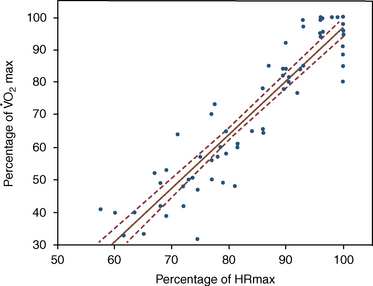
 O2max and is, therefore, a suitable measurement of fitness in treadmill tests that do not measure oxygen consumption (Evans and Rose, 1987). This correlation occurs under field conditions, as is well described by Evans (2007), Courouce et al. (2002), Marr and Burton (2010). In addition, the relative heart rate (as a percentage of HRmax) is highly correlated with relative oxygen consumption in horses during treadmill and field exercises (Evans, 2007; Evans and Rose, 1987) (Figure 11-7).
Submaximal exercise heart rates and fitness measurements
![]()
Stay updated, free articles. Join our Telegram channel

Full access? Get Clinical Tree


The cardiovascular system: Anatomy, physiology, and adaptations to exercise and training
 O2max) relative to body weight compared with most other mammals. The superior oxygen transport of the horse is attributed to its specialized spleen, which is able to add an extra volume of red blood cells (RBCs) to the circulation when it contracts after the stimuli of fear, excitement or exercise. This infusion of erythrocytes increases the oxygen-transport capacity of arterial blood and enables horses to greatly increase
  O2max during exercise. The stroke volume (SV) of blood pumped with each cardiac contraction is over 1 liter (L) in trained horses, and maximal rates of blood flow during exercise are likely to be up to approximately 400 liters per minute (L/min) in the most exceptional equine athletes. The structure and function of the equine cardiovascular system are, therefore, fundamental to the superior athletic performance of the horse.
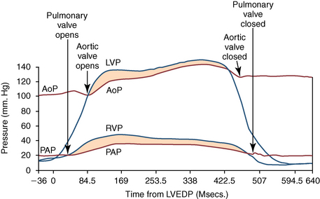
Only gold members can continue reading. Log In or Register to continue
