The autonomic nervous system

Chapter 12 The autonomic nervous system


The ANS is a diffuse system that innervates visceral structures, glandular myoepithelium, fat and vasculature throughout the body. The system has afferent, central and efferent components. Afferent fibres often use the same pathways as the efferent nerves. Afferent visceral fibres may also travel via somatic spinal nerves to reach the CNS (see also Chapter 1, p4 for introduction to the ANS).


It is an involuntary system in which effects occur in response to physiological changes within the body and do not require voluntary control. For example, exercise stimulates heart and respiratory rates, blood flow to muscles and sweating. Conversely, eating stimulates increased activity in the gastrointestinal tract, its blood flow and secretions. The ANS aims to preserve a constant internal environment.


Subdivisions


The ANS is subdivided both functionally and anatomically. The parasympathetic system, also known colloquially as the ‘rest and digest’ system, is responsible for processes that conserve and restore energy. It functions in the day-to-day control of viscera for basic ‘ticking over’ type functions, for example, breathing at rest, digestion and elimination of wastes.


The sympathetic system, also called the ‘fight or flight’ system, functions when the animal is stressed, that is, when confronted by a need to fight or flee. It enables vigorous physical activity with rapid production of energy (ATP). Thus it is responsible for increasing heart rate, respiration, diverting blood flow to active muscles and pupil dilation for increased vision.


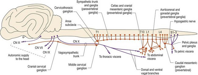
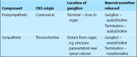
Anatomically, the two systems arise from different areas of the CNS, with the parasympathetic system arising from the brain and sacral spinal cord, and the sympathetic system from the thoracolumbar spinal cord. Hence, they are also known as the craniosacral and thoracolumbar systems, respectively (Fig. 12.1).


Despite the different origins, each organ receives both sympathetic and parasympathetic input. The balance of input from each system determines the organ’s function.


The neurotransmitter elaborated at the terminal determines the physiological effect with the parasympathetic system being cholinergic, using acetylcholine, and the sympathetic system being adrenergic, using noradrenaline (norepinephrine). Adrenaline and noradrenaline are also secreted by the adrenal medulla into the circulation enhancing the effect of sympathetic nervous system stimulation.


ANS: Central components and peripheral components


In the CNS, the main control centres for the ANS are located in the hypothalamus; these can be considered to be autonomic UMNs. The rostral hypothalamus influences the parasympathetic system and the caudal hypothalamus influences the sympathetic system. Caudally directed fibres synapse in the brain stem and sacral cord (parasympathetic system) or the thoracolumbar cord (sympathetic system). The cerebrum and limbic system can influence but not command the control centres. For example, emotional states, such as aggression or fear, cause piloerection (raising the ‘hackles’). Other parts of the CNS can also influence ANS function as exemplified by olfactory stimulation causing drooling. The hypothalamus integrates autonomic activities associated with temperature regulation, hunger, thirst, sleep, endocrine function and motility of viscera including the gut and urinary bladder. It is also connected to various autonomic brainstem centres that regulate cardiovascular and respiratory function. The cardiovascular centre of the medullary reticular formation can stimulate or depress heart rate. The respiratory centres in the pons and medulla control inspiration and expiration; dysfunction causes abnormal respiration. These areas are regulated by centres in the hypothalamus and the cerebrum.


Afferent and efferent fibres of the ANS travel via the spinal and cranial nerves to connect between the CNS and the target organ.


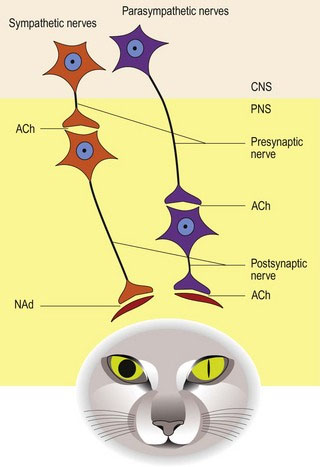
Two-neuron system in the periphery


The ANS comprises two lower motor neurons in series compared with the single LMN in the somatic nervous system. The cell body of the first neuron is in the CNS and it synapses with the second neuron in a peripheral ganglion. The postsynaptic fibre then synapses with the target organ. Presynaptic axons are myelinated and postsynaptic are non-myelinated.


The location of the second neuron is system specific. For parasympathetic fibres, the ganglion is terminal or intramural; that is it lies close to, or within the wall of, the organ being innervated. Therefore it has a long presynaptic neuron and short postsynaptic neuron.


For sympathetic fibres, the ganglion is remote from the organ being innervated. These ganglia are located ventral, or near to the vertebral column in a prevertebral or paravertebral position, respectively. Thus, the sympathetic system has a shorter presynaptic neuron and longer postsynaptic neuron (Fig. 12.2).


Note that as the presynaptic nerve can pass through a number of ganglia before synapsing, the terms ‘pre-ganglionic’ and ‘post-ganglionic’ can be misleading. Presynaptic and postsynaptic neurons are the preferred terms.


A summary of the key anatomical features of each system is given in Table 12.1.


Visceral afferent system


See also Chapter 6.


Receptors located in viscera throughout the body are sensitive to pressure, stretch and chemical changes. Most viscera are not sensitive to touch or cutting. Axons travel via local cranial nerves (CNN VII, IX, X), branches of sympathetic nerves and spinal nerves. Cell bodies are located in specific ganglia such as the geniculate ganglion of CN VII, the proximal and distal ganglia of CN X, and spinal ganglia.


Input via cranial nerves (CNN VII, IX, X) goes to the solitary tract and its nucleus in the medulla oblongata. The efferents from this nucleus go to reticular formation for reflex function (respiratory, cardiac, digestive, elimination). The solitarothalamic tract also conveys information to the thalamus and hence to the somatosensory cortex for conscious perception.


Input via segmental spinal nerves goes via the dorsal horn, synapses locally for reflex function, or enters the lateral funiculus (both ipsi- and contralateral) and travels cranially to the thalamus and somatosensory cortex.


Gut function can also occur due to local reflexes that do not involve the CNS. Some visceral afferents from the gut make local connections, in enteric plexi, with visceral motor nerves in the wall of the organ, causing local reflex activity. Most of the smooth muscle contractions such as segmentation, peristalsis and defecation, can occur in the denervated gut due to activity of local pacemakers found in the intestinal wall.


Sympathetic/thoracolumbar division


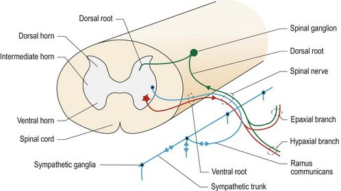
Presynaptic fibres originate in the intermediate (lateral) horn of the thoracolumbar spinal cord. Fibres may leave in the ventral root from their spinal cord segment of origin, or they may pass cranially, or caudally, a number of segments within the spinal cord before exiting it. The fibres exit the spinal cord along with the somatic motor neurons using the ventral roots of C8–L4/5 (up to L7) spinal nerves. The ventral roots fuse with the dorsal roots to form proper spinal nerve at the level of the intervertebral foramen (see Fig. 1.1). Lateral to the foramen, the proper spinal nerve splits into epaxial, hypaxial and ventral branches. The ventral branch forms the ramus communicans (ramus – L = branch). The ramus communicans conveys sympathetic efferent and visceral afferent fibres, between the spinal cord and the bilateral, sympathetic trunks that run ventrolaterally on both sides of the vertebral column (Fig. 12.3). The ramus communicans conveys both presynaptic (myelinated) sympathetic efferent fibres that are travelling to the trunk, and postsynaptic (unmyelinated) fibres that return from the trunk to rejoin the spinal nerves. This gives rise to the names of white and grey ramus communicans, respectively. These paravertebral ganglia and trunks are prominent in the thoracic region and extend into the lumbar region (see Fig. 12.4). In the caudal lumbar area, the trunks may fuse and continue caudally, ventral to the sacral and caudal vertebrae. In the abdominal region, nerves leave the paravertebral sympathetic trunks and connect to prevertebral ganglia located ventral to the vertebral column near the large abdominal arteries. Cranially, the sympathetic fibres continue into the cervical region, in conjunction with the vagus nerve forming the vagosympathetic trunk; this is located in the carotid sheath.


Stay updated, free articles. Join our Telegram channel

Aug 26, 2016 | Posted by in INTERNAL MEDICINE | Comments Off on The autonomic nervous system

Full access? Get Clinical Tree

Get Clinical Tree app for offline access