CHAPTER 3 Selected Infectious Agents
Cytologic evaluation often reveals organisms. Sometimes these organisms are the primary cause of the patient’s condition, whereas at other times they are only secondary invaders or normal flora. This chapter is intended to help in recognizing organisms that can be found during cytologic evaluation. Discussions of the conditions that they produce are found in the relevant chapters. Submission of samples for culture is covered in Chapter 1.
IDENTIFICATION OF ORGANISMS
Bacteria
Bacterial Cocci
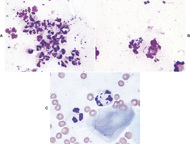
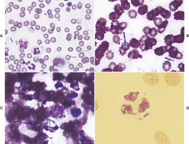
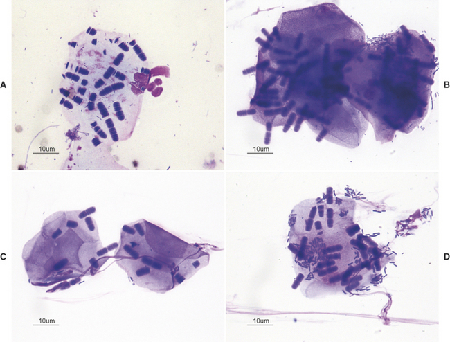
Pathogenic bacterial cocci are usually gram positive and of the genera Staphylococcus, Streptococcus, Peptostreptococcus, or Peptococcus (Figure 3-1). Staphylococci usually occur in clusters of 4 to 12 bacteria, but Streptococcus, Peptostreptococcus, and Peptococcus spp. tend to occur in short or long chains of organisms. Staphylococcus and Streptococcus spp. are aerobic, and Peptostreptococcus and Peptococcus spp. are anaerobic. When cocci are identified in cytologic preparations, aerobic and anaerobic cultures and sensitivity tests should be performed to identify the organism and the optimum antibiotic therapy. Because most cocci are gram-positive, antibiotic therapy effective against gram-positive organisms should be used when it is necessary to start therapy before culture and sensitivity results are received.
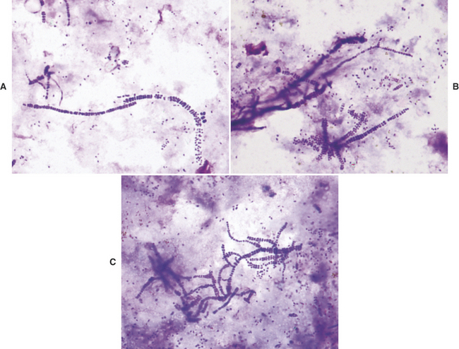
Dermatophilus congolensis replicates by transverse and longitudinal division, producing long chains of coccoid bacterial doublets that resemble small, blue railroad tracks (Figure 3-2). It infects the superficial epidermis, causing crusty lesions. Cytologic preparations from the undersurface of scabs from these crusty lesions are most rewarding in demonstrating organisms. The preparations usually contain mature epithelial cells, keratin bars, debris, and organisms. A few neutrophils may also be found.
Small Bacterial Rods
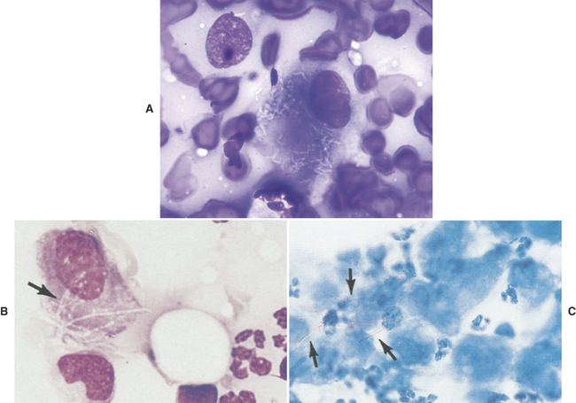
Most small bacterial rods are gram-negative. Some can be recognized as bipolar rods (Figure 3-3). All pathogenic, bipolar bacterial rods are gram-negative. Common small bacterial rods include Escherichia coli and Pasteurella spp. Infections with bacterial rods are usually associated with a marked neutrophilic inflammatory response. When small bacterial rods are recognized in cytologic preparations, the lesion should be cultured to identify the organism, and sensitivity tests should be performed to determine the optimum antibiotic therapy. If it is necessary to institute antibiotic therapy before the culture and sensitivity results are received, the therapy employed should be effective against gram-negative organisms because most pathogenic, small rods are gram-negative.
Filamentous Rods
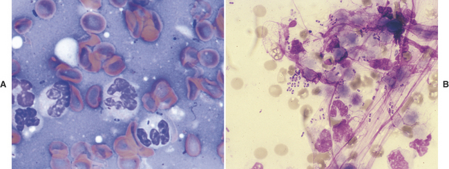
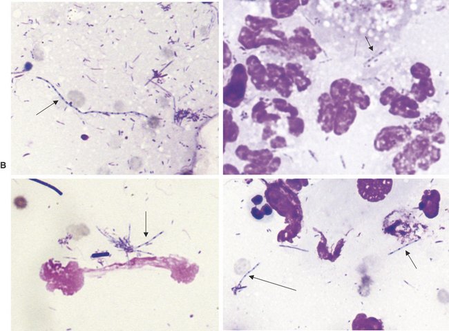
Pathogenic filamentous rods that cause cutaneous or subcutaneous lesions are usually Nocardia or Actinomyces spp. Some other anaerobes, such as Fusobacterium and Mycobacterium spp., may be filamentous, but rarely are. Nocardia and Actinomyces spp., generally have a distinctive morphology in cytologic preparations stained with Romanowsky-type stains. They are characterized by long, slender (filamentous) strands that stain pale blue and have intermittent small pink or purple areas (Figure 3-4). This morphology is characteristic of both Nocardia and Actinomyces spp. and the filamentous form of Fusobacterium spp. When these features are recognized cytologically, cultures should be performed specifically for Nocardia and Actinomyces spp. and for other anaerobes.
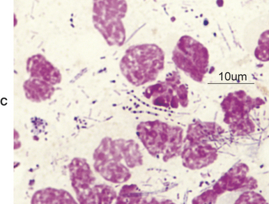
On the other hand, Mycobacterium spp. often does not stain with Romanowsky-type stains. As a result, negative images (Figure 3-5) may be observed in the cytoplasm of macrophages and/or inflammatory giant cells. When epithelioid macrophages and/or inflammatory giant cells are encountered in cytologic preparations that do not contain any obvious organisms, a careful search for negative images of Mycobacterium spp. should be made. Mycobacterium spp. stain with acid-fast stains; therefore when negative images are encountered, or when the character of the lesion suggests that Mycobacterium spp. be considered, an acid-fast stain can be performed to show the organism (see Figure 3-5, C), and/or cultures for Mycobacterium spp. can be performed for identification.
Large Bacterial Rods
Large bacterial rods that are pathogenic and sometimes infect the cutaneous and subcutaneous tissues include Clostridia spp., and infrequently, Bacillus spp. When large bacterial rods are thought to be pathogenic, both aerobic and anaerobic cultures should be performed. Also, the smears should be inspected for large rods containing spores (Figure 3-6). If spore formation is observed, Clostridium spp. is most likely present.
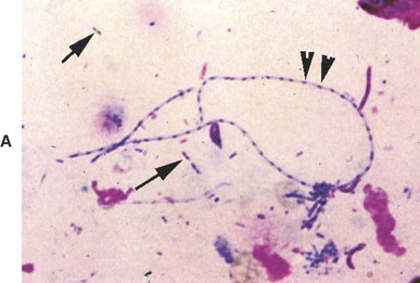
Occasionally, the extremely large nonpathogenic bacterial rods of Simonsiella spp. are observed in cytologic specimens (e.g., contaminated tracheal washes). Simonsiella spp. are bacteria that divide lengthwise, therefore yielding parallel rows of bacteria that give the impression of a single, large bacterium (Figure 3-7).
Yeast, Dermatophytes, Hyphating Fungi, and Algae
General Comments
Mycotic agents produce lesions that tend to have more macrophages present than do bacterial lesions. However, neutrophils may still be the predominant cell type, and eosinophils may be plentiful with certain hyphating fungi. Mycotic lesions often contain low numbers of lymphocytes, plasma cells, and fibroblasts. Mycotic agents tend to be phagocytized by macrophages, but may also be found in granulocytes. The infectious agent, the location of the lesion, the chronicity of the lesion, and the immune status of the animal all influence the character of the lesion. A comparison of the common fungi that form only yeasts in tissues is presented in Table 3-1.
Table 3-1 Common Fungi That Form Yeasts in Tissue
| Yeast | Distinguishing Characteristics |
|---|---|
| Small Yeasts | |
| Sporothrix spp. | Round to oval to fusiform (cigar-shaped) organisms that are about 3-9 microns long and 1-3 microns wide, and stain pale to medium blue with a slightly eccentric pink or purple nucleus. |
| Histoplasma spp. | Round to oval but not fusiform; yeasts are about 2-4 microns in diameter (one fourth to one half the size of an RBC), and stain pale to medium blue with an eccentric pink-to-purple staining nucleus that is often crescent shaped. There is usually a thin, clear halo around the yeast. |
| Medium-sized Yeasts | |
| Blastomyces spp. | Organisms are blue, spherical yeasts that are about 8-20 microns in diameter and thick-walled. Occasional organisms showing broad-based budding are found. Blastomyces organisms are differentiated from Cryptococcus neoformans by their color, lack of a clear-staining capsule, and broad-based budding. They are distinguished from Coccidioides immitis by their smaller size and lack of endospores. |
| Cryptococcus spp. | Extremely pleomorphic yeast that ranges from spherical to fusiform shape, has a smooth (encapsulated) and rough (nonencapsulated) form, and is about 4-15 microns in diameter (not including the capsule). The yeast stains pink to blue-purple and may be slightly granular. Cryptococcus spp. demonstrate narrow-based budding. The smooth form usually is easily recognized because of its thick mucoid capsule, which usually is clear and homogenous. |
| Large Yeasts | |
| Coccidioides spp. | These organisms are scarce in many cytology specimens; therefore cytologic preparations from suspected coccidioidomycosis lesions should be examined carefully. When present, the organisms are large (10-100 microns in diameter), double-contoured, blue or clear spheres with finely granular protoplasm, and will often appear folded or crumpled. Round endospores from 2-5 microns in diameter may be seen in some of the larger organisms. The tremendous variation in the size and presence of endospores differentiates Coccidioides immitis from nonbudding Blastomyces dermatitidis. |
Stay updated, free articles. Join our Telegram channel

Full access? Get Clinical Tree


