Secretions of the Gastrointestinal Tract


Secretions of the Gastrointestinal Tract


Key Points


The salivary glands


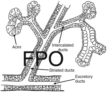
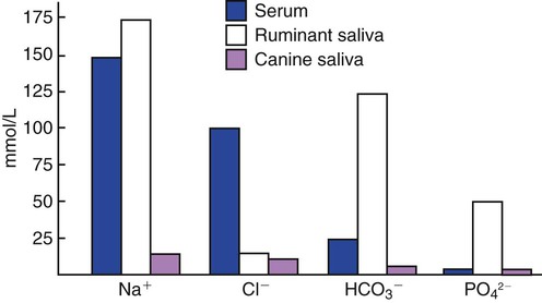
1. Saliva moistens, lubricates, and partially digests food.


2. Salivary secretions originate in the gland acini and are modified in the collecting ducts.


3. Salivary glands are regulated by the parasympathetic nervous system.


4. Ruminant saliva is a bicarbonate-phosphate buffer secreted in large quantities.


Gastric secretion


1. Depending on the species, there may be two general types of gastric mucosa: glandular and nonglandular.


2. The gastric mucosa contains many different cell types.


3. The gastric glands secrete hydrochloric acid.


4. Pepsin is secreted by gastric chief cells in an inactive form and is subsequently activated in the stomach lumen.


5. The parietal cells are stimulated to secrete by the action of acetylcholine, gastrin, and histamine.


The pancreas


1. Pancreatic exocrine secretions are indispensable for the digestion of the complex nutrients: proteins, starches, and triglycerides.


2. Acinar cells secrete enzymes, whereas centroacinar cells and duct cells secrete an electrolyte solution rich in sodium bicarbonate.


3. Pancreatic cells have cell surface receptors stimulated by acetylcholine, cholecystokinin, and secretin.


Bile secretion


1. The liver is an acinar gland with small acinar lumina known as canaliculi.


2. Bile contains phospholipids and cholesterol maintained in aqueous solution by the detergent action of bile acids.


3. The gallbladder stores and concentrates bile during the periods between feeding.


4. Bile secretion is initiated by the presence of food in the duodenum and stimulated by the return of bile acids to the liver.


Digestion and absorption can take place only in the aqueous milieu of digestive secretions. Synthesis and secretion of these fluids represent a well-controlled process regulated by endocrine, paracrine, and neural events. The total volume of digestive secretions is large, with the daily amount substantially larger than the volume of fluid ingested over a similar period. In addition, most of the digestive secretions have a relatively large concentration of electrolytes. This large outpouring of fluid and electrolyte into the gut makes reabsorption of these secretions imperative if fluid and electrolyte homeostasis of the body is to be maintained. Indeed, one of the major life-threatening ramifications of digestive diseases is the loss of water and electrolytes from the body caused by inadequate reabsorption of digestive secretions.


The Salivary Glands


Saliva Moistens, Lubricates, and Partially Digests Food


As food is chewed, it is mixed with salivary secretions that allow it to be molded into well-lubricated boluses that facilitate swallowing. In addition, saliva may have antibacterial, digestive, and evaporative cooling functions, depending on the species.


The antibacterial activity of saliva results from antibodies and antimicrobial enzymes known as lysozymes. Initially, you may think that the antibacterial properties of saliva are inefficient because the mouth normally contains a large, thriving population of bacteria. However, saliva aids in keeping this population in check, and animals with impaired salivary function are prone to infectious diseases of the oral cavity.


In omnivorous animals, such as rats and pigs, saliva contains a starch-digesting enzyme known as salivary amylase. This enzyme is usually absent from the saliva of carnivorous animals, such as cats. The saliva of some species also contains a fat-digesting enzyme known as lingual lipase. This enzyme is frequently present in young animals, such as calves, while they are on a milk diet; the enzyme disappears as they mature.


Salivary enzymes probably have their major digestive effect in the proximal stomach, because food is not retained in the mouth long enough to permit extensive digestion. The lack of mixing activity in the proximal stomach may be essential for the starch-digesting function of saliva. This is because the amylase enzyme is functional at neutral to slightly basic pH, which characterizes saliva. The low pH of the distal stomach probably inactivates the enzyme; therefore it may be important that food entering the stomach initially not be mixed with gastric secretions, so as to allow the salivary enzymes some time to work before being inactivated by gastric acid. Some birds have salivary amylase that is active in the environment of the crop.


The evaporative cooling function of saliva is covered in Chapter 53.


Salivary Secretions Originate in the Gland Acini and Are Modified in the Collecting Ducts


The salivary gland is a typical acinar gland composed of an arborizing system of collecting ducts that end in cellular evaginations known as acini (Figure 29-1). The cellular epithelium of the acini is functionally distinct from that of the collecting ducts. Saliva is initially secreted into the lumen of the acini. The glandular cells lining the acini secrete water, electrolytes, enzymes, and mucus. As the newly formed saliva progresses through the collecting ducts, its composition is modified. The duct epithelium reabsorbs electrolytes, especially sodium and chloride, in a manner similar to that in the proximal tubules of the kidneys. The final product, saliva, is hypotonic and has a sodium concentration substantially less than that of extracellular fluid. The extent to which the acinar secretion is modified in the collecting ducts depends on the rate of saliva production. At high rates of salivary flow, there is little modification, which results in higher tonicity and electrolyte concentration, in comparison to low rates of flow.


Most mammals have at least three pairs of salivary glands: the parotid glands, which lie just under the ear and behind the vertical ramus of the mandible; the mandibular glands, which are in the intramandibular space; and the lingual glands, which lie in the base of the tongue. Each of these glands drains into a main duct that has a single opening into the mouth. In addition to these major glands, there are minor glands in the tongue and buccal mucosa. These small, indistinct glands often have numerous secretory ducts emptying into the mouth. The concentration of mucus is different in the secretions of the various salivary glands. The parotid gland secretes watery, or serous, saliva, whereas many of the minor glands secrete highly mucous saliva. Other glands secrete a mixed type of saliva containing both mucous and serous material. Avian salivary glands secrete a copious amount of mucus to lubricate unmasticated food for swallowing.


Salivary Glands Are Regulated by the Parasympathetic Nervous System


Autonomic, parasympathetic nerve fibers of the facial and glossopharyngeal nerves end on the secretory cells of the salivary gland acini and stimulate the cells through cholinergic receptors. All phases of salivary activity are stimulated by this mechanism, including electrolyte, water, and enzyme secretion. The anticipation of eating can initiate a parasympathetic response that results in salivary secretion. In Pavlov’s famous experiment, parasympathetic stimulation of the salivary gland was evoked in dogs by the sound of a ringing bell. The dogs had been trained to anticipate eating after hearing the bell. This well-known experiment was one of the first demonstrations that the central nervous system (CNS) could regulate digestive functions. Chewing and stimulation of taste buds, in addition to the anticipation of eating, are afferent stimuli for salivation.


Salivary secretory cells also contain β-adrenergic receptors that are activated by sympathetic nerve stimulation or circulating catecholamines. This form of stimulation probably has little association with normal digestive activity but is related to the salivation and drooling seen in carnivores preparing to attack. Among digestive glands, the salivary glands are unique because there is no endocrine regulatory component.


Ruminant Saliva Is a Bicarbonate-Phosphate Buffer Secreted in Large Quantities


The normal composition of ruminant parotid saliva is quite different from the saliva of monogastric animals. Bovine and canine saliva are compared in Figure 29-2. Ruminant saliva is isotonic and, compared with blood serum, has high concentrations of bicarbonate and phosphate and a high pH. This well-buffered solution is necessary for neutralizing acids formed by fermentation in the rumen, and ruminants secrete it in enormous quantities. An adult cow may secrete 100 to 200 L of saliva per day. This volume is approximately equivalent to the extracellular fluid volume of most adult cattle. It is obvious that much of the water and electrolytes secreted in saliva must be reabsorbed rapidly and recirculated through the total body water, or the cow would die of dehydration. In abnormal circumstances, such as blockage of the esophagus, in which the flow of saliva is diverted from the gastrointestinal (GI) tract, cattle quickly become dehydrated and acidotic.


In general, the salivary glands of domestic animals are seldom involved in disease processes and infrequently require veterinary attention.


Gastric Secretion


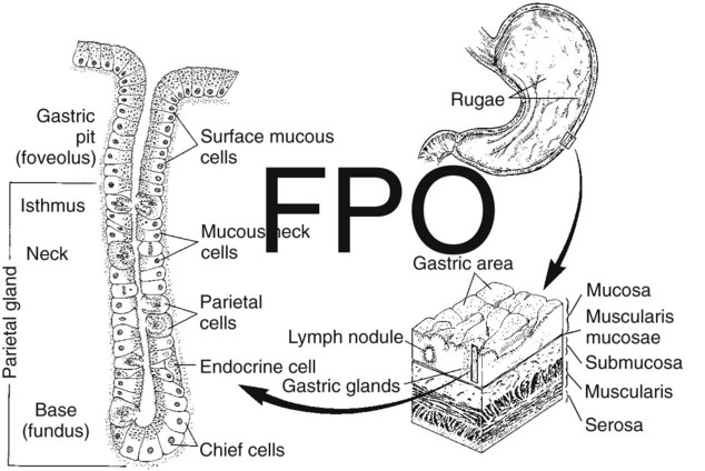
Depending on the Species, There May Be Two General Types of Gastric Mucosa: Glandular and Nonglandular


Most domestic, monogastric animals have only glandular mucosa in the stomach, but horses and rats have an area in the proximal portion of their stomachs that is covered by nonglandular, stratified squamous epithelium. This area is visibly different from the glandular area, to which it adjoins with a sharp line of demarcation. The function of the nonglandular area of gastric mucosa is unclear. The nonglandular area may serve as a place where a small amount of fermentative (rumenlike) digestion could occur. Because there is little mixing activity in the proximal stomach, food in the nonglandular area would be protected from the secretions of the gastric glands. These acid secretions kill bacteria, and thus their presence would prevent fermentation. Fermentative digestion is discussed in detail in Chapter 31.


The glandular area of the stomach is divided into three regions: cardiac mucosa, parietal mucosa, and pyloric mucosa. These areas contain glands of similar structure but with different types of secretions, as described later. In most species the cardiac mucosa forms a narrow band around the gastric opening of the esophagus. In the pig, however, the cardiac mucosa covers a substantial portion of the proximal stomach.


The Gastric Mucosa Contains Many Different Cell Types


The glandular mucosa of the stomach has frequent invaginations, or pores, known as gastric pits. The size of the pits is such that the pores leading into them can be seen with a hand-held magnifying glass. At the base of each pit is a narrowing, or isthmus, that continues into the opening of one or more gastric glands (Figure 29-3).


The major surface areas of the stomach, as well as the lining of the pits, are covered with surface mucous cells. These cells produce thick, tenacious mucus that is a special characteristic of the stomach lining. The mucous cells and their associated secretion are important for protecting the stomach epithelium from the acid conditions and grinding activity present in the lumen. When the mucous cells are injured, stomach ulcers result.


Each region of the mucosa contains glands with characteristic cell types. Within the parietal area, the glands contain parietal cells. These cells are clustered in the neck, or proximal area, of the gland. Their function is to secrete hydrochloric acid (HCl). Distributed among the parietal cells in the neck of the gland is another type of cell, the mucous neck cells. These mucous cells secrete thin mucus, less viscous than that of the surface mucous cells. The mucous neck cells, in addition to their secretory function, appear to be the progenitor cells for the gastric mucosa. They are the only cells of the stomach lining capable of division. As they divide, they migrate either down into the glands or up into the pits and onto the surface epithelium. As they migrate, the mucous neck cells differentiate into any of the several types of mature cells of the gastric surface and glands. In the base of the gastric glands is yet a third type of cell, the chief cells. These cells secrete pepsinogen, the precursor to the digestive enzyme pepsin.


The glands of the cardiac and pyloric mucosal regions resemble those of the parietal area in structure but contain different cell types. The cardiac glands secrete only mucus. Their mucus is alkaline and probably serves to protect the adjacent esophageal mucosa from the acid secretions of the stomach. The pyloric glands have no parietal cells but contain the gastrin-producing G cells. According to most reports, pyloric glands do secrete pepsinogen.


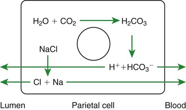
The Gastric Glands Secrete Hydrochloric Acid


When the gastric glands are stimulated maximally, the HCl solution secreted into the lumen is isotonic and has a pH of less than 1. Both the hydrogen (H+) and the chloride (Cl) ions are secreted by the parietal cells but apparently by different cellular mechanisms. H+ is secreted through an H+,K+-ATPase (adenosine triphosphatase) enzyme located on the luminal surface of the cell. This enzyme, sometimes referred to as a proton pump, exchanges H+ for potassium ions (K+), pumping one K+ into the cell for each H+ secreted into the lumen. In the exchange process, one molecule of adenosine triphosphate (ATP) is hydrolyzed to adenosine diphosphate (ADP), representing an expenditure of energy. The K+ cations that accumulate within the cells are released back into the lumen in combination with Cl anions. This allows the recycling of K+ ions as they are pumped back into the cells in exchange for H+, resulting in the net secretion of H+ and Cl, with little net movement of K+.


Hydrogen ions for secretion come from the dissociation of intracellular carbonic acid (H2CO3), leaving a bicarbonate ion (HCO3) in the cell for each H+ secreted into the lumen (Figure 29-4). Carbonic acid originates from water and carbon dioxide through the action of carbonic anhydrase, an enzyme found in high concentration in the gastric mucosa.


As hydrogen cations are secreted, bicarbonate anions accumulate in the cell. To counterbalance this accumulation, bicarbonate anions are exchanged for chloride anions at the cell’s nonluminal surface. In this manner, additional chloride is made available to the cell for secretion into the glandular lumen, and bicarbonate is secreted into the blood. During periods of intense secretion by the gastric glands, large amounts of bicarbonate are released into the blood. This transient and mild alkalization of the blood during digestion is known as the alkaline tide. Normally, the alkaline tide is reversed when bicarbonate in the blood is consumed indirectly during the neutralization of gastric secretions as they enter the intestine (see the section on pancreatic secretions later in this chapter). Thus, on a total-body basis, gastric acid production results in only small and transient changes in blood pH. In disease states, however, in which the secretions of the stomach are prevented from entering the intestine or are lost from the body because of vomiting, the pH of the blood can rise to dangerously high values.

< div class='tao-gold-member'>

Stay updated, free articles. Join our Telegram channel

Jul 18, 2016 | Posted by in PHARMACOLOGY, TOXICOLOGY & THERAPEUTICS | Comments Off on Secretions of the Gastrointestinal Tract

Full access? Get Clinical Tree

Get Clinical Tree app for offline access