19 Rabbit dacryocystitis
CLINICAL EXAMINATION
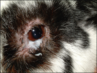
Ophthalmic examination will confirm the presence of a copious mucopurulent discharge centred around the medial canthus (Figure 19.1). Samples should be taken for bacterial culture and sensitivity (although samples from nasolacrimal flushings will be more representative – see below). The discharge can then be gently removed, including from the ventral fornix, with damp gauze swabs to facilitate examination. Conjunctival hyperaemia is normally present, often more severe at the medial canthus. Gently pressing the area will result in pus appearing from the single ventral nasolacrimal punctum. The cornea should be evaluated and fluorescein dye used – some patients will have ventromedial corneal ulceration present (presumably related to excoriation by the discharge). Intraocular contents are normally unremarkable although a mild reflex uveitis can be present.
CASE WORK-UP
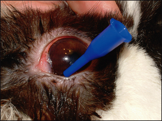
The most important diagnostic test, which is also an important part of treatment, is to perform a nasolacrimal flush in exactly the same way as in dogs. Most rabbits tolerate this conscious (Figure 19.2). This will differentiate dacryocystitis from a solely conjunctival disease and samples can be obtained for bacterial culture and sensitivity. Once the discharge has been bathed away, including pressing at the medial canthus a couple of times and removing the extra discharge thus produced, topical anaesthetic is instilled into the conjunctival sac. The ventral punctum is cannulated (rabbits do not have an upper punctum) and sterile saline is gently injected. It is likely that initially there will be marked resistance and backflow of purulent discharge around the cannula. However, with repeated gentle attempts hopefully the discharge will be loosened and milky fluid will drip from the ipsilateral nasal punctum. Further flushing and treatment is described below.
Stay updated, free articles. Join our Telegram channel

Full access? Get Clinical Tree


