13 Rabbit conjunctivitis
PRESENTING SIGNS
The owner’s usual complaint is of a red eye, but frequently it is also sore, and often quite significant discharge is present. Rabbits of any age or breed can be affected and one or both eyes can be involved. If the rabbit is housed with others these too could be affected. The rabbit is usually clinically well, although some might be eating less, and some will have a nasal discharge and the owner might report noisy breathing.
CASE HISTORY
The condition is usually sudden in onset, and often one eye is affected initially, closely followed by the other. There is frequently no relevant previous history, although sometimes the condition will coincide with a change in management – different bedding, feeding from different containers, a change in placement of the outdoor run, or the introduction of a new rabbit to the household. Some rabbits will have a history of previous ocular or respiratory infections.
CLINICAL EXAMINATION
General clinical examination can be variable. In many animals it will be normal but a significant number will have evidence of respiratory disease – nasal discharge, increased respiratory rate and effort, mild pyrexia and so on. Sometimes the rabbit will be thin. As with all rabbits which are presented to the surgery their teeth should be checked!
On ophthalmic examination the affected eye(s) will be obviously red and inflamed. Thus marked conjunctival hyperaemia will be present, usually affecting both bulbar and palpebral conjunctiva, and moderate chemosis is frequently also present. Sometimes the conjunctiva is so swollen that visualization of the eye itself is difficult. Moderate blepharospasm is present, frequently with significant lid swelling as well as hyperaemia. The lids should be checked for signs of entropion which can occur in young rabbits and will cause secondary conjunctival hyperaemia. An ocular discharge is almost always present but can be very variable in nature. Initially it is usually serous but rapidly becomes mucopurulent and can be very thick and sticky – adhering to the eyelids and medial canthal skin and sometimes causing local skin excoriation.
The cornea should always be stained with fluorescein to check for evidence of corneal ulceration but this can be difficult in very painful eyes with excessive chemosis. A slight miosis can be present but in general intraocular examination is unremarkable.
CASE WORK-UP
Although swabs for bacterial culture and sensitivity are not usually taken from rabbits on initial presentation – empirical treatment is usually tried first – it is certainly something to consider: they can always be discarded if necessary but cannot be retaken after the eye has been cleaned up! Cytology can also be considered.
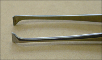
It is important to check for any foreign body – fragments of hay, straw and sawdust frequently become trapped in the conjunctival fornix or behind the third eyelid. Topical anaesthetic drops are applied to the eye and to a cotton bud. The latter can be gently rolled in the lower and upper fornix and will attract any debris. Flush copiously with sterile saline – a 5 ml syringe with a cut-off nasolacrimal cannula on the end is ideal for flushing the fornix. The third eyelid can be grasped with atraumatic forceps – von Graefe forceps (Figure 13.1) are ideal since they grip well without damaging the conjunctiva – and it is gently elevated to check behind for any foreign matter.

Stay updated, free articles. Join our Telegram channel

Full access? Get Clinical Tree