8 Prolapsed nictitans gland
PRESENTING SIGNS
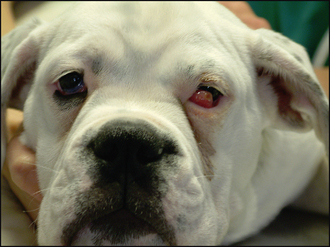
The patient will be presented due to a sudden change in appearance of the eye and the owner will have noticed a pink fleshy mass at the medial canthus (Figure 8.1). Patients are normally young dogs, 3–12 months old, although sometimes cats (e.g. Burmese) can be affected. Very occasionally an older dog will present which does not fit into the normal signalment. In this instance it is necessary to look for an underlying predisposing cause such as keratoconjunctivitis sicca which will be discussed briefly later. The condition is initially unilateral but has a definite bilateral risk. The patient is normally unconcerned about the appearance of the lump, with minimal discomfort and usually only a very mild serous ocular discharge. The mass can become very inflamed in appearance and slightly swollen within a couple of days and the discharge can become mucopurulent due to secondary bacterial infection.
CLINICAL EXAMINATION
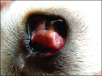
On close examination the mass can be seen extending from the bulbar aspect of the third eyelid, and the leading edge of the nictitans membrane cannot be seen. Sometimes there is kinking, or scrolling, of the cartilage of the nictitans membrane (Figure 8.2), although this might not be appreciated in the conscious patient when the gland is fully prolapsed. No ulceration is usually present. Schirmer tear tests should be performed and are usually normal or marginally reduced (presumably due to inflammation in the gland restricting the secretion of aqueous tears). Intraocular contents are also typically unremarkable.
The globe should be retropulsed to examine the nictitans membrane. Any scrolling or weakness of the cartilage can be examined. The bulbar aspect of the membrane should be examined if there is any concern about the gland’s position. A cotton bud soaked in topical anaesthetic is held against the third eyelid for 30–60 seconds and then it can be grasped with fine fixating forceps (e.g. Von Graefe forceps, Figure 13.1) and the underside examined. In addition to checking the position of the gland, its size is evaluated and the presence of any follicles or inflammatory lesions on the bulbar aspect noted.
EPIDEMIOLOGY
Stay updated, free articles. Join our Telegram channel

Full access? Get Clinical Tree


