Chronic superficial keratitis

29 Chronic superficial keratitis


CLINICAL EXAMINATION


General clinical examination is normal. On ophthalmic examination one or both eyes can be affected, but the condition is rarely symmetrical. The most striking lesion is a fleshy growth of tissue on the cornea. It normally starts in the ventrolateral or occasionally dorsolateral quadrant, with its base at the limbus growing centrally (Figures 29.1 and 29.2). The lesion is usually pink, but can be darkly pigmented. It appears vascular. The cornea at the leading edge frequently has patchy white deposits within the anterior stroma which can be cellular infiltrates or lipid. No fluorescein uptake occurs (although false positives are common on the raised fleshy tissue and copious flushing with sterile saline is required to show that these areas are not ulcerated). The conjunctiva is hyperaemic in the adjacent bulbar conjunctiva, and sometimes this can extend around the circumference of the globe. The nictitating membrane can be involved with thickening (with a cobblestone appearance) and depigmentation of the leading edge in about 10% of dogs. Vision can be reduced but unless the condition is very severe animals are not normally blinded. Schirmer tear test readings are normal. Intraocular contents are unaffected. Sometimes a mucoid or mucopurulent discharge is present.


image

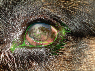
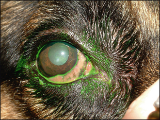
Figure 29.2 Right eye of dog in Figure 29.1 with lesions confined to the nictitans membrane (nictitans plasmacytic conjunctivitis).


EPIDEMIOLOGY


Chronic superficial keratitis (CSK), or pannus, is the subepithelial proliferation of blood vessels and connective tissue and the term implies a bilateral superficial keratitis which has an immune-mediated aetiology. Since certain breeds are predisposed to developing the condition (namely German shepherd dogs far more frequently than any other breed), a genetic predisposition is proposed. In addition, the condition is more common, and more severe, in areas of high altitude and long hours of bright sunlight. Thus ultraviolet light is a contributing factor and it is possible that the UV light alters certain corneal antigens in animals with a genetic predisposition and thus triggers the disease. Young adult dogs are affected mostly (3–5 years) although some patients are older before the symptoms develop. The condition is generally more benign in older dogs, and can be controlled with less intensive treatment than in younger animals.


Histologically, the disease is characterized by neovascularization and fibroplasia with a mixed cellular infiltrate consisting of plasma cells, lymphocytes, macrophages and melanocytes. The proportion of these cells varies and is a reflection of the clinical picture – some cases of CSK are more heavily pigmented than others for example. The corneal epithelium remains intact, with no ulceration, but it does alter in structure, becoming thin in some places and hyperplastic in others. Occasionally the pathology is confined to the nictitating membrane and it is then termed nictitans plasmacytic conjunctivitis or plasmoma, rather than chronic superficial keratitis. The underlying pathological mechanisms are identical and it is not understood why some dogs develop one form and not the other.


Sep 10, 2016 | Posted by in SMALL ANIMAL | Comments Off on Chronic superficial keratitis

Full access? Get Clinical Tree

Get Clinical Tree app for offline access