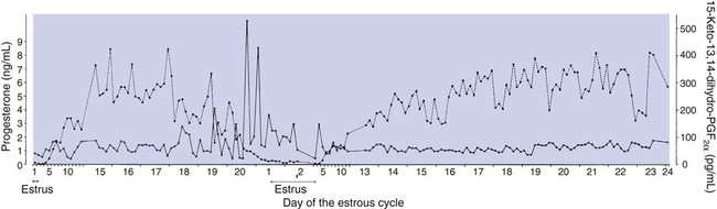
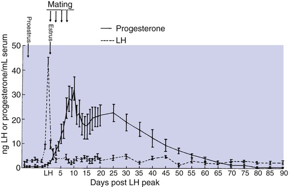
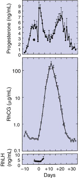
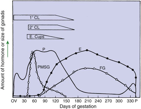
Pregnancy and Parturition


Pregnancy and Parturition


Pregnancy


The Development of an Embryo Involves Fusion of an Oocyte and Spermatozoon Within the Oviduct


The development of a new individual requires the transfer of male gametes to the female genital tract for fertilization of the female gamete(s). Spermatozoa, which have been concentrated and stored in the epididymis, gradually change from oxidative (aerobic) to glycolytic (anaerobic) metabolism as they progress through the epididymis. In this situation, spermatozoa are in a state of reduced metabolism. Mature sperm are only able to metabolize a special sugar, fructose, within the reproductive tract. Lactose, glucose, dextrose, and fructose have all been used in commercially available semen extenders.


Sperm are ejaculated usually into the vagina, although some domestic species (dog, horse, and pig) ejaculate directly into the cervix and uterus. The movement of sperm through the cervix is aided by estrogen-induced changes in cervical mucus, which result in the formation of channels that facilitate movement of sperm. This has been particularly emphasized in primates, in which the thinning of mucus occurs just before ovulation, a factor that can be used to predict the time of ovulation.


The environment of the female genital system is generally inhospitable to the survival of sperm; for example, white blood cells are quickly attracted to the uterine lumen because sperm cells are foreign to the female genital tract. Special reservoirs have evolved in the female tract to aid in the survival of sperm during transport; these include the cervix and oviduct, the latter involving areas at the uterotubule junction and within the ampulla. The reservoirs are progressively filled (from caudal to cranial in the tract), requiring hours before the oviductal reservoirs are full. Finally, the reservoir within the ampulla is able to release a few sperm on a continuous basis, so that fertilization can occur shortly after the arrival of oocytes within the oviduct.


The first studies in sperm transport emphasized the rapidity of the process, with sperm reported passing from the vagina to the fimbriated end of the oviduct within minutes. It is now known that sperm undergoing so-called fast transport are not involved in fertilization; in fact, they are damaged by the rapid transport.


Sperm need to undergo changes within the female genital tract that are a prerequisite for fertilization; the process is called capacitation. One of the effects of capacitation is the removal of glycoproteins from the sperm cell surface.


The glycoproteins, perhaps added for protective purposes, interfere with fertilization. This change allows sperm to undergo the acrosome reaction when they come in contact with oocytes. The acrosome reaction involves the release of hydrolytic enzymes from the acrosomal cap; this may be important for penetration of the sperm through the granulosa and zona pellucida to the oocyte plasma membrane. Hyaluronidase causes breakdown of hyaluronic acid, an important component of the intercellular matrix of granulosa cells that surround the oocyte. Acrosin, a proteolytic enzyme, digests the acellular coating around the oocyte. Both enzymatic events allow the sperm to penetrate to the oocyte. The acrosome reaction also changes the surface of the sperm, which allows it to fuse with the oocyte. The acrosome reaction results in tail movements that feature a flagellar beat that tends to drive sperm in a forward direction.


Because of the changes that spermatozoa must undergo within the female reproductive tract before fertilization, the deposition of sperm before ovulation is the preferred timing for producing maximal fertility. An exception to this takes place when sperm with reduced longevity are used, such as the case with chilled-extended semen or frozen semen. In these cases, deposition of semen into the female reproductive tract should occur close to the time of ova maturation associated with fertilization. Females are usually sexually receptive for at least 24 hours before ovulation and, in the natural setting (free interaction between genders), insemination usually occurs a number of hours before the occurrence of ovulation. Even with induced ovulators, such as cats, the interval from copulation to ovulation is usually 24 hours or more. In essence, the system has evolved to have ready-to-fertilize sperm at the fertilization site when oocytes arrive. This concurs with the finding that the life span of male gametes tends to be twice that of female gametes.


The presentation of male gametes before female gametes in the oviduct implies that oocytes are ready for fertilization on arrival in the ampulla; this is likely true for a majority of animals. A prerequisite for fertilization of the oocyte is that it must undergo the first meiotic division before fertilization. Although this occurs in a number of species before ovulation, in the horse and dog the first meiotic division does not occur until after ovulation (in the dog, not for at least 48 hours). In this situation, spermatozoa often wait for oocytes to mature in the oviduct before fertilization can occur. One means of adaptation to delayed completion of meiosis is that spermatozoa have a longer life span in the dog (6 to 11 days) and horse compared with other domestic species.


After fertilization has occurred, the embryo usually develops to the morula, or early blastocyst stage, within the oviduct before moving into the uterus. This period, lasting usually 4 to 5 days, affords the uterus time to finish its inflammatory response involving the removal of spermatozoa. This period also allows the endometrial glands time to secrete nutrients under the influence of progesterone from the developing corpus luteum (CL); the nutrients are essential for the development of embryos during their preimplantation stage.


An interesting finding in the mare is her ability to distinguish fertilized from unfertilized oocytes; unfertilized oocytes from previous cycles are retained within the oviduct, whereas recently fertilized oocytes (embryos) move through the oviduct to the uterus. It is likely that all animals recognize pregnancy by the presence of an embryo(s) at the early oviductal stage. However, this recognition does not necessarily result in prolongation of the CL and the continued production of progesterone, which is essential for the maintenance of pregnancy. In the bitch, despite ovulation and ova maturation spanning several hours, embryonic ages are synchronized by some mechanism inherent to the bitch’s reproductive tract.


Extension of the Life Span of the Corpus Luteum in Large Domestic Species and Cats Is Essential for Pregnancy Maintenance


For those domestic animals (cattle, goats, horses, pigs, sheep) whose luteal activity is controlled by the uterus, modification of uterine prostaglandin F (PGF) synthesis and release is critical for the establishment of pregnancy. The embryo apparently produces substances that modify uterine production of PGF. Estrogen synthesis by the embryo is one way the endometrium may be informed regarding the presence of an embryo. A specific protein of embryonic origin called trophoblastin, produced before day 14 of pregnancy (or postovulation) in both sheep and cattle, is of immunological interest for the establishment of pregnancy; it has a close structural relationship to the molecule interferon. Movement of the embryo(s) in the tract is also important for pregnancy recognition. In the mare the embryo moves throughout both horns before being fixed at day 16. In pigs a minimal number of embryos need to be present (about four), presumably to occupy a sufficiently large area of the endometrium. Litter-bearing animals also use transuterine migration to maximize the opportunity for fetal development, a procedure that aids the recognition of pregnancy process. The end result is either suppression of PGF synthesis, as seen in the cow (Figure 38-1), or modification of the secretion mode (continuous instead of pulsatile), as seen in sheep. The absence of pulsatile secretion of PGF seems to be critical for the extension of the life span of the CL and the establishment of pregnancy in large domestic species.


In the cat the CL lasts for 35 to 40 days after ovulation regardless of the presence of pregnancy, and thus early modification of luteal activity is not essential for the establishment of pregnancy. Implantation occurs at about day 13, which allows the fetoplacental unit to influence and extend luteal activity that is compatible with pregnancy maintenance. The luteotropic hormone that is responsible for luteal maintenance in the cat is not known. One hormone that likely synergizes with progesterone for the support of pregnancy is relaxin, a placental hormone produced in the cat beginning at about day 20 of gestation (see later discussion).


The dog does not extend its luteal phase during pregnancy; the luteal phase in the nonpregnant animal is often slightly longer (70 days) than in pregnant animals. Nevertheless, enhancement of luteal activity occurs through a placental luteotropin, likely relaxin, with progesterone secretion enhanced beginning at about day 20 of gestation or a few days after implantation. Early in the luteal phase, luteal function in the bitch is likely autonomous. During the second half of the luteal phase, luteinizing hormone (LH) and prolactin are likely luteotrophs (Figure 38-2).


The rescue of the CL at the onset of pregnancy in primates involves the production of a luteotropin called chorionic gonadotropin (CG; for humans, hCG), which is produced by trophoblastic cells (syncytiotrophoblasts) of the embryo (Figure 38-3). For trophoblast tissue to produce CG, it must have intimate contact with the interstitium of the endometrium. This contact occurs by a type of implantation called interstitial, in which the embryo penetrates the endometrium at about 8 to 9 days after fertilization in humans and nonhuman primates. Secretion of CG begins 24 to 48 hours after implantation, with immediate enhancement of luteal progesterone production. Rescue of the CL in human pregnancy occurs as late as 4 to 5 days before the end of the luteal phase.


As indicated, interstitial implantation is essential to the development of pregnancy in primates. Implantation is less invasive in the dog and cat, with the type termed eccentric. In the large domestic species, “invasion” of the endometrium is minimal; implantation occurs within special endometrial protrusions called caruncles in ruminants and by relatively minor villus invasion of the endometrium in horses and pigs. Domestic animals depend more on uterine secretions for the support of pregnancy than do primates. For cattle and horses the first indications of implantation begin about days 25 to 30, and another 7 to 10 days likely passes before a significant amount of embryonic nutrition is obtained through the implantation site. Subclinical uterine infections, or an inadequate number of endometrial glands, can interfere with the establishment of pregnancy in the species in which a long interval exists from fertilization to implantation. The cervix forms an important barrier to contamination of the uterine lumen in both the nonpregnant and the pregnant animal; in the latter the cervix becomes sealed.


The Placenta Acts as an Endocrine Organ


Besides the essential role of providing nutrients and oxygen for embryonic metabolism, the placenta functions as an endocrine organ. One of the most important functions of the placenta is the production of progesterone. In primates this function is established early in gestation, and the placenta likely can maintain pregnancy within 2 to 3 weeks after implantation in primates. Placental production of sufficient progesterone to maintain pregnancy occurs later in domestic animals (sheep, day 50 of 150-day gestation; horse, day 70 of 340-day gestation; cat, day 45 of 65-day gestation); in some species the placenta never produces enough progesterone to support pregnancy (cattle, goats, pigs, dogs).


The production of estrogen, in contrast to that of progesterone, requires interaction between the fetus and the placenta. This interaction has been best described in primates, in particular by the experiments of the Hungarian immigrant to Sweden Egon Dicfaluszy. He and his co-workers found that the primate placenta is unable to produce estrogen from progesterone even though the steroids are only separated by androgens in the steroid biochemical synthetic pathway. The placenta simply does not possess the enzymes necessary for the conversion of progesterone to androgens. Therefore a system has evolved in which the placenta supplies pregnenolone, the immediate precursor of progesterone, to the fetus, and the fetal zone of the adrenal cortex transforms pregnenolone to a C-19 androgen, dehydroepiandrosterone. This is returned to the placenta, which is able to convert dehydroepiandrosterone to an estrogen. In humans the primary estrogen of pregnancy is estriol. Because the fetus is involved in the production of estriol, the well-being of the fetus can be judged by determining estriol concentrations in the plasma of the mother.


The production of estrogen in the mare also involves an interaction between the placenta and fetus (Figure 38-4). From the work of Pashen and Allen, we know that the fetal gonads replace the fetal adrenals in primates as the key fetal endocrine organ involved in the cooperative synthesis of estrogen. The interstitial cells of the gonads appear to be the interactive cells, with fetal gonads enlarging to a size greater than the maternal gonads during the latter part of gestation. The production of estrogens during pregnancy in other domestic species, occurring relatively late in gestation, may involve the development of placental enzymes that allow progesterone to be metabolized to estrogens without the direct intervention of a fetal endocrine organ. (Fetal cortisol, however, is important for the induction of these placental enzymes, particularly in sheep; see next section.)


< div class='tao-gold-member'>

Stay updated, free articles. Join our Telegram channel

Jul 18, 2016 | Posted by in PHARMACOLOGY, TOXICOLOGY & THERAPEUTICS | Comments Off on Pregnancy and Parturition

Full access? Get Clinical Tree

Get Clinical Tree app for offline access