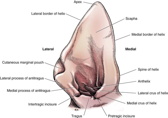
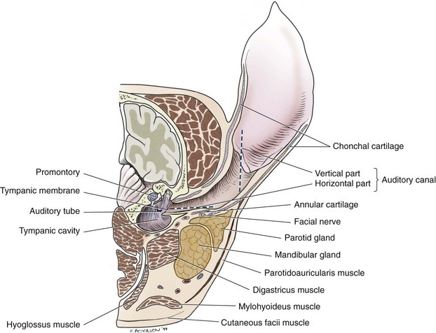
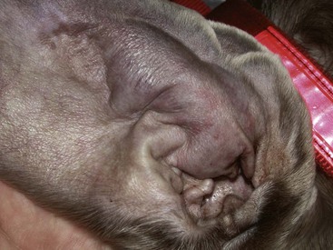
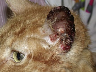
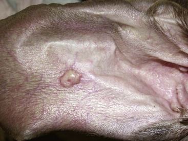
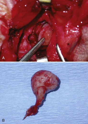
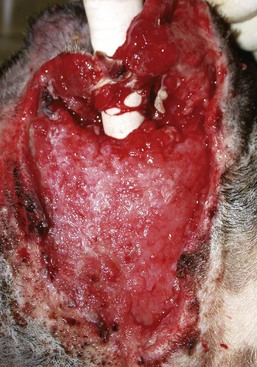
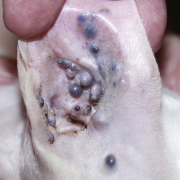
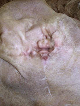
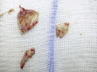
Chapter 122 The canine pinna is a wonderfully diverse structure with breed-related differences in shape, size, and conformation. Less variety is seen among feline breeds. Evolutionarily, the pinna is a focusing and localizing device, directing sound toward the middle ear, and as such has rich muscular, nervous, and vascular contributions. The auricular cartilage of the pinna (scapha) is covered by skin on both sides. The skin is closely fixed to the perichondrium on the concave surface, but some degree of skin movement is possible on the convex surface. Both surfaces have sweat and sebaceous glands.11 The cartilage broadens and thickens toward its base, where it then rolls into a coarse tube. Throughout the auricular cartilage are tiny channels, or foramina, that allow passage of blood from the convex (outer) surface to the concave (inner) surface. The anthelix is a cartilaginous protuberance on the medial auricular surface separating the flat scapha from the beginning of the funnel-shaped external ear canal. Opposite the anthelix is a roughly rectangular dense cartilage plate called the tragus that demarcates the lateral margin of the opening of the ear canal. Caudal to the tragus and delineating the caudal opening of the ear canal is the antitragus. Separating the two is the intertragic incisure. Rostral to the tragus is the helix, forming the cranial border of the ear canal. Separating these two is the pretragic incisure (Figure 122-1). There is a decreasing amount of hair from the apex of the scapha to the base of the pinna. A few fine hairs are found at the entrance to the ear canal.30 Within the rostroauricular muscles medial to the ear is a second cartilage, the scutiform cartilage. This is a small boot-shaped cartilaginous plate that was originally part of the cranial helix spine; because it becomes detached at the time of birth, it is still considered part of the external ear. The vertical ear canal begins at the external acoustic opening at the level of the tragus, antitragus, and anthelix. In a medium-size dog, it travels ventrally and slightly rostrally for 2 to 3 cm before making a medial turn into the horizontal canal. Fitting within the base of the auricular cartilage tube in the horizontal canal is a third cartilage, the annular cartilage. In medium-size dogs, this is approximately 2 cm in length and connects the horizontal canal to the osseus external auditory meatus. The annular cartilage is attached to both the auricular cartilage and the temporal bone by fibrous connective tissue. The osseous auditory meatus in dogs is an approximately 5- to 10-mm extension of the temporal bone.30 In cats, the osseous auditory meatus has a more pronounced flare than in dogs, and the vertical canal also tapers ventrally instead of forming a cylindrical tube.101 The ear canal is lined by stratified squamous epithelium and contains hair follicles and adnexal structures. The sebaceous glands are located more superficially within the epithelium and the tubular ceruminous glands (modified apocrine) deeper. Both are more numerous in the vertical ear canal than the horizontal ear canal. Normal ear secretion, cerumen, is a product of both gland types combined with desquamated epithelium.60 The facial nerve (cranial nerve [CN] VII) supplies motor innervation to the external canal. The nerve exits the cranial vault through the internal acoustic meatus with the vestibular and cochlear nerves and runs through the facial canal of the petrous temporal bone and through the middle ear on its way out of the skull,19 exiting through the stylomastoid foramen at 11 o’clock (caudodorsally) in relation to the osseous ear canal.92 It then crosses the ventral aspect of the horizontal canal (Figure 122-2). The vagus (CN X) supplies sensory innervation to the external ear canal. The great auricular artery, a branch from the external carotid, is the arterial blood supply to the ear canal. It arises medial to the dorsal apex of the parotid salivary gland, which overlies the vertical portion of the canal.54 The external carotid artery and maxillary vein lie immediately ventral to the bulla. Immediately rostral to the osseous ear canal is the retroglenoid vein; sharp dissection and debridement in this area should be avoided. Immediately medial to the bulla is the internal carotid artery; this can be damaged if the medial bulla wall collapses through excessive debridement or if it has been eroded from infection or neoplasia.92 Feline and canine aural hematomas are usually secondary to pinna trauma or a history of violent head shaking and ear scratching because of acute or chronic otitis externa. It is theorized that repetitive pinna trauma results in fracture or splitting of the auricular cartilage and shearing of the blood vessels passing from the convex to concave surface through the foramina in the scapha. The exact location of the hemorrhage is unclear. Subcutaneous, subperichondrial, and intrachondral locations have all been proposed.12,32,52 Regardless of origin, shear forces create a dead space within the pinna that fills with blood, causing the pinna to become distended, swollen, and warm to the touch (Figure 122-3). Without treatment, the clot is converted to granulation tissue by fibroblastic and capillary ingrowth,52 the hematoma cavity fibroses, and the cartilage ossifies, leading to disfigurement and chronic irritation. In cats, distortion of the shorter pinnae can lead to their deviating medially and obstructing the external acoustic opening, further exacerbating the inciting otitis externa. Exposure of poorly pigmented skin to ultraviolet radiation (ultraviolet B) may cause actinic keratoses. Lesions are initially erythematous and hyperkeratotic but become crusting plaques with chronicity. This is a premalignant change and over 3 to 4 years may progress to dysplasia; carcinoma in situ; and ultimately invasive neoplasia, typically squamous cell carcinoma. Lesions are often bilateral but not necessarily of synchronous progression.52 White cats have a 13.4 times greater risk of developing squamous cell carcinoma than colored cats,27 and some poorly pigmented dog breeds are also predisposed (e.g., Dalmatians, bull terriers, Beagles), as are sparsely haired areas of the skin (e.g., ventrum, axilla, face). Lesions are worse in animals that sunbathe.68 Treatment involves pinnectomy or laser surgery. Avoidance of the sun and use of topical sunblock should be considered. Squamous cell carcinoma of the pinna is a raised or erosive painful lesion that is locally invasive into the auricular cartilage. Cats often present with bleeding, cracked, nonhealing wounds of the ear margins (Figure 122-4). As with any epithelial malignancy, an effort should be made to rule out lymph node metastasis; thoracic radiographs are also recommended, although pinnal squamous cell carcinoma has a relatively low metastatic rate. Diagnosis may be possible with a fine-needle aspirate of an appreciable mass; otherwise, a small incisional biopsy is needed. An excisional biopsy (partial or total pinnectomy) may be used to diagnose and treat crusting, scaling, ear-tip disease in a white outdoor cat. Treatment options for pinna squamous cell carcinoma include partial pinnectomy; total pinnectomy; and, occasionally, pinnectomy plus vertical ear canal ablation when lesions are extensive. Bilateral surgeries are frequently performed under the same anesthetic. Cryosurgery, laser ablation, radiation therapy (e.g., strontium-90), and chemotherapy are other treatment options. Hemangioma and hemangiosarcoma are considered ultraviolet B induced. They are most common in light-colored cats and are rare in dogs. Hemangiomas are small, benign, dermal or subcutaneous, raised, alopecic, blue-tinged lesions. Their malignant counterparts are fast-growing, poorly circumscribed, invasive, large tumors.68 Metastasis to the lungs and parenchymal organs is more likely than to local lymph nodes. After surgical excision of hemangiosarcoma in four cats, tumor regrowth occurred in all cats, with a median disease-free interval time of 9.5 months.73 Basal cell carcinoma is the most common feline cutaneous neoplasm and is frequently found on the pinna. Basal cell carcinomas are small, slow-growing, well-demarcated, raised, white to hyperpigmented nodules. Some are so pigmented that they are grossly mistaken for melanoma. Diagnosis is by fine-needle aspirate. Adult Siamese, Himalayan, and Persian cats may be predisposed, and an association with ultraviolet B exposure has been proposed.36 Surgical excision with a few millimeters of skin margin is usually curative, although a much more aggressive phenotype has been described that invaded adjacent structures and demonstrated early lymphatic and vascular invasion.24 Mast cell tumors are the second most commonly diagnosed cutaneous tumor in cats. Those involving the pinna account for 59% of all cutaneous mast cell tumors arising in the head region.72 Siamese cats are overrepresented. Mast cell tumors are usually benign and well-circumscribed, discrete, cutaneous lesions. Excision with a narrow skin margin is normally curative, although this may still necessitate pinnectomy if the mass is located at the base of the pinna. Tumors incompletely excised were not associated with a higher recurrence rate compared with completely resected feline mast cell tumors in one study.75 Mast cell tumors on the pinnae in dogs can present as firm or fluctuant, dermal or subcutaneous, centrally or peripherally located, concave or convex pinna surface, and solitary to multiple (Figure 122-5). Diagnosis is most easily achieved by fine-needle aspirates; lymph node aspirates for metastatic disease are recommended even if nodes are palpably normal. In one study of 24 cutaneous aural mast cell tumors, the incidence of regional lymph node metastasis was reported to be 42.8%, suggestive of a more aggressive biologic behavior than cutaneous mast cell tumors elsewhere in the body.45 Histiocytomas on the pinna in the dog are raised, hairless, erythematous cutaneous masses and appear as malignant round cells on fine-needle aspirate. They are most often seen in young dogs (mean age, 3.5 years) with a male : female ratio of 2.5 : 1.103 Every attempt should be made to differentiate pinna histiocytoma from other malignant round cell tumors. Most histiocytomas resolve spontaneously over several months but become superficially ulcerated during regression, causing pruritus and scratching. Surgery should be avoided for this benign disease of juvenile dogs because of the lifelong cosmetic impact of pinnectomy. If necessary for palliation, conservative surgical resection or cryotherapy could be considered. Sebaceous adenomas are benign skin tumors of the canine pinna. They are common around the head, neck, and feet in older dogs and appear as raised, hairless, white-yellow masses that are often pedunculated, cauliflower-like, and usually less than 5 mm in size. Fine-needle aspiration will yield a diagnosis, and surgical or laser excision is curative.68 Their malignant counterpart, sebaceous adenocarcinoma, tends to be more invasive and metastatic and needs more aggressive surgical excision and potentially adjunctive therapies. Reported infectious diseases that can cause lesions on the pinna include canine leproid granuloma syndrome (mycobacterium), dermatophytosis (Microsporum spp. and Trichophyton spp.), Malassezia dermatitis, feline cowpox virus, Leishmania spp., sarcoptic mange, and Demodex spp. Common inflammatory conditions include allergic contact dermatitis, atopic dermatitis, food hypersensitivity, pemphigus, discoid lupus erythematosus, systemic lupus erythematosus, and vasculitis.68 These are medically managed conditions, and surgery is rarely indicated unless palliative excision of severely affected pinnae is required.76 The clinical signs of otitis externa include head shaking, ear scratching, odor, otorrhea (aural discharge), aural hematomas, swelling, pain, and depression. In long-standing cases, these are largely a result of proliferative changes in the epithelial lining of the canal, characterized by hyperkeratinization, hyperplasia of the sebaceous and ceruminous glands, and infiltration with inflammatory cells,103 leading to stenosis, fibrosis, mineralization, and ultimately occlusion. In a canalographic study of 222 canine ears, 70 were diagnosed as stenotic and underwent total ear canal ablations. Subsequent histopathology of the excised tissue showed hyperplasia of the epidermal layer in 56 of 70 ears and stenosis from otitis externa in the remaining 14.29 Tympanic membrane rupture is often a sequela to severe pathology, with progression to otitis media and, finally, otitis interna with vestibular or auditory damage. The pathophysiology of otitis externa is a combination of primary causes, predisposing causes, and perpetuating causes. It should be noted early on that bacteria and yeasts found in otitis externa are opportunistic organisms, often commensals, and are rarely the primary cause. Treating the infection alone is therefore unlikely to offer long-term resolution of ear disease in the individual.83 Any case of bilateral or recurrent ear disease should raise suspicion for atopic dermatitis or food allergy. Although skin disease and ear disease are commonly found concurrently, 3% to 5% of dogs with atopy present solely with ear disease,40 and 25% of dogs with food allergy have otitis externa as the only clinical sign.84 Altered keratinization and cerumen gland production is a consequence of endocrine disorders such as hypothyroidism, hyperadrenocorticism, and sex hormone imbalances. Consequentially, a ceruminous or seborrheic otitis externa results. Perpetuating factors allow the disease to continue and need to be addressed (in addition to the primary cause) for resolution to occur. The most significant factor is proliferation and overcolonization of bacteria. Culture of normal ears will yield Staphylococcus, Pseudomonas, Streptococcus, and Proteus spp.86 Pathologic populations of bacteria include Staphylococcus intermedius or pseudintermedius, Pseudomonas aeruginosa, Proteus mirabilis, Escherichia coli, Corynebacterium spp., Enterococcus spp., and Streptococcus spp. Overgrowth is typically polymicrobial. In one study examining 100 ears in 50 dogs with bilaterally affected ear canals, the most commonly isolated pathogen (70% of ears) was S. intermedius.77 Because of advances in microbial identification, many of these S. intermedius isolates may actually have been S. pseudintermedius. Most animals were affected with multiple pathogens: a single microorganism was present in 18% of the samples, 62% had two microorganisms, and 20% had three or more microorganisms. Importantly, a statistical difference was observed in the isolation pattern between the left and right ears in 68% of dogs, meaning the practice of obtaining a sample from the more affected ear in a bilaterally affected patient may result in the contralateral ear’s being inappropriately medically treated.77 Anaerobic bacteria were not isolated in any sample in that study77 but have been isolated in one study to date.44 Yeast infection is normally an overpopulation of the commensal Malassezia pachydermatis, which is present in 15% to 49% of normal canine ear canals and in up to 73% to 83% of ears with otitis externa.7,22,39,77 Candida spp. is occasionally isolated (3% of ears with otitis externa). M. pachydermatis has also been recovered from 34.2% of middle ears of dogs with chronic otitis.16 A study in normal Beagles examined the rates of bacterial and yeast isolation in three parts of the ear: the pinna, anthelix, and vertical ear canal. Rates of bacterial isolation were 91.8%, 70.5%, and 48.8%, respectively. The corresponding rates of yeast isolation were 48.8%, 81.4%, and 83.7%, respectively, showing that physiologic factors affecting these three parts must be significantly different and that depth from which any cultures or impression smears are taken may influence the outcome.2 Progression of otitis externa to otitis media through a ruptured tympanic membrane will also establish a perpetuating cycle of recurrent and chronic infection and re-infection of the external ear canal. Rupture of the tympanic membrane was seen in 18% of dogs with bilateral otitis externa in one study.77 A polyp growing into the external ear canal from the tympanic bulla is easily identified as a spherical red proliferation. Inflammatory polyps confined to the middle ear in five dogs have also been described as causing otitis media and externa. All dogs were male and were between 4 and 13 years old. Polypoid disease was unilateral or bilateral and was treated successfully with ventral bulla osteotomy in one dog and total ear canal ablation and bulla osteotomy in four dogs (Figure 122-6).79 The majority of tumors found in the external ear canal of dogs are epithelial in origin,103 and 60% are malignant. There is no significant difference in distribution of tumors between the vertical and horizontal canals.63 Malignant differential diagnoses include ceruminous carcinoma, squamous cell carcinoma, anaplastic carcinoma, and occasionally soft tissue sarcoma and melanoma.63 Cocker spaniels may be overrepresented in the development of aural tumors, constituting up to 16% of patients with ceruminous gland adenocarcinoma and 39% of ceruminous gland adenoma.37,74 Bilateral ear canal neoplasia in dogs is rare, with only three cases reported—one bilateral ceruminous gland carcinoma and two bilateral squamous cell carcinomas—but it should be considered in cases of nonresponsive bilateral proliferative aural pathology.111 In cats, ear canal tumors are almost exclusively epithelial,4,103 and 87.5% are malignant, with ceruminous gland adenocarcinoma diagnosed most commonly and squamous cell carcinoma, sebaceous adenocarcinoma, and anaplastic carcinoma also seen (Figure 122-7).63 Cats often present with bilateral external canal carcinomas; this should be ruled out before definitive treatment is performed.4,97 Figure 122-7 Extensive multifocal ceruminous carcinoma in a cat affecting the opening of the ear canal and extending up the pinna. Ceruminous adenocarcinoma accounts for most tumors arising in the external ear canal. These arise from the modified apocrine sweat glands underlying the superficial epithelial lining of the external ear canal. They are locally invasive, with almost 50% of malignant tumors in dogs and almost 60% in cats invading the cartilage.63 Invasion across the auricular or annular cartilage into periaural tissue is rare, however, so compartmental excision through total ear canal ablation offers the best chance for local cure. Rarely, ceruminous carcinomas invade the deep auditory structures and temporal bone. Computed tomography1 is useful to facilitate treatment planning.97 Benign epithelial tumors in dogs and cats are more often pedunculated compared with the broad-based malignancies.63 Types include papilloma, ceruminous adenoma, ceruminous cystadenoma, sebaceous adenoma, basal cell carcinoma, and histiocytoma. Non-neoplastic diseases that result in a mass effect include ceruminous hyperplasia, inflammatory polyps, ceruminal gland cysts, and nodular hyperplasia of the sebaceous gland.80 With traumatic injury to the ear, the auditory canal can rupture at the junction between the auricular and annular cartilage and, if left untreated, can lead to obstruction of the proximal vertical canal by a pseudotympanic membrane and creation of external auditory canal atresia (Figure 122-8).8,18,70 The auditory canals are lined by glandular epithelium, and external auditory canal atresia results in accumulation of ceruminous septic inflammatory exudate behind the site of obstruction in the deeper ear canal. Subsequently, animals may present with a facial swelling, otic discharge, head tilt, or periaural pain and irritation. In dogs, it may be possible to isolate the exudate-filled horizontal canal remnant, debride the end, and suture the open end of the auricular cartilage to the skin.8 The blind-ending vertical ear canal does not need to be dissected or removed if asymptomatic. Traumatic separation of the auricular/annular cartilage junction has been described in dogs and cats. These injuries have been managed with total ear canal ablation and lateral bulla osteotomy, horizontal canal ablation and lateral bulla osteotomy with preservation of the vertical ear canal, or primary repair of the separation through a caudal approach to the ear.14,58,90,100 In one cat, the separation was found between the annular cartilage and the external auditory meatus.90 Congenital external auditory canal atresia is a result of improper development of ectodermal cells of the first branchial and pharyngeal clefts. Congenital external auditory canal atresia in dogs has been associated with haired skin covering the external auditory meatus, blind termination of the vertical canal midway along its length, or atresia at the junction between the auricular and annular cartilages.10,49,85,89 With atresia confined solely to the vertical canal, it may be possible to salvage the ear by performing a pull-through of the horizontal canal remnant to the overlying skin;85 otherwise, a total ear canal ablation and lateral bulla osteotomy is indicated.89 Congenital external auditory canal atresia has also recently been described in the cat and was treated by debridement of the two blind ends of the vertical canal and re-anastomosis.20 When external auditory canal atresia is discovered in the absence of clinical signs, no attempt at surgical repair should be made. A blind-ending atretic epithelial-lined ear canal, however, will accumulate ceruminous debris. The ceruminous material and cellular debris will be absent of bacteria and inflammatory cells if the external ear canal failed to ever open and will have remained sterile.85 Facial pain and discomfort can be seen and can be alleviated with decompression and drainage of the distended blocked canal.20 Extension of infection outside the external ear canal can arise from trauma,8,18 neoplasia,80 cat bites to the neck or ear,4 foreign bodies, and (rarely) erosive otitis externa. Trauma usually results in separation of the otic cartilages (annular and auricular), resulting in clinical signs of swelling at the base of the ear, periotic fistulation, head tilt, and pain on opening the mouth.8 Obstruction of the canal by neoplasia may result in an accumulation of suppurative secretions and discharge that may break through the otic cartilage into the surrounding soft tissues, with subsequent abscess formation in the parotid region.80 The most common cause of para-aural abscess, fistulation, and recurrent otitis media is incomplete debridement of the epithelial lining of the tympanic bulla during previous total ear canal ablation (Figure 122-9). This complication can arise in 6% to 10% of dogs after ear canal ablation and bulla osteotomy;66,91,91a an incidence much greater than this should prompt surgeons to review their techniques of bulla debridement. Exploratory surgery of the bulla is indicated for treatment through either a lateral or ventral approach to remove residual epithelium (Figure 122-10). Ventral bulla osteotomy in dogs is a challenging surgery, and the lateral approach is theoretically simpler because the bulla is more superficial. An absence of the external ear canal landmark, however, increases the risk of complications such as facial nerve neurapraxia or paralysis or retroglenoid vein hemorrhage. Figure 122-9 A granulation-lined fistula at the junction of the T-shaped incision after total ear canal ablation. Figure 122-10 Epithelial lining removed from the dog’s bulla in Figure 122-9 after exploratory surgery of the middle ear. This amount of residual epithelium easily explains the chronic fistulation in this case. In the majority of patients, otitis externa is a manifestation of concurrent generalized disease. There may be evidence of inflammation or pruritus in other areas, notably the muzzle, periorbital area, feet, inguinal or ventral abdominal skin, axillae, and flexural surfaces of the elbow and carpus. Other findings may include lymphadenopathy, concurrent conjunctivitis (suggestive of an allergic component), gynecomastia, or asymmetric testes; the latter two findings are likely related to other conditions.83 Early ear-specific clinical signs include head rubbing and shaking and scratching. Mild erythema is noted initially but progresses to reddened, inflamed skin around the opening of the canal, purulent smelly exudate, and aural pain, often manifested by the patient’s becoming head shy or aggressive when the face is approached. Some animals present with neurologic signs such as facial nerve paralysis, vestibular signs, and head tilt when the disease extends to the middle and inner ears.69 Severe aural disease with tympanic bulla osteomyelitis may extend to affect the temporomandibular joint, resulting in pain when opening the mouth. Otoscopy, either with a hand-held otoscope or video-otoscope, should be performed in every animal presenting for aural disease. The ears are typically painful, and heavy sedation or general anesthesia may be required. Both ears should be examined even if the disease appears overwhelmingly unilateral, with examination starting with the lesser affected ear. The ears should be assessed for inflammation, exudate, stenosis and proliferation.15 The normal ear canal is light pink and smooth and contains minimal exudate. The lumen diameter at the junction of the vertical/horizontal ear canals is 5 to 10 mm in most breeds.35 In some dogs, a tuft of hair is present in front of the tympanic membrane. Identifying an intact tympanic membrane does not rule out otitis media because it can be intact in up to 71.1% of ears with proven otitis media.16 If there is any suspicion of otitis media, a myringotomy and tympanic bulla cytology should be attempted. A common finding in animals with otitis externa is stenosis of the external ear canal from edema, inflammation, and fibrosis. Ulcerations are uncommon but, when present, are usually associated with a gram-negative organism such as P. aeruginosa. In most cases of chronic otitis externa, it is virtually impossible to visualize the tympanic membrane on the initial examination without some degree of otic flushing. In one study, the tympanic membrane could not be visualized with otoscopic examination in 70 of 222 ears, even after deep cleansing. This was hampered by the narrow diameter of the proximal annular cartilage (mean diameter, 2.6 mm) in these stenotic canals.29 Some cats with otitis media may have a dark, dried, crumbly exudate mimicking an ear mite infestation or a visible polyp in the ear canal.38 Polyps grow from the middle ear into the external canal and are usually associated with copious mucus and pus overlying the mass. This liquid exudate must be suctioned before tissue biopsy to ensure the center of the mass is biopsied and damage to the ear canal epithelium is avoided.38 Although the majority of aural neoplasms can be seen otoscopically, collapse of the ear canal from extraluminal compression may be the only sign.110 Material for cytology should be obtained preferably from the deeper horizontal, rather than the vertical, canal; this may require the swab to be shielded during collection. In larger dogs, this is most easily achieved with an otoscopic cone. If otitis media is present, material should be cultured from the bulla preferentially because isolates from the bulla may differ from isolates in the horizontal canal in up to 89.5% of cases.16 Any collected material should be rolled onto a dry slide and stained with a modified Wright’s stain (e.g., Diff-Quik, Baxter Scientific Products, McGraw Park, IL). Bacteria and yeast tend to stain blue-purple. Gram stains are often unnecessary because most morphologically coccoid bacteria found in the ear canal are gram positive and most rod bacteria are gram negative. Leukocytes are not found in the normal ear canal; their presence on otic cytology indicates exudative inflammation and epithelial ulceration, supporting a diagnosis of infection. Identification of bacterial phagocytosis also indicates active infection.1
Pinna and External Ear Canal
Anatomy
External Ear Canal
Nerves
Vessels
Conditions Affecting the Pinna
Neoplasia
Squamous Cell Carcinoma
Hemangioma and Hemangiosarcoma
Basal Cell Tumors
Mast Cell Tumors
Histiocytomas
Sebaceous Adenomas
Infectious and Inflammatory Conditions
Conditions Affecting the External Ear Canal
Clinical Signs
Pathophysiology
Primary Causes
Perpetuating Factors
Polyps Originating in the Middle Ear in Cats and Dogs
Neoplasia
Trauma and Avulsion
Developmental and Congenital
Para-aural Abscess
Investigation of External Ear Conditions
Otoscopy
Normal Findings
Abnormal Appearance
Otic Cytology and Biopsy
< div class='tao-gold-member'>
![]()
Stay updated, free articles. Join our Telegram channel

Full access? Get Clinical Tree


