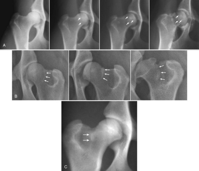
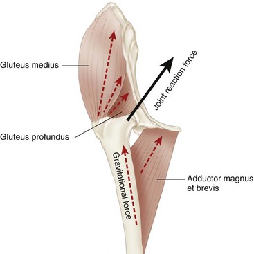
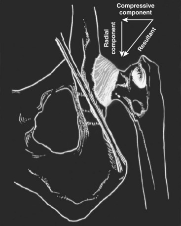
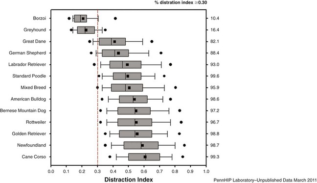
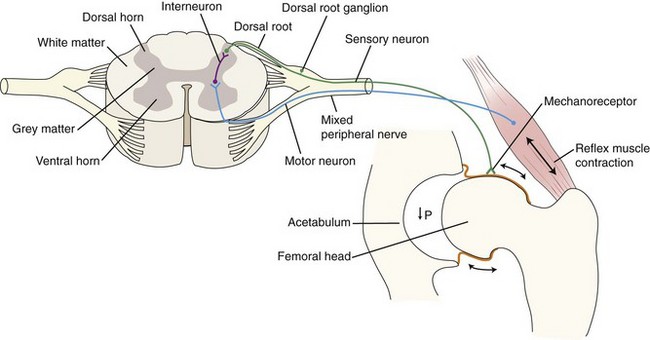
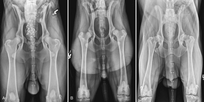
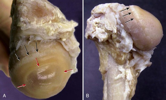
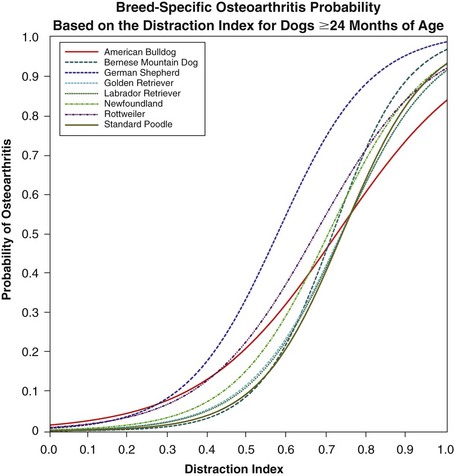
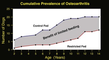
Chapter 59 Hip dysplasia is the most common orthopedic condition of the dog, causing joint inflammation and secondary osteoarthritis, which lead to variable degrees of clinical discomfort.66 Genetically, it is a disease of complex inheritance, meaning that multiple genes, combined with environmental influences, ultimately cause expression of the condition. It was first described in 1935 by Gerry Schnelle,161 and since that time, numerous investigators have reported on an array of potential causes.* To date, the underlying etiology and pathogenesis of the condition remain unclear. A central theme of most studies, however, is that hip joint laxity somehow plays a role in the development of osteoarthritis of canine hip dysplasia. See Figure 59-1 for degrees of hip dysplasia diagnosed from the hip-extended radiograph. Historically, the understanding that hip dysplasia has a genetic basis, coupled with the empirical observation that hip laxity plays a role in disease expression, led to diagnostic (screening) methods aimed at assessing hip laxity early in life with the hope that selecting the best candidates for breeding would lower the frequency of this common disease. Screening methods have ranged from palpation7–9,132 to radiography† and, more recently, ultrasound, computed tomography (CT), and magnetic resonance imaging (MRI).‡ Despite 75 years of observation and investigation, the diagnosis and treatment of hip dysplasia remain controversial. This chapter will attempt to compile available information on the role of hip laxity in the diagnosis of hip dysplasia and will cover genetic and other nonsurgical strategies used to lower the frequency of the disease, delay its expression, or ameliorate its severity. Special importance will be given to studies that were designed and conducted using the scientific method and contribute to evidence-based medicine. The study of the etiology and pathogenesis of canine hip dysplasia has been long and circuitous, and a comprehensive description of this history is not within the scope of this chapter. Multiple factors have been linked to the expression of canine hip dysplasia—some well studied, and others empirically associated with the disease.§ The true cause of canine hip dysplasia remains unclear; however, it has been accepted that the disease reflects the interaction of multiple genes with environmental influences. The manifestation of the disease phenotype occurs in genetically predisposed animals exposed to environmental (nongenetic) factors that enhance expression of the genetic weakness. Probably the most descriptive definition of the disease was put forth by Olsson et al.59 in 1966: “Hip dysplasia is a disease that stems from a ‘varying degree of laxity of the hip joint, permitting subluxation during early life, giving rise to varying degrees of shallow acetabulum and flattening of the femoral head, finally inevitably leading to osteoarthritis.’ ” The early phenotype of canine hip dysplasia therefore has been defined empirically as hip joint laxity with or without radiographic evidence of osteoarthritic joint changes. In the sections to follow, we will show how this empirical definition of hip dysplasia continues to confound progress in unraveling the mysteries of hip dysplasia. At birth, canine hip joints are normal,112,153 and they are thought to continue normal development if complete congruity between the femoral head and the acetabulum is maintained.150,182,183 During development of the hip, the earliest dysplastic joint changes are observed at 30 days of age: an edematous ligament of the head of the femur with torn fibers and capillary hemorrhage at the tearing sites.131,151,156 Increased volume of the ligament of the head of the femur and increased synovial fluid volume have been considered the earliest findings of canine hip dysplasia.17,100,167 In one study, the volume of the ligament of the head of the femur was significantly greater in puppies already displaying overt ostoarthritis but also in puppies at high risk for osteoarthritis based on parental phenotype.100 Another study showed that Labrador Retrievers at high risk for osteoarthritis (as determined by a high distraction index) had an edematous ligament of the head of the femur and edematous cartilage in the respective lesion areas of the femoral head.17 During the first month of life, the ligament of the head of the femur is thought to be primarily responsible for maintaining hip joint stability, such that in the first 2 weeks of life, the ligament is so short that the femoral head fractures at the fovea (attachment of the ligament) if forced to luxate.151,156 After the initial 2 weeks, the ligament slowly begins to lengthen, and it has been posited that in dysplastic dogs it is this excessive lengthening that permits lateral subluxation of the adult hip joint.151,156 The first radiographic signs of canine hip dysplasia, seen as early as 7 weeks of age, are subluxation of the femoral head and underdevelopment of the craniodorsal acetabular rim.151,156 At this time, the joint capsule is stretched but is not otherwise structurally altered, and the ligament of the head of the femur is lengthened. From 60 to 90 days of age, the degree of subluxation increases and significant radiographic changes are evident. Gross pathology reveals thickening and stretching of the joint capsule, permitting the femoral head to displace laterally and, in the most severely affected cases, dorsally. When subluxation occurs, the articular cartilage is worn and roughened on the dorsal surface of the femoral head at its point of contact with the acetabular rim (Figure 59-2). Evidence of palpable or radiographic laxity appears prior to degenerative structural changes. It has been reported that in a group of 48 Labrador Retrievers followed longitudinally to the end of life (i.e., the lifespan study), coxofemoral subluxation as seen on the hip-extended radiograph occurred by 2 years of age and not thereafter.174 In a healthy, congruent hip joint, forces during weight bearing are distributed across the entire cartilaginous surface of the acetabulum. The forces crossing the joint, the so-called joint reaction force, represent the vector addition of gravitational forces (the shared weight of the dog proximal to the hip joint) coupled with the muscle forces necessary to balance the moments of standing and locomotion (Figure 59-3). The muscle forces usually exceed the gravitational forces by a large margin, particularly during exertion. For example, in man, jogging may impose a peak hip joint reaction force of 4.3 to 5.0 times body weight (gravitational force) and stumbling 7.2 to 8.7 times body weight.67 In the canine subluxated (lateralized) hip, the transarticular muscle forces must substantially increase to generate higher moments necessary to compensate for lateralization of the center of rotation of the joint. Additionally, cartilage stress (force divided by area of contact) is vastly increased in the subluxated (lateralized) hip because forces acting on the articular cartilage are spread over a markedly reduced surface area, namely the dorsal labrum of the acetabulum. Therefore, two destructive events accompany functional subluxation: (1) the forces crossing the joint increase, and (2) the area over which the forces are transmitted decreases. The associated increase in cartilage stress beyond its failure limits causes cartilage damage, joint inflammation, and ultimately osteoarthritis. Figure 59-3 Cranial view of transarticular musculature during weight-bearing. The illustration shows the major muscles contracting during weight-bearing. The illustration is of a cranial view of the pelvis. The large gluteal muscle group acts to extend and abduct and internally rotate the hip whereas the adductor magnus et brevis muscles have compensatory adduction and external rotation (from insertion on linea aspera of femur). The illustration shows the lines of action of each muscle and the gravitational force (red dashed lines), which sum to make up the joint reaction force (black line). The co-contraction of these muscles, along with the biceps femoris, semimembranosus, and semitendinosus (not shown), sum to form a large resolved force tending to reduce (and stabilize) the femoral head into the acetabulum during weight-bearing. During the swing phase, however, the transarticular muscles acting to advance the hind limb in preparation for foot strike are the rectus femoris, sartorius, and iliopsoas. These muscles have long muscle bellies with lines of action more parallel to the axis of the femur. Although they generate much lower loads than the muscles of weight-bearing, their orientation makes them prime candidates to cause subluxation in a lax hip joint. We propose that the femoral head subluxates during the swing phase of gate and upon foot strike the larger hip extensor muscles cause catastrophic reduction of the femoral head producing the characteristic cartilage erosion shown (see Figure 59-2). (Modified from Evans HE: Miller’s anatomy of the dog, ed 3, Philadelphia, 1993, Saunders/Elsevier.) For dogs with functional subluxation, it is not known whether the hip is seated properly during the swing phase and then subluxates under the combined load of weight bearing and locomotion or, more likely, whether the hip is subluxated during the swing phase when the limb is not weight bearing, and the femoral head moves medially (and traumatically) toward a reduced position under the abrupt application of weight bearing and locomotor forces (see Figure 59-3). Although this was not described previously, mechanical justification exists for the latter mechanism for two important reasons. First, when the canine hip is weight bearing, the large and powerful muscles necessary for weight bearing and locomotion (propulsion) are acting. These muscles, particularly the gluteal and opposing adductor muscles, are oriented about the hip in such a way that each co-contraction creates a large resolved force, tending to reduce the femoral head into the acetabulum (see Figure 59-3). During the swing phase, on the other hand, the muscles necessary to advance the hind leg in preparation for the next foot strike generate relatively lower peak contractile forces, but, critically important, these muscles (e.g., the rectus femoris, iliopsoas and sartorius) are oriented more parallel to the femur, making the orientation of the net load on the hip vertical and therefore more conducive to subluxation. If the hip happens to be in the subluxated position when foot strike occurs, traumatic hip reduction ensues. Second, the characteristic location of cartilage wear on the femoral head and acetabulum (see Figure 59-2) supports the latter theory that subluxation occurs during the swing phase. If subluxation, leading to cartilage wear, primarily occurred during weight bearing, one would expect the site of cartilage wear to be codirectional with the summation of those propulsive and weight-bearing forces crossing the coxofemoral joint (i.e., along the weight-bearing axis) and thereby more cranial and in line with the axis of the ilial shaft. The characteristic position of cartilage wear, directly dorsal to the fovea capitis and not in line with maximum propulsive forces, however, suggests that catastrophic reduction at foot strike is the cause, rather than catastrophic subluxation at the point of maximal contractile muscle forces during weight bearing. Admittedly, this is a theory based on observed anatomy and assumed mechanics and is in search of definitive proof. The timing of functional subluxation within the gait cycle may be of no importance for hips having the most extreme forms of laxity, the so-called luxoid hips. For these hips, reduction cannot occur and the hip is permanently subluxated. At the origin of the cascade leading to hip osteoarthritis is functional hip joint laxity; accordingly, all diagnostic tests aim to assess hip joint laxity in a variety of positions. Heyman et al.,60 from mechanical testing of cadaver hips, reported that passive hip-joint laxity is at its maximum when the joint is placed in a neutral weight-bearing position. This neutral stance position is defined as 15 degrees of extension, 10 degrees of abduction, and 0 degrees of internal/external rotation, relative to the plane and axis of the pelvis.17 Pulling the hindlimbs into extension as typically performed for radiographic hip screening, was shown to produce a windup of the coxofemoral joint capsule, which severely limited the lateral movement of the femoral heads (Figure 59-4),60 thereby limiting observable hip laxity. Furthermore, mechanical testing of cadaver canine hips in this neutral position indicated that the degree of lateral femoral head displacement is not directly proportional to the applied force, as some have proffered. In fact, the hip joint can be modeled as a ball on a rope (biphasic behavior) rather than a ball on a spring (so-called hookean behavior). The understanding that displacement of the femoral head from the acetabulum was maximized in the neutral position and was largely independent of the distraction force (Figure 59-5)173 formed the research basis for a diagnostic method (the University of Pennsylvania Hip Improvement Program [PennHIP] method). The relative independence of lateral displacement from applied force also suggested that multiple examiners performing this method should expect high method repeatability both within and between examiners..60,70,104,168,171 Figure 59-4 Coxofemoral joint capsule windup during extension. Figure shows the resultant force of capsular tightening with the hip in full extension resolved into two orthogonal components: the radial component prevents further rotation (extension) of the coxofemoral joint and the compressive component forces the femoral head into the acetabulum. The compressive component is absent in the neutral distraction radiographic position, which explains the 2.5 to 11 times more passive hip laxity that can be measured.74 (Modified from Smith GK, Biery DN, Gregor TP: New concepts of coxofemoral joint stability and the development of a clinical stress-radiographic method for quantitating hip joint laxity in the dog. J Am Vet Med Assoc 196:59, 1990.) Similar slow progress has been observed in molecular genetic studies of quantitative traits in man. Such traits include cancer and Alzheimer’s disease, among others.196 Some progress toward unraveling the genetic underpinnings of canine hip dysplasia has been made, however. Quantitative trait loci (QTL) for hip dysplasia or related phenotypes have been found in a few breeds. A QTL is a region on a chromosome that contains a gene or a group of genes that influences the phenotypic expression of a quantitative trait such as hip dysplasia. A QTL for acetabular osteophyte formation has been mapped to chromosome CFA03 (canine familiaris autosome 3) in Portuguese Water Dogs.23 Susceptibility loci for canine hip dysplasia have been mapped to several chromosomes, including chromosomes 4, 9, 10, 11, 16, 20, 22, 25, 29, 30, 35, and 37.22,113,192 QTL intervals associated with the distraction index on CFA11 and CFA29 have been refined using single nucleotide polymorphism genotyping. Linkage analysis showed that the QTL on CFA11 in the 16.2 to 21 cM region explained 15% to 18% of the total variance in distraction index. Evidence for an independent QTL on CFA29 was weaker than that on CFA11.207 Most recently, a study identified four susceptibility single nucleotide polymorphisms associated with canine hip dysplasia and two single nucleotide polymorphisms associated with hip osteoarthritis. Three of the single nucleotide polymorphisms are adjacent to human genes previously associated with human osteoarthritis. Clearly there is a long way to go to equal or surpass the clinical utility of a phenotype like the distraction index or even the Norberg angle or the subjective hip-extended score. Continued progress in identifying candidate genes depends on the accuracy of the phenotype to which genomic association is made. Next steps in molecular biology include fine mapping of regions of interest, followed by screening for recognized candidate genes within these regions. It is conjectured that identifying a mutation or a genetic marker such as a SNP or a haplotype of single nucleotide polymorphisms could be combined with the pedigree and other phenotypic information in a complex mathematical formula, suitably weighted, to arrive at more accurate estimates of breeding value related to hip conformation.208 However, it is doubtful that all phenotypic variation will be explained by focusing on association or candidate gene identification because not all phenotypic variation originates from the genes. It is a sobering recognition that epigenetics, which is the study of inherited changes in phenotype or gene expression caused by mechanisms other than changes in the underlying DNA sequence, could be responsible for 50 to 100 times more phenotypic variation than that produced by the genes themselves.24,116 Joint laxity as measured by distraction index has been shown to be the primary risk factor for the development of coxofemoral osteoarthritis in all breeds studied (Figures 59-6 and 59-7).159,169,175,177 Passive hip laxity, an estimation of functional hip laxity, permits subluxation of the femoral head during the gait cycle, resulting in abnormal force distribution across the joint, leading to premature wear of the articular cartilage and microfractures in the subchondral bone and ultimately progressing to osteophyte formation and osteoarthritis.157 Dogs with higher degrees of joint laxity are at increased risk for developing osteoarthritis compared with those with lower degrees of laxity.159,169,175 Laxity measurements by distraction index are predictive of osteoarthritis susceptibility. The risk of osteoarthritis increases as the distraction index increases beyond 0.30, and dogs with distraction index below 0.30, such as most Greyhounds and Borzois, are not susceptible to acquiring osteoarthritis, even later in life (Figure 59-8).70,169,175 Subluxation, a subjective assessment of hip laxity, derived from the hip-extended radiograph is another risk factor for osteoarthritis. Data from a lifespan study showed that dogs with coxofemoral subluxation developed osteoarthritis on average 9 years earlier than those without subluxation;174 however, 98% of dogs in that study developed osteoarthritis by end of life, whether or not they showed subluxation. This was explained by the fact that all dogs in the study had hip laxity in the osteoarthritis-susceptible range as measured by distraction index (DI > 0.30).174 Figure 59-6 Metrics of hip laxity estimation. These laxity measurements are calculated from radiographs of a 25-month-old intact female Golden Retriever, with the Norberg angle and % femoral head coverage measurement made from the hip-extended radiograph and the distraction index measured on the distraction radiograph taken at the same radiographic session. Each of these metrics is consistent with hips of increased passive laxity. A, Norberg angle (NA) is a measurement of femoral head displacement from the acetabulum. NA is calculated by drawing a line connecting the centers of the femoral heads and one from the center of each femoral head to the craniolateral acetabular rim on the same side. NA ≥105 degrees is considered normal by one source.201 This dog’s NAs of 96 degrees and 93 degrees on left and right hips, respectively, are consistent with increased hip laxity. B, The distraction index is the measurement of maximal femoral head displacement from the acetabulum when the legs are placed in a neutral position and a distractive force is applied. The distraction index is calculated by dividing the distance between the geometric center of the femoral head and the geometric center of the acetabulum by the radius of the femoral head. Risk of osteoarthritis increases as the distraction idex exceeds 0.3. This dog’s distraction index of 0.71 is consistent with increased laxity and increased risk of osteoarthritis. C, Percentage of femoral head coverage (% FHC) is a measurement of femoral head displacement from the acetabulum. Normal FHC (i.e., good hip joint congruity) is defined as ≥50% coverage; therefore the % FHC of 30.87% shows increased laxity and poor femoral head coverage. Figure 59-8 Box and whisker plots of the PennHIP distraction index in 13 breeds of dogs, including the eight breeds in Figure 59-7. Box indicates 25th to 75th percentiles; vertical line indicates the median; square indicates the mean, with circles indicating the outliers. Figure indicates the vastly different breed laxity profiles, particularly comparing Greyhounds and Borzois versus breeds with increasing susceptibility to canine hip dysplasia. In breeds of dogs in which all members have a distraction index of ≥0.30, it is possible that canine hip dysplasia susceptibility is genetically fixed. The numbers of dogs used to generate each breed-specific box plot are 67 Borzois, 55 Greyhounds, 847 Great Danes, 9659 German Shepherd Dogs, 21,148 Labrador Retrievers, 1875 Standard Poodles, 5453 Mixed-breed dogs, 2503 American Bulldogs, 1692 Bernese Mountain Dogs, 2199 Rottweilers, 13,524 Golden Retrievers, 1636 Newfoundlands, and 1090 Cane Corsos. Significantly increased passive joint laxity and incidence of canine hip dysplasia have been reported in dogs with significantly higher volumes of synovial fluid and a thickened ligament of the head of the femur.96,100 In a cadaver study, the experimental addition of fluid to the hip joint caused an increase in passive laxity; similarly, removal of excessive joint fluid reduced joint laxity.173 It is still uncertain whether these changes are the primary cause of hip laxity, or if they occur as secondary changes caused by the disease of hip dysplasia. Homeostatic mechanisms to regulate hip synovial fluid volume have not been identified. Synovial production is primarily mediated by dialysis of blood from the intracapsular vessels whereby the endothelium, connective tissues, and synoviocytes modify the plasma for synovial fluid production. Equilibrium between new formation and removal of synovial fluid is maintained by drainage through the intracapsular veins and lymphatic vessels; however, the mechanism for volume control is not understood. This equilibrium becomes impaired when inflammatory processes occur in the form of capsular edema and leakage of proteins from the synovial vasculature.3,100,146 Inflammatory, effusive, and degenerative changes may obscure the specific nature of the disease, making it impossible to distinguish between primary and secondary alterations.109 However, the finding that joint laxity as measured by distraction index (itself a reflection of synovial fluid volume) is essentially constant during development (ri > 0.82)169 and for the periods measured (up to 3 years) provides evidence to suggest a genetic basis for the regulation of synovial fluid volume.33 A positive correlation between pelvic muscle mass and the prevalence of hip dysplasia has been reported.152 Investigators stated that a disparity between strength of the pelvic muscles and rapid weight growth in the young dog led to joint instability (laxity) and ultimately to hip dysplasia. It was found that the muscle mass of dysplastic breeds of dog was less than that of nondysplastic breeds, with Greyhounds having large thigh muscles and a low incidence of hip dysplasia compared with the small thigh muscle mass of German Shepherd Dogs with a high incidence of hip dysplasia. Various hormones, including estrogen and relaxin with their synergistic effects, contribute to the relaxation of pelvic and coxofemoral ligaments during parturition.6,200 Administration of high doses of exogenous estrogen to young pups and pregnant bitches resulted in increased coxofemoral joint laxity in the pups, along with other skeletal malformations.54,137,200 Estrogen levels in the physiologic range, however, have not been shown to cause changes in hip laxity or dysplasia in the dog. Hassinger et al.55 followed nine bitches through a single estrous cycle, measuring serum hormone concentrations and hip laxity (distraction index and Norberg angle) during each phase of the estrous cycle. No association was found between serum hormone levels and hip laxity. Laxity measurements by distraction index remained constant throughout the entire cycle with intraclass correlations of 0.93 and 0.92 for left and right hips, respectively. Variability of Norberg angle measurements was slightly greater than that of distraction index measurements throughout this study, but the measurements could be considered constant throughout the cycle having high intraclass correlations of 0.86 and 0.82 for the left and right hips, respectively. Relaxin, however, found in high concentrations during the last trimester of pregnancy and in the milk of lactating bitches, has been associated with increased peripheral joint laxity in human beings and dogs. Higher levels of relaxin were found in a group of Labrador Retrievers than in a group of Beagles. The authors concluded that this might contribute to the relatively higher incidence of canine hip dysplasia in Labrador Retrievers.180 Hip dysplasia development has been linked to the age and weight of the animal. In 1964, it was reported that rapidly growing pups had a higher incidence of canine hip dysplasia at maturity than those with slower weight gain. In a group of German Shepherd Dogs, it was observed that the heaviest males and the heaviest females at 60 days of age had the highest incidence of canine hip dysplasia at maturity.149 A contribution to the development of canine hip dysplasia was attributed to the rate of acetabular growth plate fusion, with earlier fusion seeming to lead to dysplastic joints.99 Body weight has proved to be an influential environmental factor through several studies.79,176 Although increased body weight does not cause canine hip dysplasia, it plays an instrumental role in the manifestation of the disease phenotype in dogs having genetic susceptibility for the disease. In a life span study following 48 Labrador Retrievers, it was reported that heavier dogs developed radiographic osteoarthritis on average 6 years earlier than their thinner littermates. See Figure 59-9 for the benefits of restricted feeding to slow the onset of hip osteoarthritis.176 Heavier dogs required long-term treatment for osteoarthritis 3 years earlier on average than slimmer dogs.75 Weight reduction has been accepted as a highly effective preventive strategy for delaying or preventing the onset of osteoarthritis in susceptible dogs.58,73,149 Puppies delivered by cesarean section and raised at drastically reduced rates of weight gain developed lower frequency of canine hip dysplasia at adulthood. Although radiographically negative for hip dysplasia, these dogs are known to be susceptible to canine hip dysplasia because their offspring tested positive for the disease.98 No dietary deficiencies have been shown to “cause” hip dysplasia, but dietary excesses have been found to contribute to development of the disease.78,121–123,148 Vitamin C has a role in collagen synthesis; which is diminished in states of extreme deficiency. The resulting disease is known as scurvy. An early theory was that megadoses of vitamin C given to pregnant bitches and puppies for 2 years after birth prevented canine hip dysplasia.11 However, this hypothesis was never rigorously investigated. Furthermore, megadoses of vitamin C can be harmful, potentially leading to hypercalcemia in puppies and resulting in delayed bone remodeling and cartilage maturation.148 High dietary calcium and excessive vitamin D increased calcium absorption in the intestinal tract, causing delayed endochondral ossification and skeletal remodeling and ultimately leading to canine hip dysplasia.56,57,108 High dietary anion gap was associated with a corresponding increased osmolality in synovial fluid, higher synovial fluid volume, and increased joint laxity as measured by the Norberg angle on the ventrodorsal hip–extended radiographic projection.78 Protein and carbohydrate quality and concentrations were investigated as causal for hip dysplasia; however, valid evidence to support these theories does not currently exist. Hip dysplasia is known to be a complex trait (also known as a quantitative or polygenic trait); therefore by definition, it has both a genetic and an environmental basis to explain the observed variation in expressed phenotype. For example, in gender-matched cohorts of Labrador Retriever littermates, food restriction (an environmental influence) to maintain a lean body mass was shown to have a profound effect on delaying the onset and lowering the severity of hip dysplasia.* Because genetic variation in the two feeding cohorts was minimized by pairing littermates, the variation in observed hip phenotype could be attributed to diet restriction—an environmental (nongenetic) factor. This life span study followed earlier studies, leading to similar conclusions.58,73,149 One additional environmental factor shown to delay the expression, or lessen the severity, of canine hip dysplasia in puppies is injectable polysulfated glycosaminoglycan. Twice weekly intramuscular injections of polysulfated glycosaminoglycan administered from 6 weeks to 8 months of age to puppies derived from dysplastic parents reduced hip subluxation scores and reduced histopathologic evidence of arthritis at 8 months of age when compared with untreated controls.103 Other potentially ameliorative candidate strategies await discovery, including the use of pharmaceuticals, nutraceuticals, omega-3 fatty acid diets, or specific exercise and rehabilitation regimens, to mention a few. Adequate longitudinal trials imposing these strategies in cohorts of puppies matched for osteoarthritis susceptibility have not been carried out; however, this is an exciting area for future investigation. It has been hypothesized that the ratio of collagen type I to type III was related to the development of canine hip dysplasia. It was initially believed that lack of type I collagen (responsible for strength) was found in the joint capsule in dysplastic animals; however, when the ratios were compared between dysplastic and nondysplastic dogs, no significant difference was found. It was theorized that the amount of procollagen type III (PIIINP) in serum and synovial fluid would be indicative of dysplasia; however, these levels stay the same throughout adulthood, and no differences between dysplastic and nondysplastic dogs have been found.109 Pectineal muscle myopathy was associated with hip dysplasia. Tight pectineal muscles generated a pull on the femur that pushed the femoral head dorsally onto the acetabular rim. Histologic changes were found in the myofibers of the pectineus; however, neither myectomy nor tenotomy reduced the incidence of canine hip dysplasia.18,19,63 In toto, none of the proposed etiologies in this section fully explains the development of canine hip dysplasia, and none has led to a reliable diagnostic test that provides more predictive value than the measurement of hip joint laxity. The most compelling pathogenic factor is the synovial fluid volume theory168 to explain observed early joint laxity in puppies. Of course it is possible that a combination of causes could be acting in concert.35,96,168 Some have argued that synovial fluid volume could be simply joint effusion resulting from the trauma of instability of hip dysplasia—an effect rather than a causal factor.197 However, if such were the case, the volume of synovial fluid would not be expected to be so tightly biologically regulated as demonstrated by the constancy of joint laxity in dogs from 16 weeks to 3 years of age.169 This theory is central to the distraction diagnostic method that is gaining increasing acceptance for detection of susceptibility to canine hip dysplasia. With the discovery in the 1980s that mechanoreceptors exist within the joint capsule and other fibrous constraints of the joint, a new awareness of the complexity of joint function followed. The joint was an organ—not just a system of ropes, pulleys, gliding surfaces, and lubrication. The joint was constantly sending out signals to the brain and surrounding tissue as to its position, location in space, and state of loading. By this reasoning, a cruciate ligament under conditions of high strain or load might send a signal (by pathways as yet not fully understood) to the semitendinosus muscle, recruiting contraction to assist the anterior cruciate ligament in its function and protecting it from dangerously high loads.92 Although the knee has been the subject of much more investigation than the hip in this regard, we can for this exercise assume the two joints to be more similar than different in terms of their neurobiology and feedback mechanisms. A theoretical pathogenic mechanism to explain how increased synovial fluid volume might lead to destructive joint forces in the hip is illustrated (Figure 59-10). Under ideal conditions, the synovial fluid volume of the hip is fixed and minimal, such that when the joint tends to subluxate during the swing phase (also theoretical, see Figure 59-3), the low intracapsular pressure generated severely limits possible translation. With low synovial fluid volume, critical pressure differentials would cause the capsule to invaginate, thereby stretching the capsule.167 This stretching early in the translation of the femoral head would trigger capsular mechanorecepters to recruit appropriate periarticular muscles into protective roles. The protective muscle action would serve to position the femoral head close to the acetabulum, such that when the much larger loads of foot strike and weight bearing are initiated, little or no abrupt reduction of the hip occurs. In contrast, with increasing synovial fluid volume, these protective mechanisms are triggered progressively later during hip subluxation of the swing phase (i.e., it takes greater subluxation of the femoral head to cause the same stretching of the capsular mechanoreceptors), and it is conceivable that in the extremely lax joint, no muscle recruitment whatsoever occurs. With the hip subluxated at the point of foot strike, increasingly catastrophic reduction of the femoral head into the acetabulum would ensue. The degree of functional subluxation would correspondingly increase with the increasing volume of synovial fluid, resulting in greater and greater impact of the femoral head on the acetabular labrum upon foot strike, producing cartilage damage and erosion at the characteristic sites and ultimately leading to osteoarthritis. Although this description is admittedly theoretical, it does begin to piece together our evolving understanding of the anatomy, mechanics, and neurobiology of the hip joint. Other joints in a dog diagnosed with hip dysplasia may have similar increased synovial fluid volume, but by reason of anatomic design, these joints (such as the knee or elbow) may be more geometrically constrained and less dependent on synovial fluid volume for stability. However, high synovial fluid volume may still be detrimental. Evidence of this possibility can be derived from a study showing that 8-year-old Labrador Retrievers having radiographic hip osteoarthritis had a greater likelihood of showing radiographic osteoarthritis of the shoulder and elbow than dogs without hip osteoarthritis at 8 years of age.76 Figure 59-10 Theorized hip-joint capsule mechanoreceptor feedback loop. Under ideal conditions, the synovial fluid volume of the hip is fixed and minimal, such that when the joint tends to subluxate during the swing phase (also theorized), the low intracapsular pressure (P) created is one factor tending to limit possible translation. A second factor is an active factor, a feedback loop triggered by this capsular stretching. With lower synovial fluid volume, critical pressure differentials would occur earlier in the subluxation process, causing the capsule to invaginate and thereby stretching it. This stretching early in the translation of the femoral head may activate capsular mechanorecepters to recruit appropriate periarticular muscles into protective roles. The protective muscle action would serve to position the femoral head closer to the acetabulum such that when the much larger loads of foot strike and weight bearing are initiated, little or no abrupt reduction of the hip occurs. With increasing synovial fluid volume, these protective mechanisms are triggered later and later during the subluxation of swing phase (i.e., it takes greater subluxation of the femoral head to trigger stretching of the capsular mechanoreceptors). In dogs having the most severe hip laxity, it is possible that recruitment of protective mechanisms fails to be successfully initiated prior to foot strike. As proposed earlier (see Figure 59-2), this would lead to catastrophic hip reduction at foot strike. Canine hip dysplasia can affect any breed of dog, but it is most commonly reported in large and giant breeds, such as German Shepherd Dogs, Rottweilers, Labrador Retrievers, Golden Retrievers, Saint Bernards, and many more.26,28,29,45,202 In contrast, sight hounds, such as Greyhounds and Borzois, have a very low risk of canine hip dysplasia, and it is almost nonexistent in these breeds. See Figure 59-8 for how Greyhound and Borzoi distraction index distribution compares with breeds with higher canine hip dysplasia prevalence.70,154,169 The clinical signs of canine hip dysplasia can vary extensively from slight discomfort to severe acute or chronic pain. Although disease onset has a linear progression over time,176 the clinical signs can be divided into two forms. In the juvenile or severe form, dogs typically present between 5 and 12 months of age.151 Young dogs often present with sudden onset of unilateral or bilateral hindlimb lameness, bunny-hopping, difficulty rising after rest, reluctance to walk, run, jump, or climb stairs, exercise intolerance, or pain/soreness of the hindlimbs. These early acute clinical signs are thought to be the result of extreme joint laxity. Histologic examination of such juvenile dogs with canine hip dysplasia has shown that stretching and tearing of the joint capsule (synovitis), ligaments, and muscles, along with microfracture of the dorsal acetabular rim from overloading, are the most likely causes of pain in young patients.151 As dogs become older, the capsular strain and inflammation induced by joint laxity cause periarticular fibrosis. This phase of the disease is often associated with reduction or even elimination of clinical signs. It had been thought that fibrosis helps to stabilize the joint.10,28 However, another study51 showed that hips develop increased laxity as chronic osteoarthritis progresses. A complete general physical examination should be performed both to rule out other disease processes and to determine the health of the patient for sedation and/or general anesthesia. Orthopedic and neurologic examinations are necessary to localize clinical signs to the coxofemoral joint and to eliminate other orthopedic or neurologic conditions that may have a similar presentation to canine hip dysplasia. Because many breeds at high risk for canine hip dysplasia have a high incidence of other causes of hindlimb lameness, such as panosteitis, osteochondrosis, or rarely, hypertrophic osteodystrophy in the juvenile dog, and cranial cruciate disease, lumbosacral disease, or neoplasia in the mature dog, it is imperative to accurately localize the clinical signs to avoid inappropriate diagnosis and treatment of canine hip dysplasia. For instance, one study141 found that 32% of dogs referred to a teaching hospital for management of canine hip dysplasia actually had clinical signs attributed to cranial cruciate ligament rupture. Within some breeds of dogs, hip dysplasia may affect more than 90% of the breed in later life, so it is important to rule out coincident orthopedic diagnoses before arriving at canine hip dysplasia as the source of clinical signs.141,174 On palpation, pain sometimes can be elicited when pressure is applied over the hip or during range of motion, most notably during extension, particularly in dogs having the severe juvenile form of the disease. Coxofemoral range of motion may be decreased and typically is most limited during extension. As the disease advances, crepitus can be palpated during range of motion. In the young dog, semiquantitative palpation maneuvers may be used to assess joint laxity, including the Bardens, Barlow, and Ortolani tests.7–9,132 The most widely adapted maneuver is the Ortolani test (Figure 59-11). When the Ortolani test is performed, the patient can be placed in dorsal or lateral recumbency according to practitioner preference. In the lateral position with the hip in neutral orientation, one hand is placed on the distal aspect of the stifle, and the other is placed on the dorsal pelvis and spine to provide stabilization. The first part of the Ortolani is the Barlow test, in which the limb is in an adducted position and a force is directed through the femur toward the dorsum of the dog via the hand grasping the dog’s stifle. This force causes dorsal subluxation of the femoral head in dogs with hip laxity. The Barlow test is considered to be a provocative test in that it creates subluxation of the lax hip. In the second part of the Ortolani test, the limb is abducted, maintaining force up the axis of the femur, and a click or clunk can be heard and/or palpated as abrupt reduction of the hip occurs. This clunk is interpreted as a positive Ortolani sign and suggests laxity of the coxofemoral joint. The Ortolani test is considered a reduction maneuver. Although the presence of a positive Ortolani sign is consistent with joint laxity, this has not been shown definitively to be a predictor of later development of clinical signs or osteoarthritis; conversely, Ortolani-negative dogs have not been shown to be normal throughout life. For example, the authors of one study reported that although distraction index was significantly associated with positive Ortolani, such that for every 0.1 increase in distraction index the risk of a positive Ortolani increased by 3.1 times, 50% of Ortolani-negative dogs, and therefore assumed to be normal, had hip laxity indicating osteoarthritis susceptibility by distraction index.143 Subjective tests, like the Ortolani sign, need to be combined with complete assessment of the patient before an accurate diagnosis and a potential treatment plan can be implemented. These palpation techniques are further reviewed later in the chapter. The ventrodorsal hip-extended radiograph has been used to evaluate the canine hip since the first case of canine hip dysplasia was reported by Schnelle in 1935162 with the first hip-extended radiograph of the pelvis published in 1937. The animal is positioned in dorsal recumbency, and the hindlimbs are pulled into extension with femora parallel and slightly pronated (internally rotated), such that the patella appears superimposed centrally over the trochlear groove (see Figure 59-1, A).155 By convention, this hip-extended positioning has been accepted and used worldwide for nearly 50 years to screen for canine hip dysplasia.4,12,155,168,198 Recently, it has come under closer scrutiny and has been reported to be an insensitive diagnostic tool having poor precision and predictive accuracy for diagnosis of canine hip dysplasia,72,195,204 such that even among highly skilled examiners, radiographic interpretation of the hip-extended view varies greatly.72,135,155,195 In a biomechanical study of the hip joint, a windup of the coxofemoral joint capsule was observed when the hind legs were pulled into extension (see Figure 59-4), as they are for the hip-extended radiograph, forcing the femoral head to become more tightly seated in the acetabulum and thereby masking observable joint laxity (Figure 59-5). This phenomenon is thought to be one factor contributing to the high rate of false-negative diagnoses of canine hip dysplasia: dogs having joint laxity may appear normal via this radiographic technique.60,168 Nonetheless, hip laxity, subjectively or objectively interpreted as subluxation on the hip-extended radiograph, has been empirically accepted as the earliest visible radiographic change associated with canine hip dysplasia.59,157 In addition to subluxation, radiologists assess the radiographic presence of osteoarthritis to arrive at a confirmed diagnosis of canine hip dysplasia when evaluating the hip-extended projection (Box 59-1). It is established that the development of radiographic signs of osteoarthritis lags behind the early structural changes associated with osteoarthritis. Early changes typical of osteoarthritis can, however, be found on arthroscopy or gross pathology and histopathology.61,155 Because the characteristic radiographic signs of osteoarthritis take time to develop, radiologists have empirically accepted the presence of apparent joint laxity (subluxation) on the hip-extended radiograph to be diagnostic of canine hip dysplasia. As mentioned previously, radiographic signs of osteoarthritis and therewith definitive diagnosis of canine hip dysplasia are strongly influenced by the age of the animal at the time of evaluation.65,176,203 Jessen and Spurrell reported that at 6 months of age, 16% to 32% of the dogs examined were correctly diagnosed as dysplastic; at 1 year of age, correct diagnoses increased to 63% to 69%, and at 2 years of age, 92% to 95% of dogs were correctly diagnosed as dysplastic based on characteristic radiographic signs of canine hip dysplasia on the hip-extended radiographic evaluation performed at 5 years of age.65 Their plot of cumulative canine hip dysplasia prevalence versus age of the dog suggested that no new cases of canine hip dysplasia develop after 5 years of age, although the dogs were not followed beyond 5 years of age. The Orthopedic Foundation for Animals (OFA) decided to limit the earliest age of hip screening in the United States to dogs 2 years of age or older on the basis of this Jessen and Spurrell study. A similar association between age and canine hip dysplasia diagnosis was reported by Smith et al., but in this study it was determined that osteoarthritis prevalence increased linearly with age (r2 = 0.987) in a fixed cohort of Labrador Retrievers followed longitudinally for life.174,176 Other hip-extended screening methods around the world such as the British, European, and Australian systems specify dogs 1 year of age or older to be screened for canine hip dysplasia, with the rare exception of a few specific large and giant breeds of dogs.16,40,46,201 Based on available evidence, this practice is associated with a minimum error in diagnosis of 30%. Radiographic changes associated with osteoarthritis have recently been under investigation. Over the past decade, three radiographic features on hip-extended radiography that were hitherto ignored have been considered to have clinical significance.114,139,158,186 These are the caudolateral curvilinear osteophyte (CCO), the circumferential femoral head osteophyte (CFHO), and, for lack of a better term, what has been described as a “puppy line” in the area of the femoral neck. Evidence is fairly conclusive that the caudolateral curvilinear osteophyte and the circumferential femoral head osteophyte represent early radiographic osteophytic signs that predict later development of the characteristic (and well-accepted) radiographic signs discussed earlier in this chapter (Figure 59-12).114,118,139,158,186 Adoption and application of these radiographic features are not yet standardized among veterinarians and veterinary radiologists.
Pathogenesis, Diagnosis, and Control of Canine Hip Dysplasia
Etiology and Pathogenesis
Hip Development
Biomechanics
Genetics
Joint Laxity

Joint Fluid
Pelvic Muscle Mass
Hormonal Factors
Weight and Growth
Nutrition
Environmental Factors
Other Causes
Proposed Pathogenesis of Hip Dysplasia
Signalment and History
Physical Examination
Imaging Examination
Hip-Extended Radiography
< div class='tao-gold-member'>
![]()
Stay updated, free articles. Join our Telegram channel

Full access? Get Clinical Tree


Pathogenesis, Diagnosis, and Control of Canine Hip Dysplasia
Only gold members can continue reading. Log In or Register to continue


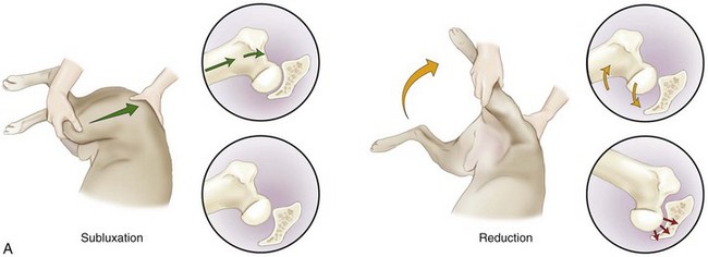
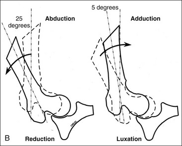
 inch suggests laxity of the joint. In the mature dog, a positive Ortolani sign or Bardens test is rarely palpated, even in dysplastic dogs, most likely because of the presence of periarticular fibrosis, remodeling of the dorsal acetabular rim, or the presence of a shallow acetabulum. The older dog’s physical examination findings likely will be consistent with the chronic pain of degenerative joint disease, and, for the typical pet-quality dog, it is often difficult to find any clinical signs associated with mild osteoarthritic changes.