Chapter 45 Management of fractures can be accomplished with internal fixation, external skeletal fixation, and external coaptation. Objectives of fracture management include anatomic reduction; prevention of displacement, angulation and, rotation of bone fragments; preservation of soft tissues; prevention of fracture disease; the use of as many joints as possible during the healing period; and early return to full limb function.35,38 External coaptation as a fracture management technique, when appropriate, does offer advantages. Disruption to the fracture site is minimal, and the blood supply is not further compromised.35,46 With no implants, the risk of infection is decreased, and complications such as loosening of the implants are eliminated.46 The cost of utilizing external coaptation fracture management can be much lower than that of surgical repair.26 Disadvantages include the need to immobilize the joint proximal and distal to the fracture, as prolonged immobilization can result in severe disuse atrophy and fracture disease.26 Selection of an effective fracture management technique begins with patient and fracture assessment.35 Components of patient assessment include signalment, history, and physical examination.35 Animals younger than 1 year of age generally heal quickly, limiting time in coaptation, and therefore are ideal candidates for external coaptation as a primary method of fracture repair.26,35,38,46 Breed is also an important factor. Although in general, young patients can heal well in external coaptation, small and toy-breed dogs have up to an 83% incidence of malunion and nonunion when external coaptation is utilized as primary repair for fracture of the antebrachium.18,31,32,46 Patient confirmation should be considered as well. Constructing an adequate external coaptation device for chondrodystrophic breeds or obese animals can present a challenge.35 Thorough physical examination of a patient with a fracture includes orthopedic and neurologic evaluation. Soft tissue injury associated with the fracture plays a significant role in repair selection.35 Concurrent disease and associated treatments should be considered. Animals presenting with multiple limb injury may not function well in external coaptation. Immunosuppressive therapy can significantly slow healing, requiring prolonged time in external coaptation, which can lead to additional complications. Patient temperament should also be considered. Internal fixation may be a more suitable option for particularly aggressive patients.12 Radiographic evaluation is necessary to accurately categorize the fracture and should include mediolateral and craniocaudal views. Stress views may be necessary to assess joint stability and ligament integrity. Fracture repair selection must consider the fracture environment (open versus closed), the type and location of the fracture, the degree of displacement, if multiple fractures are present, and the fracture forces present.35 Four mechanical forces may be present at a fracture site: bending, rotation, compression or shear, and distraction.12 Forces present at a particular fracture site must be adequately neutralized by the repair method for healing to occur. External coaptation can effectively counteract bending and rotational forces, provided that the joints proximal and distal to the fracture are immobilized.12 If adjacent bones (e.g., ulna, fibula) are intact, they can act as anatomic functional splints and help reduce bending, rotation, and shearing forces.35,46 Typically, fractures subject to compression, shear, and/or distractive forces require internal fixation, as external coaptation alone cannot counteract these forces.35 The “50% rule” states that cortical positioning of fracture ends should have 50% contact to expect fracture healing. This means that at least 50% contact must occur for healing to be possible, not probable. The goal of any closed reduction should be 100%. Radiographs should be performed after closed reduction and external coaptation. If fracture reduction cannot be maintained, surgical intervention must be considered.12,35,38,46 Proper rotational alignment between the joints proximal and distal to the fracture is imperative to limb function.12,38 If the bone fragments of the fracture are not aligned with respect to the joints, rotational or angular malunion may result and can cause functional gait abnormality and lameness due to secondary osteoarthritis.12 Care must be given during initial placement of coaptation to prevent this outcome.12 Joint stiffness is not uncommon after trauma, surgery, and immobilization of a limb. In prolonged immobilization, adhesions between muscles, tendons, and bone may limit range of motion in affected joints. If coaptation is performed at an abnormal angle, loss of function may result. Maintaining a neutral standing position during application of bandages, splints, and casts will encourage weight bearing during and after coaptation. Utilizing the limb after coaptation can often relieve symptoms. Most important is joint mobilization as early as possible during recovery.12 Immobilization of the joints proximal and distal to a fracture is a basic principle in external coaptation.12,26,35,46 Splints and casts can generally meet this criterion for fractures distal to the elbow and stifle joints.12,26,35,38,46 Metacarpal and metatarsal fractures with minimal displacement and affecting only one or two bones, particularly if there is an intact weight-bearing bone, may be managed utilizing external coaptation.22,35 Fractures involving the humerus and the femur are largely unsuitable for external coaptation as a primary method of repair. Consideration may be given only for young patients (<1 year old) with a stable, nonarticular, nondisplaced fracture of the humerus or femur.35 A spica splint may be utilized for this specific situation; however, applying this splint to the hindlimb is challenging, especially in the male dog, and ambulation can be difficult.35,38 Closed, minimally displaced fractures of the scapula may be treated conservatively with a Velpeau sling and limited activity.22,35 Use of a below-the-knee cast (Sarmiento-type) has been described for certain tibial fractures and, if applied properly, allows almost full range of motion of the stifle joint.26,34 For fractures distal to the elbow and stifle joints temporary splintage can offer improved patient comfort, decrease or prevent edema, and minimize additional trauma if reduction and fixation is delayed. Temporary splintage may be beneficial for open fractures to allow wound treatment, to reduce the risk of infection, and to decrease soft tissue swelling before definitive repair.46 For most other types of fractures, cage rest and sedation/analgesia are all that is necessary until definitive repair is performed. However, if travel is required before fixation, splintage of these fractures may be indicated.38 Appropriate selection of external coaptation as a primary method of fracture repair is based on numerous patient, fracture, and environmental factors.12,35 Beyond this, external coaptation serves many other functions. Bandages, splints, or casts may be used for temporary stabilization when definitive fracture repair is delayed, as adjunctive therapy in managing swelling and edema, and in providing support before and/or after internal fixation. These coaptation devices can also be beneficial after removal of external skeletal fixation devices, for stabilization and support of orthopedic soft tissue injuries or repairs, and in wound management.12,26,35,46 Slings may be utilized as a primary method of management of certain fractures and dislocations. Some slings and the carpal flexion bandage can also be utilized when stabilization is not necessary and weight bearing is not desired. The level of joint mobility desired influences selection of non–weight-bearing coaptation for this purpose. Selection of orthotic devices is outlined later in this chapter. Carefully considered use of external coaptation can be rewarding; however, improper case selection, incorrect application, and mismanagement of the patient during external coaptation use can have devastating repercussions.12,35,46 Complications arise from poor patient and fracture assessment, improper device application, case mismanagement, and patient and/or owner compliance issues.12,35,46 Complications can range from minor issues such as skin irritation and mild dermatitis to limb- and life-threatening conditions such as fracture disease or necrosis secondary to swelling and vascular compromise. Utmost consideration must be given to injury, patient, and environmental factors in selection of external coaptation management. Coaptation must be applied and monitored appropriately. Communications with owners must convey the gravity of home monitoring for potential complications, proper coaptation care, and diligent follow-up. The carpal flexion bandage immobilizes and maintains moderate flexion of the carpus and prevents weight bearing of the forelimb, while allowing passive motion of the shoulder and elbow joints (Figure 45-1). The most common use of this bandage is to relieve the tension of flexor tendons after repair. Otherwise, it is used primarily to prevent weight bearing after other orthopedic repair.12 Permanent contracture can result if the carpus is maintained in flexion for a prolonged period (i.e., typically >1 month, although even within 1 to 2 weeks, decreased range of motion may require rehabilitation therapy). Limited extension should begin within 2 to 4 weeks with successive lengthening of the 1 inch tape loop.43 Some dogs will walk on the dorsal surface of the carpus; those dogs may require a Velpeau sling to prevent weight bearing. Otherwise, the bandage is generally well tolerated with few complications. The Robert Jones bandage (Figure 45-2) and its modifications are the most commonly utilized bandages in orthopedic external coaptation.43 Use of these bandages is limited to injuries at or distal to the elbow or stifle joints. Injuries may include wounds, fractures, dislocations/luxations, and strains and sprains. Variations involve wound dressing, padding, and rigidity. Wound dressings are numerous and are detailed elsewhere in this textbook. The amount and type of padding and reinforcement materials are determined by the type and location of injury and the compression and support needed. The Robert Jones bandage is a bulky bandage indicated as a temporary method of stabilization for fractures or dislocations present at or distal to the elbow or stifle joint until definitive repair is performed, or after surgical intervention to address postoperative swelling.12,21,38,43,46 An appropriately applied Robert Jones bandage provides support and immobilization for traumatized soft and bony tissues, reduces and prevents edema, and limits effusion and hemarthrosis before or after surgical intervention.5,12,21 Its bulk and evenly applied mild compression achieve these objectives without compromise of vascularity of the soft tissues.21 The bandage is well tolerated in most patients; however, its bulk can hinder ambulation. Although highly effective for temporary stability and immobilization of the extremity, this bandage is unsuitable for primary fracture fixation; the thick cotton padding loosens and can contribute to instability at the fracture site.12,38 The materials required to apply a Robert Jones bandage include 1 inch white adhesive tape, one to three rolls of 1 pound 12 inch cotton, one to three rolls of 3 to 4 inch roll gauze, one to two rolls of 3 to 4 inch elastic adhesive tape or self-adherent stretch tape, and wound dressing if indicated.43 Before application, all wounds should be appropriately treated and covered with a dressing. Strips of adhesive tape (stirrups) of appropriate width ( Prepare the cotton by unrolling, removing the paper backing, and rerolling. Tear the cotton to a narrower width of either one half or one third, depending on the size of the patient. The narrower width allows for better conformation to the limb.12 Apply the cotton beginning at the level of the second and fifth digits, while leaving the toenails of digits three and four exposed. Apply the cotton tightly with no wrinkles or twists. Apply from distal to proximal, while overlapping the previous layer by 50% with each successive layer. Continue to the midhumerus or midfemur, and then return distally in the same fashion. If the injury is just distal to the level of the elbow or stifle joint, the bandage should extend proximally to the axilla or groin. Repeat until sufficient bulk is achieved—4 to 8 cm thick.35 Rolled gauze is applied next in the same manner as the cotton. To achieve a smooth bandage with uniform tension, at least two to three layers of gauze should be applied with even pressure so that the cotton is compressed by 40% to 50%.12 The cotton is sufficiently compressed when tapping the bandage with a finger reproduces the sound of tapping a ripe watermelon.4,34 The tape stirrups should be separated, inverted, and applied to the gauze layer of the bandage. The outer layer of elastic tape or self-adherent tape is applied in the same manner as the two previous layers. Care must be taken to avoid applying this layer with too much tension, causing overcompression and vascular compromise.12 The completed bandage should be smooth in appearance and should reveal the distal aspect of the toes and toenails of the third and fourth digits, allowing evaluation after bandage placement.12,43 Loosening of the cotton layer will occur within several hours or days; therefore, bandage evaluation should be performed every 4 to 6 hours, and bandage change is recommended every few days or sooner, as dictated by wound treatment or observation of complications.12 Less padding is utilized in the modified Robert Jones bandage (Figure 45-3). Cast padding is used in place of cotton. A modified Robert Jones bandage is indicated for light compression and partial immobilization of the limb at or distal to the elbow or stifle joint.43 It is useful for reduction or prevention of posttrauma or postoperative swelling, soft tissue protection and limb support after surgical fixation, or removal of orthopedic devices.38 However, the modified Robert Jones bandage is contraindicated if severe inflammation is expected. Because it does not provide rigidity and complete immobilization of the limb, the modified Robert Jones bandage is not appropriate for temporary support of fractures or dislocations.12 Figure 45-3 The modified Robert Jones bandage applied to the hindlimb of a dog after stifle surgery. The modified Robert Jones bandage is constructed in the same manner as the Robert Jones bandage; however, the padding is applied to a thickness of A reinforced Robert Jones bandage (Figure 45-4) incorporates a rigid material into the modified Robert Jones bandage to enhance immobilization and support of the limb and joints. Offering the stability of the Robert Jones bandage without the bulk, this bandage is often utilized in place of the conventional Robert Jones bandage for temporary stabilization of a fracture before surgery or after a tenuous internal repair.12 A reinforced Robert Jones bandage may be utilized as primary fixation for stable fractures in young animals.46 Preformed splints are available for the front limbs and hindlimbs and are primarily utilized for stabilization of fractures of the distal radius and ulna, fracture-dislocations of the carpus or tarsus, and fractures of the metacarpal or metatarsal bones or phalanges.26 However, the use of pre-formed splints can be counterproductive. Although they are available in multiple sizes, exact fit is rare. Copious padding, which typically is required, can result in increased occurrence of soft tissue complications and loss of rigid immobilization.38 Therefore, it is the authors’ opinion that these splints should not be used for primary fracture stabilization. Molded splints are more efficient stabilizers of bones and joints, and because they are custom-fitted, fewer soft tissue complications occur and patient tolerance is improved as compared with preformed splints.38 The spica splint is used to immobilize the shoulder or hip joint (Figure 45-5). The bandage material is applied beginning at the toes and continuing proximally around the torso. The splint extends over the scapula or hip to the dorsal midline down to the toes. Distal joints are also immobilized by this splint. It may be used as temporary splintage for humeral and femoral fractures or in adjunctive stabilization after internal fixation. It is rarely used for primary management of these fractures. Most humeral and femoral fractures are displaced, and manipulation to reduce them can create further damage. Cage rest until definitive repair is performed is preferable. However, the spica splint may be utilized for prolonged delay of surgery or for patient travel before fixation. The spica splint may also be considered in young animals with a greenstick fracture of the humerus or femur.12,21,38 Placement of the splint on the hindlimb can be challenging, especially in male dogs.38 Figure 45-5 Spica splint applied to the front limb of a dog for shoulder joint immobilization. Stirrups are not necessary because slippage is prevented by attachment of the bandage to the torso. Cotton padding is applied in the same fashion as the modified Robert Jones bandage. At the level of the torso, the padding is wrapped around the animal’s torso several times, caudal to the contralateral limb and alternating cranially and caudally to the affected limb. Follow with gauze wrapped in the same pattern with mild tension. A splint rod or synthetic casting tape may be used to construct the splint. These materials should be conformed to the bandaged limb from the toes to the dorsal midline. Four to eight layers of casting tape is sufficient to provide the immobilization and support required. Gauze followed by elastic tape is applied to hold the splint in place and cover the bandage completely. Care must be taken in applying a forelimb spica splint to avoid compression of the thorax and compromising of respiratory function, especially when thoracic trauma is present. Construction of a hindlimb spica splint in a male dog requires avoiding the prepuce.12,21,26,38 Few complications are noted with the spica splint; however, abrasion may develop within the axilla or the inguinal region. Patients may require assistance with ambulation.12 The Schroeder-Thomas splint is a traction device constructed of a wire frame and soft bandage material. The limb is suspended within the frame, and when constructed and applied properly, this splint can counteract muscle forces and immobilize joints and certain fractures. Its use has been largely superseded by splints and casts.38 It may be used as primary fixation for selected, minimally displaced, midshaft fractures of the radius, ulna, and tibia. Fractures of the distal third of the humerus, excluding condylar fractures, have been successfully managed with this splint.26 The splint does not adequately immobilize the shoulder or hip joint and is contraindicated for most humeral and all femoral fractures.12,26,38 Careful and accurate construction is required and is detailed in other publications. Casts are custom-made, molded, external coaptation devices. Molded coaptation devices are efficient stabilizers of the bones and joints.38 Previously, casts were primarily constructed with plaster of Paris or plaster-impregnated casting tape. After the advent of other moldable materials, plaster is rarely used today. Although inexpensive and easily moldable, plaster has many disadvantages. It is heavy, loses strength when wet, and retains moisture, which can cause moist dermatitis and tissue maceration. Its radiopaque qualities may require cast removal for radiographic assessment of the limb. Synthetic casting tapes are significantly more expensive than plaster but offer many advantages. Made of thermomoldable and fiberglass/resin materials, these products are lightweight, radiolucent, and impact resistant and offer adequate strength for coaptation in small-animal orthopedics. They conform well to the limb and are easy to apply.12,38 General anesthesia is generally necessary for closed fracture reduction and cast application. Once the fracture has been reduced and aligned, the limb must be carefully supported. Tape stirrups followed by one layer of cast stockinet are applied to the limb. The stockinet should exceed the cast both proximally and distally by 1 to 2 inches. To help prevent pressure ulcerations, bony prominences and protuberances should be covered with doughnuts constructed from cast padding or rolled stockinet, or covered with materials made for this specific purpose. Cast padding is applied to the limb in the same fashion as the modified Robert Jones bandage; however, the cast padding should extend both proximally and distally to the cast by 1 cm. Additionally, for optimal fracture stability, padding should not exceed two layers. Select the widest width of casting tape appropriate for the animal’s size. Gloves should be used when handling the casting tape. The tape is immersed in cold water. To facilitate water penetration, squeeze the roll several times while immersed. Squeeze and shake the roll to remove excess water. Begin application of the casting tape at the toes, leaving the bottom open for evaluation. Toes should be enclosed by the cast, as any portion of the toes extending beyond the cast may suffer maceration during weight bearing. While moving quickly but with even, moderate pressure, continue applying the casting tape proximally with 50% overlap of the preceding layer. Tension is increased to compress the larger muscles of the upper extremities; however, the bandage should be loose enough to place a finger between the soft tissues and the primary layer. Wrinkles must be avoided. After one layer has been applied up to the proximal limit of the cast, the ends of the stockinet are reflected back over the cast, and the tape stirrups are inverted onto the cast. A second layer of casting tape is now applied in the same fashion, covering the stockinet and tape stirrups. When applied with 50% overlap in this manner, four layers of casting tape are created. For small to moderately sized dogs, four layers is sufficient for weight bearing. Large dogs require four to six layers. After application, radiographs should be taken to ensure no loss of reduction of the bone fragments.12 Small windows may be made in the cast to allow access to wounds or to relieve pressure from an area. Large windows can affect the integrity of the cast.12 Bivalving the cast allows access to large wounds without requiring a new cast at each evaluation/treatment.12,38 Note that if a bivalved cast (see section on bivalved cast) is planned, cast tape application should be completed without incorporating the stockinet and tape stirrups.12 An oscillating cast saw is used to remove the cast. Significant noise and vibration are generated by the saw, requiring the use of a mild tranquilizer/sedation for some patients. The cast should be cut medially and laterally or cranially and caudally. Use an up-and-down motion rather than a side-to-side motion when using the cast saw to minimize the risk of skin laceration.12,38 If properly applied and maintained, a cast can remain in place for up to 1 month in an adult animal. Growing juveniles will require cast changes at least every 2 weeks. Owners must be diligent in providing care and monitoring and must immediately seek veterinary care if signs of complications (e.g., tissue swelling, cast movement, pain, lameness, odor) are noted. The cast must be kept clean and dry and must be covered with a waterproof material when the animal is taken outside. Acute dermatitis will develop quickly if the cast or bandage material becomes wet. Skin along the proximal and distal ends of the cast should be evaluated frequently for signs of abrasions. Toes should be monitored closely for any evidence of swelling indicating vascular obstruction. Pressure ulcerations manifest with lameness and a fetid odor. Radiographs should be performed monthly in adult animals and at cast changes in young animals.12 A full-leg cast extends from the toes to the axilla or groin and can sufficiently immobilize the elbow and stifle joints, the radius and ulna, and the tibia and fibula.38 The shoulder and hip joints are not immobilized by a full-leg cast; therefore, use of a cast in fracture management is limited to fractures distal to the elbow and stifle joints. Minimally displaced stable fractures of the radius, ulna, tibia, and fibula in young animals respond well to cast coaptation. Casts can also be used to treat displaced fractures if the fracture is properly reduced and aligned before application. However, if fracture reduction cannot be maintained, surgical intervention is indicated. Casts can also be useful for additional fixation of a tenuous internal fixation.12,38 The half-cast does not extend proximal to the elbow or stifle joint and is indicated for minimally displaced fractures of the metacarpus and metatarsus. Half-casts have been used for fractures of the radius and tibia that are relatively stable and nondisplaced, with the ulna or fibula intact. Use of this cast for these fractures relies on muscle compression rather than immobilization of the joints to stabilize the fracture site.12 To prevent direct weight bearing on the toes, a walking bar can be applied to a cast. This may be useful for injuries of the metacarpals or metatarsals or for prevention of maceration of the toes if the cast is too short. To apply, contour an aluminum splint rod to the end of the cast, and tape into place.12 Wound care may necessitate frequent cast changes. When frequent cast change is expected, casts can be bivalved by cutting through the cast with the oscillating cast cutter along the medial and lateral or cranial and caudal sides of the cast. Nonelastic adhesive tape is applied to the cast to hold it in place. At the time of the cast change, the halves of the cast are removed intact. When reapplying, slight modification of the original cast may be needed to improve fit over the replaced cast padding. Additionally, cutting sharp corners can enhance fit.12
Orthopedic Coaptation Devices and Small-Animal Prosthetics
Principles of External Coaptation for Fracture Management
Patient Assessment
Fracture Assessment
Basic Guidelines for External Coaptation as Primary Fracture Management
Fracture Alignment
Standing Position
Joints Proximal and Distal
Temporary Splintage
Guidelines for Selecting Appropriate External Coaptation
Complications and Postapplication Care
Bandages, Splints, and Casts
The Robert Jones Bandage and Its Modifications
Robert Jones Bandage
 inch for a small dog or cat; 1 inch for a large dog) are applied to the dorsal and palmar (or plantar) aspects, or medial and lateral aspects, of the limb, extending from the carpus or tarsus 3 to 6 inches beyond the toes.12 These stirrups will help prevent bandage slippage. The ends of the tape can be tabbed and a tongue depressor applied to the distal tape ends to facilitate separation of the tape later in the bandaging process. Circumferential bands of tape should not be applied, as obstruction of the vascular supply may occur.34
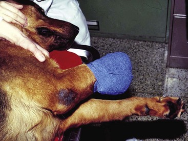
Modified Robert Jones Bandage
 to 2 cm.35
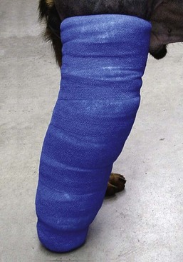
Reinforced Robert Jones Bandage
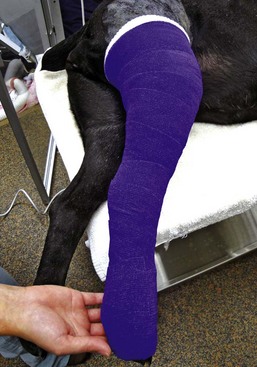
Spica Splint
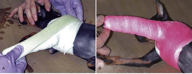
A, Cast padding and stretch gauze are applied to the limb and around the thorax. Fiberglass casting material is used to construct a splint that extends from the toes to beyond the dorsal midline. B, Self-adherent stretch tape is applied to secure the splint in place. (Courtesy Dr. Sean Aiken.)
Schroeder-Thomas Splint
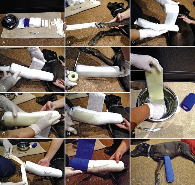
Casts
Application
Cast Removal
Complications and Postapplication Care
The Full-Leg Cast
Half-Cast
Walking Bar
Bivalved Cast
![]()
Stay updated, free articles. Join our Telegram channel

Full access? Get Clinical Tree


Orthopedic Coaptation Devices and Small-Animal Prosthetics
Only gold members can continue reading. Log In or Register to continue
