Optic Nerve

Chapter 12 Optic Nerve


INTRODUCTION


The optic nerve enters the eye through the lamina cribrosa of the sclera. In cats the normal optic nerve loses its myelination as it enters the eye. In dogs the nerve retains some degree of myelination as it passes through the sclera and loses the myelin as it radiates into the nerve fiber layer of the retina. The shape of the optic nerve in cats is normally fairly circular, whereas in dogs the extent of myelination determines the shape—which may be quite irregular, especially in some breeds.


Lesions of the nerve may be developmental or acquired. The most common developmental conditions are optic nerve hypoplasia (aplasia is rare), in which the nerve leaving the eye has reduced or absent nerve fibers and retinal ganglion cells (resulting most often in blindness), and coloboma, in which a defect of fusion of the developing optic cup results in a deep fissure or hole in the nerve head (which may be abnormally large) and occasionally adjacent sclera, choroid, and retina. The extent of visual impairment depends on the size of the defect. Colobomas are most frequently seen in dogs with collie eye anomaly.


Acquired lesions of the optic nerve include papilledema or optic disc edema, optic neuritis, and optic atrophy. These conditions can be divided by clinical appearance into those in which the disc appears swollen (papilledema, disc edema, and neuritis) and optic atrophy, in which the disc loses its myelination and vascularity and develops a dark gray color. The various entities in which the disc is swollen may be difficult to differentiate clinically.


Optic disc edema is swelling of the optic nerve head and may be associated with conditions such as acute glaucoma, uveitis, and very low intraocular pressure after surgery or optic nerve tumors. The disc appears fluffier and raised, with indistinct edges and with edema that radiates into the peripapillary retina. Vision may or may not be affected, depending on the cause.


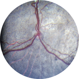
Papilledema implies bilateral edema of the optic nerve head seen in the funduscopic view. It develops from stasis of axoplasmic flow, often resulting from elevated cerebrospinal fluid pressure associated with central nervous system (CNS) tumors. The eyes retain sight unless central blindness occurs because of the lesion in the brain.


Optic neuritis is inflammation of the optic nerve that may be associated with infectious diseases (e.g., distemper or cryptococcosis) or may be idiopathic and presumedly an autoimmune disease. The optic nerve head and disc appear edematous and raised with irregular indistinct borders and edema that radiates into the retina. Hemorrhage may be present in the nerve and peripapillary retina or preretinal vitreous, and vitreal exudate may partially obscure the nerve. Optic neuritis results in loss of vision in the affected eye.


Optic atrophy most commonly develops secondarily to optic neuritis, glaucoma, and retinal degenerations.


< div class='tao-gold-member'>

Stay updated, free articles. Join our Telegram channel

Jul 31, 2016 | Posted by in INTERNAL MEDICINE | Comments Off on Optic Nerve

Full access? Get Clinical Tree

Get Clinical Tree app for offline access